 Fibrous scaffolds for tissue engineering: from conceptualization to implementation
Abstract
To reduce reliance on transplants in cases of disease or trauma caused by accidents, biotechnologies in regenerative medicine and tissue engineering have emerged. The primary goal of tissue engineering is to fabricate devices that mimic the extracellular matrix of injured tissues, thereby facilitating their repair. The synthetic polymer poly(ε-caprolactone) (PCL) is recognized for its favorable mechanical properties, including load-bearing capacity, biocompatibility, and controllable biodegradability. Gelatin, derived from collagen, can replicate the natural extracellular matrix and is rich in amino acids that promote cell adhesion, proliferation, and differentiation, all of which contribute to tissue repair. This study aims to review the fabrication of fibrous scaffolds for tissue engineering, covering process from biomaterial conception to production and application. Conceptual challenges are discussed using gelatin as an example of a natural polymer and PCL as an example of a synthetic polymer.
Keywords:
biotechnology; gelatin; poly (ε-caprolactone); regenerative medicine; spinning techniques
1. Introduction
According to the Ministry of Health, Brazil is currently the second largest transplant-performing nation in the world, after the United States, and has the largest global public organ, tissue and cell transplant program. However, more than 60,000 people nationwide are presently on the transplant waiting list, which includes 406 for hearts, 2,278 for livers, 171 for lungs, and 38,258 for kidneys, among others. To reduce dependence on transplants, biotechnologies in tissue engineering and regenerative medicine have emerged. These technologies focus on repairing and maintaining the patient’s own tissue rather than replacing it[1].
Two primary clinical procedures are used for the functional restoration of damaged tissue: transplants and implants[2]. In the first scenario, tissues or organs are provided by donors (living persons, cadavers, or even animals). Transplants are frequently associated with infections and rejection, a fact that requires the use of immunosuppressive drugs that, in turn, increase the risk of recurrent infections by microorganisms. Ethical and religious concerns present additional barriers related to transplants[2,3]. The second procedure involves using implants for tissue repair. This approach offers significant advantages over transplants[4-7].
To provide lower-risk treatments to the patient, tissue engineering emerged with the purpose of solving some of the main problems related to tissue and organ transplantation. The term was officially introduced in 1988 by the United States National Science Foundation using the following definition: Application of the principles and methods of engineering and the life sciences toward the fundamental understanding of structure/function relationships in normal and pathological mammalian tissues and the development of biological substitutes to restore, maintain, or improve tissue functions[8].
As the name implies, tissue engineering is an interdisciplinary field that combines life sciences and biotechnology with engineering principles, seeking safer and more efficient regenerative medicine solutions for the treatment of tissue injuries that are beyond the body’s natural repair capacity. Various tissue engineering techniques, combined with numerous types of biomaterials, are available. Both must be selected according to the physicochemical and biological characteristics of the tissue to be treated, as these properties can vary substantially[5,9,10].
When developing scaffolds for tissue engineering, the goal is to repair damaged tissue by implanting a device that either replaces it or facilitates its regeneration. To achieve this, scaffold fabrication typically proposes to mimic the extracellular matrix (ECM) of the tissue to be treated. Although the ECM varies in composition and morphology, its structure contains a complex mesh of fibers that support local cells[11]. Fibrous biomaterials are therefore important and have gained prominence in tissue engineering because they not only morphologically resemble the ECM but can also be produced using various available methodologies. Moreover, depending on the fabrication method and biomaterial used, it is possible to control fiber organization, including fiber orientation and distribution patterns, thickness, and scaffold pore size[10]. Consequently, this study aims to review fibrous scaffolds in tissue engineering, covering the development of the biomaterial from its conception through production to application. Conceptual challenges are discussed using gelatin, derived from collagen, as an example of a natural polymer, and poly(ε-caprolactone) (PCL) as an example of a synthetic polymer.
2. Tissue Engineering
Tissue engineering relies on the integration of three fundamental components: scaffolds, cells, and signaling molecules[7]. Additionally, cell culture conditions (whether static or dynamic) must also be taken into consideration[12]. Thus, porous scaffolds are designed to mimic damaged tissue, more specifically the ECM, providing a suitable environment for tissue regeneration. The properties of the scaffold are critical to the success of the device: these matrices must ensure a suitable environment for cell adhesion, differentiation (when stem cells are used), and proliferation. To achieve this, the material must be biocompatible and have appropriate architecture with interconnected pores that allow cell incorporation and the flow of fluids for nutrient delivery and waste removal[13,14].
Figure 1 provides an overview of the steps involved in tissue engineering. The cells used can be autologous or allogeneic, differentiated (e.g., fibroblasts, chondrocytes, or keratinocytes) or stem cells, and originate from different sources[1]. Regardless of the source, cells can be cultured in vitro, either as isolated cells or in cultures, and then inoculated onto a matrix (scaffold) for subsequent administration to the patient or for in vivo implantation at the affected site. Cell adhesion to materials can be enhanced by incorporating signaling molecules, such as growth and differentiation factors. Additionally, bioreactors can be utilized to optimize these conditions. Once properly established, the cells begin to perform their physiological functions by secreting ECM compounds, thereby creating functional tissue. This system is then implanted at the site of injury so that it can eventually be replaced by the patient’s own regenerated tissue[1,8,15].

Overview of the steps of tissue engineering. I. Cells collected from the source. II. Inoculation of cells onto the scaffold together with signaling molecules. III. Use of a bioreactor. IV. Implantation into the patient.
2.1 Extracellular matrix
Tissue engineering is based on the concept that an artificially synthesized matrix can replace and mimic the ECM and also perform its primary functions, thereby ensuring the integrity and regeneration of the affected tissue[11]. Therefore, to develop biomaterials capable of performing this function, it is essential to understand ECM and its interactions with cells. The ECM is a natural scaffold, filling the intercellular space. It is composed of molecules secreted by local cells, mainly proteins and polysaccharides, which vary depending on the tissue type. The ECM is a significant element of connective tissues, where its components are mainly synthesized and secreted by fibroblasts[11,16,17].
The ECM participates in diverse events such as morphogenesis and tissue homeostasis by regulating cell physiology, as well as in cell adhesion, growth, differentiation, migration, and apoptosis, all of which are mediated by cell signaling pathways[11]. Furthermore, as previously mentioned, the composition of the ECM varies according to the type of tissue, with distinct physical and chemical characteristics that result in different functions. For example, the ECM provides interconnection, support, and nutrition in loose connective tissues; mechanical support, lubrication, and elasticity in cartilage; hardness and mechanical strength in bone, and transparency and refraction in the cornea[16].
Cellular receptors are essential for the transmission of information between cells, tissues, and organs. Among their diverse functions, these receptors capture extracellular signals and convert them into a cascade of reactions that elicit a cellular response. The receptors (generally integrins) recognize ECM molecules and enable interactions between this matrix and cells. In addition to integrins, other proteins are involved in ECM recognition, such as the arginine-glycine-aspartic acid (RGD) amino acid sequence, fibrinogen, collagen, and fibronectin; these molecules can be incorporated into scaffolds designed to replace the ECM[11,16].
2.2 Biomaterials and biocompatibility: history and definitions
In 1987, the European Society for Biomaterials defined a biomaterial as a non-biological material used in medical devices with the specific purpose of interacting with a biological system. However, the concept of biomaterials has been updated over the years. Currently, biomaterials are defined as materials that actively interact with a biological system in order to evaluate, treat, or replace a tissue or the function of an organ[18-20]. The evolution of biomaterials is closely associated with the functions they have served over time. Between the 1960s and 1970s, the concept of biomaterials focused on developing inert devices primarily intended to provide mechanical support for hard tissues. The second generation (1980-1990) introduced a new perspective of biomaterials, utilizing chemical interactions between tissues and bioactive materials. Later, to solve rejection issues caused by bioactive materials, bioresorbable biomaterials were developed. These materials can be degraded and eliminated by the body’s natural physiological processes, excluding the need for surgical removal of implants. Finally, the current generation of biomaterials, also referred as “smart” materials (which respond to external stimuli such as temperature and pH) and “biomimetic” materials (inspired by natural structures and capable of simulating tissues like bone and cartilage), is designed to mimic natural organization and functionality, promoting repair and regeneration of damaged tissue[19].
Biocompatibility is one of the most relevant criteria of the acceptance of a biomaterial. The definition of biocompatibility has undergone several changes over time, and there is still no universal consensus on its precise meaning. The most widely accepted definition describes biocompatibility as the ability of a material to perform with an appropriate host response in a specific application. However, various authors have proposed updates to this interpretation, such as emphasizing that the material must interact with living systems without posing risks to the host’s health[21,22]. Chen and Thouas[23] define biocompatibility as a factor that can be assessed based on parameters such as cell viability, tissue response, tumor formation, genetic integrity, immune reaction, and blood clotting potential. According to Crawford et al.[20], biocompatibility is the ability of the material to locally activate and guide host proteins and cells towards tissue reconstruction without the formation of a fibrous capsule and to enable vascularization and integration of the damaged tissue.
Numerous factors must be considered when evaluating a material’s biocompatibility: the physical and chemical properties, the duration of exposure to the tissue, the release of residues, and the characteristics of the affected tissue itself[8]. However, Williams[24] suggests that biocompatibility should not be viewed as an inherent property of the material alone but rather of the biomaterial-tissue system, since the same material can elicit different responses depending on the site of application. Generally, all definitions of biocompatibility involve beneficial interaction between the tissue and the material, varying according to the desired performance or function of the biomaterial[18]. Biocompatibility encompasses not only chemical compatibility with cells but also factors such as design (e.g., architecture, topology, electrical and mechanical properties). Also, research indicates that, although chemical composition is important, surface roughness plays a more significant role in influencing cellular responses[18,24-26].
2.3 Cell Adhesion and cellular response
The human body has a complex defense system against foreign substances from the external environment, whether living or not-living. Medications such as immunosuppressants are often used to prevent the rejection of implants or transplants. However, the resulting weakened immune system significantly increases the patient’s susceptibility to diseases and infections. To control these undesirable reactions, tissue engineering seeks to develop scaffolds with physical, chemical, and biological properties like those of the specific tissue. These scaffolds not only help prevent rejection but also promote cell adhesion, differentiation, and proliferation, ensuring the maintenance of the original function of the affected tissue[6,24].
The primary requirement for cells to survive on a scaffold is cell adhesion. This initial stage of interaction between the material and the cell critically influences cell viability, growth, and differentiation. Cells generally adhere to surfaces through specific proteins (e.g., integrins) and those unable to adhere generally undergo cell death. Additionally, cell adhesion plays an important role in cellular communication, regulation, as well as in organ formation, and tissue maintenance[17,27]. Therefore, studying mechanisms of cell adhesion to the scaffold surface requires an understanding of cell adhesion to the ECM during tissue formation[8,28]. The adhesion of cells to a scaffold is directly related to the physicochemical properties and topology of the material.
2.4 Effect of scaffold topology and architecture on cell adhesion
As previously mentioned, each tissue possesses unique characteristics. Scaffold morphology, for example, can influence cell behavior; at the microscale, it affects cell morphology, while at the nanoscale, regulates subcellular recognition mechanisms. Furthermore, the material’s surface plays an important role by influencing the initial sequence of adsorbed proteins, as well as interaction with blood, inflammatory responses, and other vital cellular activities[27]. Among the topological properties, surface roughness and porosity should be highlighted[17,18].
Roughness is a key factor in the success of cell adhesion, with studies indicating that it is even more influential than the cell type or the biomaterial of the scaffold. Surface roughness can be categorized into different scales: macroscopic, microscopic, submicron, and nanometer roughness. Macroscopic roughness (>100 μm) does not significantly affect cell adhesion, as there is sufficient space for cells to spread in the free spaces; however, it can affect the arrangement of cells within a colony (a set of cells that form the tissue). The micro and submicron scales (0.1-100 μm) can affect individual cells and play a more significant role in cell adhesion and growth. Nevertheless, these characteristics are even more susceptible to nanoroughness (1-100 nm) because this scale closely matches that of cell receptors, directly impacting protein adsorption, cell proliferation, and differentiation[17,27].
The architecture of the device also plays a relevant role in biocompatibility. Scaffold morphology can range from spongy to fibrous, as long as it contains interconnected pores that create an adequate environment for cell infiltration, proliferation, and differentiation. These pores should also enable the flow of nutrients, gases, and cellular waste. For temporary and bioresorbable implants, the pores must allow cells to produce ECM replacement as it degrades. Additionally, the pore size and distribution affect macrophage infiltration, neovascularization, ECM remodeling, and the overall healing process[27,29,30].
In principle, a more porous matrix with larger pores may be advantageous for cell migration, distribution, nutrient flow, and neovascularization. However, smaller pores provide a greater specific surface area formed by the internal walls of the scaffold, which also promotes cell adhesion[6,27,29]. Therefore, a balance between these properties is necessary, as each tissue or cell type requires specific pore sizes. It is important to note that all the aforementioned prerequisites depend directly on the material used and the fabrication process, which will be discussed below.
2.5 Effect of physical properties of the scaffold on cell adhesion
To produce an effective scaffold, it is essential to consider its mechanical properties, as the structure must provide characteristics like those of the original tissue, even if only temporarily. The ECM of most soft tissues exhibits nonlinear elasticity, i.e., it stiffens in response to increased tension. This mechanism helps prevent the loss of tissue structural integrity and permanent deformations[31,32]. Nevertheless, developing a scaffold with mechanical properties corresponding to those of the affected tissue is challenging because different tissue types have specific properties, in addition to being subjected to different mechanical stresses. For example, cardiac tissue requires a material with high resilience, resistance, and durability, as well as electrical conductivity to facilitate the propagation of electrical impulses. In contrast, bone tissue demands a material with high rigidity and resistance, particularly to withstand compression, traction, and flexural forces[29,33].
The stiffness of the ECM can vary widely, ranging from approximately 0.1 kPa (brain) to 100 GPa (bone) and is primarily determined by its composition of collagen and elastin. Matrix stiffness is not only structurally important but also directly influences cellular activities such as adhesion, differentiation, cytoskeletal remodeling, and intercellular interactions. Generally, the looser the bonds and the lower the ECM stiffness, the less mechanical feedback is required to recruit integrin complexes for cell signaling and adhesion; conversely, this process tends to increase with greater matrix stiffness[17,34]. Additionally, scaffolds must possess sufficient pores and interconnectivity to facilitate cell migration, adhesion, proliferation, and flow of nutrients and waste. Therefore, an optimal pore number, distribution, and size are essential without compromising the scaffold’s mechanical integrity. Finally, the mechanical properties of the scaffold should generally last until tissue regeneration period, which varies depending on the tissue type, as well as the patient’s age and other conditions[6,29].
The first parameter to consider when analyzing cell adhesion to a scaffold is the wettability of the material, which determines its hydrophilicity (Figure 2). Although hydrophilic materials generally promote cell adhesion, proteins tend to adsorb more readily onto hydrophobic surfaces; therefore, a balance between these properties is essential. Like superhydrophobic materials (θ > 150°), superhydrophilic materials (θ < 5°) are also unable to support cell adhesion and growth[11,17]. Physical adsorption is mediated by weak intermolecular forces, such as Van der Waals and electrostatic interactions, and occurs more rapidly when there is charge difference between cell membrane molecules and the material surface[8,11].

Schematic representation of wettability. The higher the wettability index, the smaller the water contact angle and the more hydrophilic the material.
The water layer formed on the surface of the biomaterial mediates its hydroxylation (the formation of −OH groups) through the dissociation of water molecules upon contact with a hydrophilic surface. This process enables the biomaterial to interact with proteins present in body fluids. First, small proteins (e.g., albumin) reach and adsorb onto the biomaterial. Subsequently, larger proteins with a higher affinity for the molecular groups on the scaffold surface force desorption of smaller proteins (Vroman effect) and can influence cell adhesion. This competition among proteins for surface sites is dynamic and complex, depending on various factors such as protein concentration in the fluid[11]. Additional important factors influence protein adsorption, including temperature, pH, ionic strength, and electrostatic interactions[27].
2.6 Effect of chemical properties of the scaffold on cell adhesion
The chemical properties of a biomaterial play a fundamental role in cell adhesion. One of the most important properties is surface energy (also called interfacial free energy or surface free energy), which relates to the number of ruptured intermolecular bonds present on the surface of a solid. The higher the surface free energy, the more easily cells adhere and spread; in other words, the greater the wettability of the biomaterial. However, it should be noted that certain proteins preferentially adsorb onto surfaces with lower free energy[17]. In addition to surface energy, we highlight the importance of the surface charge (determined mainly by functional chemical groups), as it can also affect cell adhesion. Generally, cells adhere more readily to positively charged surfaces[35]. However, research suggests that negative charges can enhance protein adsorption, thereby promoting cell adhesion. It is important to note that, as with other properties, the type of cell influences adhesion success in relation to chemical properties, and no universal model applies to all cell types[17].
2.7 Types of biomaterials for tissue engineering
Biomaterials can be classified into three groups: bioinert, bioactive, and bioresorbable. The primary characteristic of bioinert materials is the minimal interaction at the interface between the material and biological tissue. Generally, the tissues exhibit a minimal response, which often form a fibrous capsule around the interface. Bioactive materials can significantly interact with tissue through chemical bonding. Finally, bioresorbable materials do not require surgical removal because since they degrade over time, their residues are naturally absorbed and eliminated by the body. Generally, bioinert materials are metals or ceramics, while bioresorbable materials are commonly polymers[13,36,37].
The most widely used classes of biomaterials in tissue engineering are ceramics and polymers. Ceramics are commonly employed in applications involving hard tissues due to their equivalent mechanical properties. Hydroxyapatite and tricalcium phosphate are examples of ceramics used for bone tissue repair; they exhibit bioactivity that stimulates the differentiation and proliferation of osteoblasts[6,38]. Polymeric materials, on the other hand, are more versatile and have broader applications.
Polymeric materials can be classified into two categories: synthetic and natural. Synthetic polymers are laboratory-synthesized materials typically derived from non-renewable sources. Their primary advantages include ease fabrication and greater versatility, as they can be custom-made and produced on a large scale. Additionally, their production costs are generally lower. These materials exhibit more standardized characteristics, and their manufacturing processes can be more easily manipulated to improve their properties. However, synthetic polymers may exhibit cytotoxicity-related problems, increasing the risk of rejection[39,40].
Natural polymers, in contrast, often exhibit greater similarity to the ECM because they are derived from biological sources, such as proteins or polysaccharides. Consequently, their biocompatibility is generally higher than that of synthetic polymers. However, natural polymers can be more difficult to manipulate and less homogeneous, i.e., their properties and composition vary depending on the source material. Natural polymers typically have inferior mechanical properties compared to synthetic polymers[39,41,42].
Despite their disadvantages, natural polymers such as collagen and gelatin present promising solutions for tissue engineering. Collagen is a high-molecular-weight protein and the most abundant biopolymer in mammals, particularly types I and II. This protein is found in the ECM of both hard and soft connective tissues, where it provides structural stability[6,39,41]. Collagen is converted into gelatin through partial hydrolysis, which results in the loss of its tertiary and quaternary structures. Since gelatin is derived from collagen, it shares many similar properties[29,43,44]. For these reasons, collagen and gelatin hold significant potential as biomaterials and could be widely utilized in biomedical and pharmaceutical applications. These biopolymers can be obtained from a variety of animal tissues, including those of cattle, pigs, and birds. However, from a commercial perspective, collagen is primarily obtained from the skin and tendons of cattle and pigs[45,46].
3. Collagen
Collagen is produced by various cell types, including chondroblasts, osteoblasts, epithelial cells and, mainly, fibroblasts. It is an abundant protein present in the ECM that directly promotes cell anchorage and new tissue formation or indirectly influences these processes through collagen receptors such as fibronectin and laminin found on cells[47]. Like all proteins, collagen is composed of amino acids linked by covalent (peptide) bonds, which organize into a helical α-helix structure consisting of three molecular chains (tropocollagen) stabilized by secondary (weaker) bonds. These α-chains aggregate to form collagen fibers[39,48].
The primary structure of collagen consists of approximately 1,000 amino acids that form either identical or non-identical polypeptide chains. The most common repeating sequence in the collagen molecule is glycine-X-Y, where X and Y are predominantly proline and hydroxyproline[43,49]. Glycine is a small amino acid, typically located in the inner part of the helix. The composition and distribution of glycine are therefore responsible for the protein’s native state. Proline and hydroxyproline have rigid side rings, which result in steric hindrances that help maintain structural stability. Intramolecular hydrogen bonds also contribute to the stability of the secondary structure, while crosslinks are essential for preserving the fibrous structure[39,48,50].
The helical conformation of collagen fibers, determined by the physicochemical properties of their constituent amino acids, ensures the tissue’s strength. Collagen molecules within the fibers are stabilized by various inter- and intramolecular forces, such as hydrogen bonds between NH and C=O groups oriented perpendicular to the fiber axis. These bonds promote strong interactions between neighboring molecules; thus, when a force is applied, it is distributed across the fiber to adjacent collagen molecules. Furthermore, the fibers present in tissues can naturally form crosslinks, ensuring high stability and tensile strength. There is a wide variety of collagen types, including type I (the most common), abundant in skin, tendons, and bones; type II, found in cartilage; and type III, present in skin and blood vessels[39,48,50].
Although collagen is a stable molecule that is generally insoluble in water, like other proteins, it can undergo denaturation and may revert to its primary structure under certain conditions. Temperature-induced denaturation also depends on factors such as water content, pH of the medium, and the density of crosslinks. However, once the denaturation temperature is reached, only the weak intramolecular bonds are broken, while the covalent intermolecular bonds remain intact. Consequently, the three chains that form the α-helix separate and disperse into the aqueous medium, resulting in a colloidal system. When the temperature is lowered again, the collagen chains rearrange, and hydrogen bonds form between water molecules and the collagen, leading to the formation of gelatin[45,48,50].
4. Gelatin
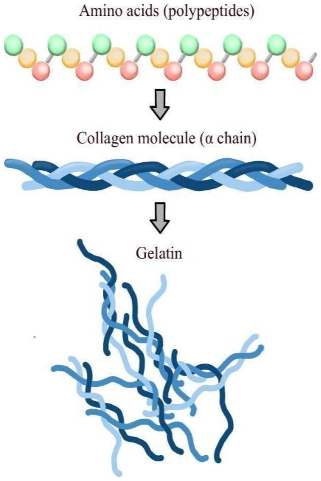
Gelatin is a biopolymer produced by the partial hydrolysis of collagen, primarily involving the loss of its tertiary and quaternary structure, generating a colloidal system (Figure 3). Gelatin is derived from type I collagen-rich tissues of animals such as cattle, pigs, birds, and fish. Its extraction properties vary not only according to the animal species but also with age, tissue of origin, and collagen type. Furthermore, the amino acid distribution differs depending on the animal source; for example, gelatins derived from pigs and cattle lack cysteine residues, while those obtained from fish contain less glycine compared to mammalian gelatins. Gelatin is produced by pretreating collagen in acidic, alkaline, or enzymatic medium; the first two being the most common. When an acidic medium is used, type A gelatin is formed, which has an isoelectric point between 8 and 9, whereas gelatin B with an isoelectric point between 4 and 5, is produced in an alkaline medium. The pretreatment of collagen breaks non-covalent bonds such as hydrogen bonds, hydrophobic interactions, and crosslinks, leading to the disruption of the triple-helix structure. This loosens the chains, facilitating their swelling and the solubilization of collagen, which is necessary for the gelatin extraction. The pretreated collagen is then immersed in a saline or acid solution, followed by filtration of the biopolymer, evaporation, drying, grinding, and sieving until the gelatin becomes powder[29,43,44,48,51].
Schematic illustration of the partial hydrolysis (denaturation) of collagen into gelatin (colloidal system in aqueous medium).
Regarding its biological properties, gelatin, being derived from collagen, retains several notable characteristics relevant to cell-biomaterial interactions, such as biocompatibility, bioresorption, and the ability to mimic the ECM. Consequently, it is a promising material for scaffold fabrication[44,51,52]. Gelatin contains integrin-binding sites that facilitate cell adhesion. Additionally, gelatin exhibits lower antigenicity and immunogenicity compared to collagen. Its production costs are also lower, making gelatin a preferred choice over collagen scaffold manufacturing. Other advantageous properties include its natural abundance, biodegradability, and the aforementioned biocompatibility[53-59].
Unlike collagen, gelatin dissolves spontaneously in aqueous solutions and can undergo a sol-gel transition depending on its type, concentration, and temperature. This process is reversible and involves the transition between a random colloidal system and the partial restoration of collagen triple helices. However, this sol-gel transition can pose challenges for certain biological applications because gelatin in the gel phase loses its structure at body temperature, reverting to the fluid state (sol phase). To maintain gelatin in the gel phase, crosslinking (the formation of crosslinks) is necessary. Crosslinks enhance the material’s mechanical properties, making it insoluble in water and stable under biological conditions. Crosslinking can be achieved through three main methods: chemical, physical, and enzymatic[44,51,52].
Physical crosslinking is a technique that employs irradiation, plasma, or dehydrothermal treatment. A specific physical stimulus induces the separation of polymer chains, generating free radicals that bind to each other, resulting in crosslinks. The primary advantage of this method is its lower cytotoxicity, as it does not produce potentially toxic compounds within the chains and eliminates the need for solvents. Additionally, irradiation can simultaneously sterilize the material while promoting crosslinking. However, these physical methods are less efficient at forming crosslinks and yield polymers with inferior mechanical properties compared to those produced by chemical methods. Enzymatic crosslinking utilizes transglutaminase; an enzyme found in certain plants and animals. This enzyme facilitates crosslinking through an acyltransferase reaction between the glutamine residue of one chain and the amine group of another[44,51,52].
Chemical crosslinking, currently the most widely used method, offers a broader range of options compared to other techniques. This process involves forming covalent bonds between polymer chains, resulting in more stable structures with better-controlled physicochemical properties than those achieved by physical crosslinking. Chemical crosslinking agents are classified into two categories: zero-length and non-zero-length crosslinkers. Zero-length crosslinking creates direct bonds between peptide chains, with the reagent being completely removed after the reaction; thus, the crosslinker acts as a catalyst and is not incorporated into the final gelatin structure. The advantage of this approach is the preservation of the gelatin structure, ensuring good biocompatibility and high conversion efficiency. In contrast, non-zero-length method forms crosslinks by incorporating the crosslinker molecules into the final gelatin structure[51]. This technique generally provides good crosslinking efficiency, which correlates with enhanced hydrogel stability, but it may also cause cytotoxicity-related issues. Notable examples of method include the use of glutaraldehyde (GA), as well as the combination of N-ethyl-N’-(3-dimethylaminopropyl)carbodiimide with N-hydroxysuccinimide (EDC/NHS)[44,52].
Although GA is an effective crosslinking agent for fibrous scaffolds derived from natural proteins, its use presents significant challenges due to its inherent cytotoxicity. If GA residues are not completely neutralized or removed, they can compromise cell viability in vitro and induce harmful inflammatory responses in vivo. Consequently, tissue engineering research has increasingly focused on less toxic alternatives, which can be broadly categorized as chemical or physical methods. Chemical alternatives include the EDC/NHS system (a zero-length crosslinker that does not incorporate into the final bond), PEGDE (Poly(ethylene glycol) diglycidyl ether, known for its high biocompatibility), and genipin (a naturally derived crosslinker). Physical methods include Dehydrothermal Treatment (DHT) and controlled application of UV/Gamma radiation, thereby ensuring the necessary safety and biocompatibility for the clinical application of these scaffolds[51,52].
5. Poly(ɛ-caprolactone)
Poly(ɛ-caprolactone) (PCL) is an aliphatic, bioresorbable, linear synthetic polyester distinguished by its ability to be molded into various shapes, setting it apart from other biomaterials used in scaffold development. It exhibits excellent thermal stability, and it is susceptible to surface modifications. Its physicochemical, mechanical, and biocompatibility properties can be significantly altered, while its hydrophobicity and degradation behavior are influenced by surface and structural modifications of the scaffolds[60]. PCL has a glass transition temperature of -60 °C, a melting point ranging from 59 to 64 °C, and a hydrophobic, and semicrystalline nature that tends to decrease with increasing molecular weight[61]. This polymer can be synthesized via ring-opening polymerization of the lactone or by condensation of 6-hydroxyhexanoic acid through catalyzed reactions under appropriate conditions[62].
PCL is easy to process and characterize, also being soluble in various organic solvents. Additionally, it exhibits properties such as high toughness, biocompatibility, and bioresorbability. The versatility and safety of this polymer enable its use in controlled drug release applications, blend scaffold fabrication, bone regeneration, and vascular grafting. Consequently, PCL has been approved by the United States Food and Drug Administration (FDA) for clinical and therapeutic use[61]. When in contact with body fluids, PCL degrades on its surface through non-enzymatic hydrolytic cleavage of the ester groups in its structure. This reaction produces lower molecular weight fragments by diffusion of oligomers from the polymer matrix (which does not imply a loss of molecular mass), generating soluble and non-toxic oligomers. Upon exposure to the body’s metabolism, the carboxylic acids released during hydrolysis are further degraded and converted into CO2 and H2O, which are eliminated through the body’s natural mechanisms. As a result of these reactions, the polymer’s molecular weight and crystallinity decrease[62,63].
The kinetics of the polymer-organism interaction during bioresorption promote cell proliferation and the secretion of the ECM, which occupies the space previously filled by the polymer. Thus, tissue repair occurs gradually as the material degrades, which is the desired outcome in clinical practice. However, the ability of PCL to stimulate cell adhesion and proliferation is limited, making its combination with other polymers particularly relevant[61].
6. Fiber Fabrication Techniques
Given the need for a biocompatible environment that supports the establishment of cells and tissues, polymers are particularly attractive for scaffold design due to their versatility and broad applicability in soft tissues, along with a variety of fabrication techniques. These techniques enable the development of structures that closely mimic the original tissue; for example, fibrous polymeric structures exhibit a high morphological similarity to the ECM[10,64,65]. In this context, both the choice of polymer and the fabrication method can be optimized by considering the relationship between the specific physicochemical properties of each material and the characteristics of the processing techniques used.
Since PCL is a polymer with a semicrystalline structure, high viscosity, and heat resistance, it can be easily manipulated and spun into continuous, uniform fibers using rotary jet spinning. This technique is particularly effective for producing micrometer-scale fibers with superior mechanical properties, making them suitable for applications requiring strong structures such as bone regeneration. On the other hand, gelatin is a natural protein with low thermal stability that tends to form low-viscosity solutions, characteristics that make it more suitable for electrospinning. This process employs a strong electric field to stretch a polymer solution into ultrafine fibers. Due to its ability to create nanoscale fiber networks, electrospinning is ideal for materials such as gelatin, which benefit from highly porous structures with a large surface area. These structures are especially advantageous in tissue engineering applications, where promoting cell adhesion and proliferation, along with nutrient diffusion, is critical[66].
As previously mentioned, since gelatin is derived from collagen, it possesses biologically advantageous properties as a biomaterial. Gelatin is used in bulk form, as fibers, and as a carrier for drugs and cells, in addition to several other applications across various tissues such as bone, skeletal muscle, and neural tissues[67-69]. Furthermore, different scaffold fabrication techniques have been well described in the literature, including electrospinning, rotary jet spinning, and solution blow spinning.
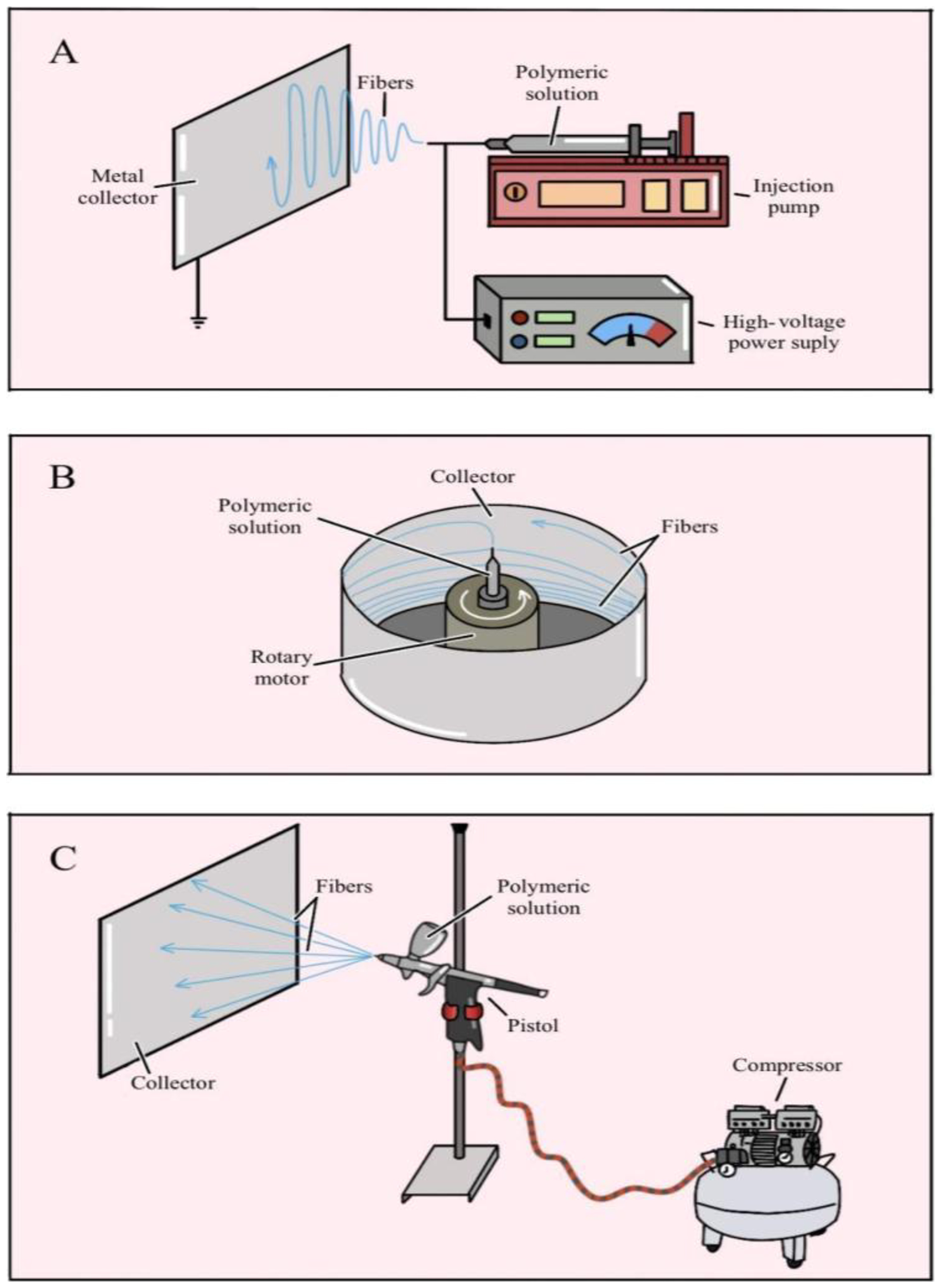
Electrospinning is one of the most widely used techniques, employing a high-voltage power supply to generate an electric field that facilitates the extrusion of material through a Taylor cone[70,71]. The process involves charging the polymer using a potential difference and consists of three main components: a high-voltage power supply, a syringe with a capillary, and a grounded collector (typically a metal plate or a rotating mandrel). This setup allows the material to be accelerated toward the oppositely charged collector, with the final fiber thickness controlled by the deposition time. Electrospinning enables the production of fibers with various interconnected porous structures, facilitating drug incorporation, enhanced mechanical properties, and improved chemical stability[71]. Although the technique is highly versatile and widely used, it depends on the solution’s conductivity and the application of a high-voltage electric field, and its use is limited by low yield[10,71,72]. Electrospinning has been used to fabricate PCL-gelatin nanofibers combined with bone marrow-derived mesenchymal stem cells (BMSCs). Studies show that this scaffold, when applied to skin wounds, enhances cell adhesion and proliferation in vitro. In vivo, the material promotes improved wound contraction and accelerates re-epithelialization[73]. Furthermore, when applied to bone regeneration, the PCL-gelatin mesh serves as a robust three-dimensional niche capable of modulating and optimizing the therapeutic performance of BMSCs, thereby promoting osteogenesis[74].
Rotary jet spinning enables the production of anisotropic fiber matrices by extruding material through centrifugal forces via capillaries on the lateral surface of a rotating reservoir. This technique is efficient, low-cost, and insensitive to the dielectric constant of the materials. It does not require high-voltage electric fields and demonstrates high reproducibility[70,71]. The rotary jet spinning system consists of a reservoir that retains the solution flow, projecting it against the collector wall along a curvilinear trajectory (due to rotational inertia). The final characteristics of the fibers are closely linked to the fabrication process. Factors such as the choice of solvent, solution concentration, surface tension, orifice diameter and geometry, and rotation speed (which varies depending on the material) can control the porosity and diameter of the polymeric fiber. Additionally, the location where the fibers are collected within the device is an important factor[10,70,71,75,76].
Solution blow spinning is an alternative technique that combines electrospinning and melt blowing. It involves the controlled spraying of a polymer jet, which is accelerated by a flow of compressed gas. The resulting fibers are then deposited onto a collector. This method has been adapted for applications such as drug delivery; however, the reproducibility of the formed fibers still requires further evaluation[10,77]. The airbrush is commonly used in solution blow spinning; it features a concentric nozzle that extrudes the polymer jet with the assistance of compressed gas. The injection rate can be regulated using an injection pump, while the pressure is controlled via an air compressor pressure regulator[78,79]. Although blow spinning is a good alternative to techniques such as electrospinning, it requires more sophisticated equipment, such as an injection pump. This technique employs a pistol fed with a polymer solution, which is extruded through a concentric nozzle using a stream of compressed gas (Figure 4). When the pistol is triggered, the system opens to supply both the polymer solution and compressed gas, forming a jet of polymer solution. The high-velocity gas flow generates shear forces at the gas/solution interface, deforming the polymer solution from a droplet into a conical shape[80,81]. The solvent in the solution evaporates along the jet path from the nozzle to the collector, resulting in fiber formation depending on the material and its viscosity. This technique can produce micro-, and nanofiber meshes with diverse characteristics, including variations in fiber diameter, morphology, alignment, and porosity[10,82]. It is also worth noting that the process uses compressed air at room temperature, preventing thermal degradation of the polymer[81]. Several parameters must be considered when airbrushing is employed: (a) solution properties: viscosity, concentration, surface tension, solvent nature; (b) processing conditions: pressure of the compressed gas and distance from the pistol nozzle to the collector (screen); (d) system characteristics: nozzle diameter and type of collector; and (d) environment factors: temperature, pressure, and humidity.

Schematic illustration of spinning techniques. (A) electrospinning; (B) rotary jet spinning; (C) airbrushing.
A comparison of the mentioned fiber formation methods, covering fiber diameter ranges, yield (g/relative smallest), solvents, polymer examples, advantages, limitations, and target applications, can be seen in Table 1.
Among the parameters listed above, the concentration of the solution is one of the key features for fiber production, as it directly influences the material’s viscosity. The distance from the nozzle to the collector screen is also crucial for fiber formation. Generally, polymers with low intermolecular chain interactions lack sufficient fiber-forming capacity; they tend to appear more diluted, often forming droplets that deposit as a film on the collector and may promote bead formation on the fibers. However, this can also arise if the solvent fails to evaporate upon extrusion, causing the material to reach the collector screen in a fluid state. Successful fiber production requires higher material concentration (and consequently greater viscosity) to ensure polymer chain cohesion without interruption[82]. Conversely, excessively viscous materials can hinder processing by clogging the nozzle. Therefore, it is essential to determine the optimal concentration and viscosity of the material when using airbrushing.
7. Barriers to the Clinical Translation of Tissue-Engineered Constructs
The successful translation of tissue-engineered constructs into clinical practice represents a significant challenge in regenerative medicine. As emphasized in analyses of personalized scaffolds and perspectives on clinical translation, and consistent with the identified barriers to translation and commercialization, transitioning from a laboratory concept to a commercial product requires overcoming critical, interconnected regulatory and technical obstacles.
The regulatory complexity and challenges of clinical translation present significant issues, with the regulatory system serving as the primary and most crucial barrier. In the United States, the Food and Drug Administration (FDA) classifies tissue-engineered medical products (TEMPs) as devices, biologics, drugs, or combination products. This classification determines the approval pathway, requiring comprehensive preclinical testing to demonstrate both safety and efficacy, along with stringent ethical considerations regarding cell sources, donor consent, and the conduct of clinical trials[83,84].
Scalability, reproducibility, and adherence to manufacturing standards are essential. Transitioning to large-scale production requires implementing a rigorous quality management system, primarily governed by Good Manufacturing Practices (GMP), which is a complex challenge for products developed from laboratory-scale processes. Similarly, maintaining manufacturing reproducibility is crucial, as consistent replication and assurance of batch quality are vital. In the context of personalized bioengineered implants, which utilize techniques such as 3D biofabrication, the challenge lies in balancing anatomical and functional customization (essential for clinical success in applications like craniofacial reconstruction) with process standardization. Any variability in scaffold composition, mechanical properties, or fibrous architecture can affect host integration and long-term performance[83,85].
Furthermore, scalability in industrial production requires the development of standardized workflows for all stages, from the rational selection of biomaterials to final fabrication. This approach eliminates the artisanal nature of bench research, enabling the volume and cost-effectiveness necessary for commercial viability. Additionally, critical technical challenges must be addressed, including sterilization and biocompatibility (two essential factors for clinical acceptance of the product). Many biomaterials, particularly polymers sensitive to temperature or radiation and constructs incorporating growth factors or cells are vulnerable to conventional sterilization methods. The challenge lies in developing and validating sterilization protocols that effectively eliminate microorganisms without compromising the material’s structural integrity, mechanical properties, or bioactivity. Moreover, demonstrating biological safety is mandatory and must comply with international standards, such as the ISO 10993 series. Testing must confirm the absence of cytotoxicity and genotoxicity, ensure an appropriate inflammatory response, and verify predictable scaffold degradation within the physiological environment[83,86].
In summary, clinical translation requires bridging biological innovation with engineering precision. Success depends on the ability to integrate an optimal scaffold design with reproducible, sterilizable, and scalable manufacturing processes, all within a stringent regulatory framework that ensures long-term safety and efficacy.
8. Conclusions
A series of technical and scientific challenges must be addressed to ensure the viability of scaffold production and its application in tissue engineering. Controlling and standardizing operational parameters are essential for producing fibrous scaffolds from polymeric materials with properties suitable for biotechnological applications. The optimal scaffold design also depends on the characteristics of the target tissue, including whether the fibers are thin or thick, and whether their orientation is aligned (as in tendons) or random (as in skin). The polymer’s bioactivity and resorption time are critical factors, as is the consideration of tissue characteristics during repair. Additionally, given the high demand, the scalability of scaffold method used for the production methods must be carefully evaluated. There are many challenges to overcome; however, by mimicking the tissue environment, fibrous materials play a key role in biotechnological procedures used in tissue engineering.
10. Acknowledgements
The authors wish to thank the National Council for Scientific and Technological Development (CNPq), process number 404701/2023-0, for financial support.
-
Data Availability:
All data supporting the findings of this study are included in this article and its supplementary materials.
-
How to cite:
Iguma, T. S., Nascimento, V. A., Giorno, L. P., Malmonge, S. M., & Santos Jr., A. R. (2026). Fibrous scaffolds for tissue engineering: from conceptualization to implementation. Polímeros: Ciência e Tecnologia, 36(1), e20260006. https://doi.org/10.1590/0104-1428.20240116
11. References
-
1 Brasil. Ministério da Saúde. (2025). Relatórios de lista de espera. Retrieved in 2025, August 13, from https://www.gov.br/saude/pt-br/composicao/saes/snt/relatorios/lista-de-espera-serie-historica
» https://www.gov.br/saude/pt-br/composicao/saes/snt/relatorios/lista-de-espera-serie-historica -
2 Santos, A. R., Jr. (2010). Bioresorbable polymers for tissue engineering. In D. Erbeli (Ed.), Tissue engineering (pp. 225-246). London: IntechOpen. https://doi.org/10.5772/8580
» https://doi.org/10.5772/8580 -
3 Silva, A., Arora, S., Dhanani, S., Hornby, L., Luctkar-Flude, M., Ross-White, A., Lotherington, K., Rochon, A., Wilson, L., Latifi, M., Giorno, L., & Silva e Silva, V. (2022). Quality improvement tools to manage deceased organ donation processes: a scoping review protocol. Nurse Education in Practice, 61, 103322. https://doi.org/10.1016/j.nepr.2022.103322 PMid:35306317.
» https://doi.org/10.1016/j.nepr.2022.103322 -
4 Sanjanwala, D., Londhe, V., Trivedi, R., Bonde, S., Sawarkar, S., Kale, V., & Patravale, V. (2024). Polysaccharide-based hydrogels for medical devices, implants and tissue engineering: a review. International Journal of Biological Macromolecules, 256(Pt 2), 128488. https://doi.org/10.1016/j.ijbiomac.2023.128488 PMid:38043653.
» https://doi.org/10.1016/j.ijbiomac.2023.128488 -
5 Cao, L., Su, H., Si, M., Xu, J., Chang, X., Lv, J., & Zhai, Y. (2021). Tissue engineering in stomatology: a review of potential approaches for oral disease treatments. Frontiers in Bioengineering and Biotechnology, 9, 662418. https://doi.org/10.3389/fbioe.2021.662418 PMid:34820359.
» https://doi.org/10.3389/fbioe.2021.662418 -
6 Marsudi, M. A., Ariski, R. T., Wibowo, A., Cooper, G., Barlian, A., Rachmantyo, R., & Bartolo, P. J. D. S. (2021). Conductive polymeric-based electroactive scaffolds for tissue engineering applications: current progress and challenges from biomaterials and manufacturing perspectives. International Journal of Molecular Sciences, 22(21), 11543. https://doi.org/10.3390/ijms222111543 PMid:34768972.
» https://doi.org/10.3390/ijms222111543 -
7 Baudequin, T., & Tabrizian, M. (2018). Multilineage constructs for scaffold‐based tissue engineering: a review of tissue‐specific challenges. Advanced Healthcare Materials, 7(3), 1700734. https://doi.org/10.1002/adhm.201700734 PMid:29193897.
» https://doi.org/10.1002/adhm.201700734 - 8 Oréfice, R. L., Pereira, M. M., & Mansur, H. S. (2012). Biomateriais: fundamentos e aplicações Rio de Janeiro: Guanabara Koogan.
-
9 Sun, W., Gregory, D. A., Tomeh, M. A., & Zhao, X. (2021). Silk fibroin as a functional biomaterial for tissue engineering. International Journal of Molecular Sciences, 22(3), 1499. https://doi.org/10.3390/ijms22031499 PMid:33540895.
» https://doi.org/10.3390/ijms22031499 -
10 Iguma, T. S., Malmonge, S. M., & Santos, A. R., Jr. (2019). Natural fibrous polymers for tissue engineering. Stem Cell and Regenerative Medicine, 3(1), 1-4. https://doi.org/10.33425/2639-9512.1037
» https://doi.org/10.33425/2639-9512.1037 -
11 Bandzerewicz, A., & Gadomska-Gajadhur, A. (2022). Into the tissues: Extracellular matrix and its artificial substitutes: cell signalling mechanisms. Cells, 11(5), 914. https://doi.org/10.3390/cells11050914 PMid:35269536.
» https://doi.org/10.3390/cells11050914 -
12 Kaasi, A., & Jardini, A. L. (2016). Bioreactors. In S. Hashmi. (Ed.), Reference module in materials science and materials engineering. Amsterdam: Elsevier. https://doi.org/10.1016/B978-0-12-803581-8.04140-0
» https://doi.org/10.1016/B978-0-12-803581-8.04140-0 -
13 Williams, D. F. (2019). Challenges with the development of biomaterials for sustainable tissue engineering. Frontiers in Bioengineering and Biotechnology, 7, 127. https://doi.org/10.3389/fbioe.2019.00127 PMid:31214584.
» https://doi.org/10.3389/fbioe.2019.00127 -
14 Ameer, J. M., Pr, A. K., & Kasoju, N. (2019). Strategies to tune electrospun scaffold porosity for effective cell response in tissue engineering. Journal of Functional Biomaterials, 10(3), 30. https://doi.org/10.3390/jfb10030030 PMid:31324062.
» https://doi.org/10.3390/jfb10030030 -
15 Santos, A. R., Jr., & Zavaglia, C. A. C. (2016). Tissue engineering concepts. In S. Hashmi. (Ed.), Reference module in materials science and materials engineering. Amsterdam: Elsevier. https://doi.org/10.1016/B978-0-12-803581-8.04141-2
» https://doi.org/10.1016/B978-0-12-803581-8.04141-2 -
16 Karamanos, N. K., Theocharis, A. D., Piperigkou, Z., Manou, D., Passi, A., Skandalis, S. S., Vynios, D. H., Orian-Rousseau, V., Ricard-Blum, S., Schmelzer, C. E. H., Duca, L., Durbeej, M., Afratis, N. A., Troeberg, L., Franchi, M., Masola, V., & Onisto, M. (2021). A guide to the composition and functions of the extracellular matrix. The FEBS Journal, 288(24), 6850-6912. https://doi.org/10.1111/febs.15776 PMid:33605520.
» https://doi.org/10.1111/febs.15776 -
17 Cai, S., Wu, C., Yang, W., Liang, W., Yu, H., & Liu, L. (2020). Recent advance in surface modification for regulating cell adhesion and behaviors. Nanotechnology Reviews, 9(1), 971-989. https://doi.org/10.1515/ntrev-2020-0076
» https://doi.org/10.1515/ntrev-2020-0076 -
18 Sánchez-Bodón, J., Diaz-Galbarriatu, M., Pérez-Álvarez, L., Moreno-Benítez, I., & Vilas-Vilela, J. L. (2023). Strategies to enhance biomedical device performance and safety: a comprehensive review. Coatings, 13(12), 1981. https://doi.org/10.3390/coatings13121981
» https://doi.org/10.3390/coatings13121981 -
19 Festas, A. J., Ramos, A., & Davim, J. P. (2020). Medical devices biomaterials: a review. Proceedings of the Institution of Mechanical Engineers. Proceedings Part L, Journal of Materials: Design and Applications, 234(1), 218-228. https://doi.org/10.1177/1464420719882458
» https://doi.org/10.1177/1464420719882458 -
20 Crawford, L., Wyatt, M., Bryers, J., & Ratner, B. (2021). Biocompatibility evolves: phenomenology to toxicology to regeneration. Advanced Healthcare Materials, 10(11), e2002153. https://doi.org/10.1002/adhm.202002153 PMid:33829678.
» https://doi.org/10.1002/adhm.202002153 -
21 Ghasemi-Mobarakeh, L., Kolahreez, D., Ramakrishna, S., & Williams, D. (2019). Key terminology in biomaterials and biocompatibility. Current Opinion in Biomedical Engineering, 10, 45-50. https://doi.org/10.1016/j.cobme.2019.02.004
» https://doi.org/10.1016/j.cobme.2019.02.004 -
22 Williams, D. F. (2009). On the nature of biomaterials. Biomaterials, 30(30), 5897-5909. https://doi.org/10.1016/j.biomaterials.2009.07.027 PMid:19651435.
» https://doi.org/10.1016/j.biomaterials.2009.07.027 -
23 Chen, Q., & Thouas, G. A. (2015). Metallic implant biomaterials. Materials Science and Engineering R Reports, 87, 1-57. https://doi.org/10.1016/j.mser.2014.10.001
» https://doi.org/10.1016/j.mser.2014.10.001 -
24 Williams, D. F. (2017). Biocompatibility pathways: biomaterials-induced sterile inflammation, mechanotransduction, and principles of biocompatibility control. ACS Biomaterials Science & Engineering, 3(1), 2-35. https://doi.org/10.1021/acsbiomaterials.6b00607 PMid:33429689.
» https://doi.org/10.1021/acsbiomaterials.6b00607 -
25 Williams, D. F. (2008). On the mechanisms of biocompatibility. Biomaterials, 29(20), 2941-2953. https://doi.org/10.1016/j.biomaterials.2008.04.023 PMid:18440630.
» https://doi.org/10.1016/j.biomaterials.2008.04.023 -
26 Chen, P.-R., Kang, P.-L., Su, W.-Y., Lin, F.-H., & Chen, M.-H. (2005). The evaluation of thermal properties and in vitro test of carbodiimide or glutaraldehyde cross-linked gelatin for PC 12 cells culture. Biomedical Engineering : Applications, Basis, and Communications, 17(2), 101-107. https://doi.org/10.4015/S1016237205000160
» https://doi.org/10.4015/S1016237205000160 -
27 Luo, J., Walker, M., Xiao, Y., Donnelly, H., Dalby, M. J., & Salmeron-Sanchez, M. (2021). The influence of nanotopography on cell behaviour through interactions with the extracellular matrix: a review. Bioactive Materials, 15, 145-159. https://doi.org/10.1016/j.bioactmat.2021.11.024 PMid:35386337.
» https://doi.org/10.1016/j.bioactmat.2021.11.024 -
28 Kanchanawong, P., & Calderwood, D. A. (2023). Organization, dynamics and mechanoregulation of integrin-mediated cell–ECM adhesions. Nature Reviews. Molecular Cell Biology, 24(2), 142-161. https://doi.org/10.1038/s41580-022-00531-5 PMid:36168065.
» https://doi.org/10.1038/s41580-022-00531-5 -
29 Zhang, F., & King, M. W. (2020). Biodegradable polymers as the pivotal player in the design of tissue engineering scaffolds. Advanced Healthcare Materials, 9(13), e1901358. https://doi.org/10.1002/adhm.201901358 PMid:32424996.
» https://doi.org/10.1002/adhm.201901358 -
30 Sussman, E. M., Halpin, M. C., Muster, J., Moon, R. T., & Ratner, B. D. (2014). Porous implants modulate healing and induce shifts in local macrophage polarization in the foreign body reaction. Annals of Biomedical Engineering, 42(7), 1508-1516. https://doi.org/10.1007/s10439-013-0933-0 PMid:24248559.
» https://doi.org/10.1007/s10439-013-0933-0 -
31 Arzash, S., Shivers, J. L., & MacKintosh, F. C. (2021). Shear-induced phase transition and critical exponents in three-dimensional fiber networks. Physical Review. E, 104(2), L022402. https://doi.org/10.1103/PhysRevE.104.L022402 PMid:34525571.
» https://doi.org/10.1103/PhysRevE.104.L022402 -
32 Storm, C., Pastore, J. J., MacKintosh, F. C., Lubensky, T. C., & Janmey, P. A. (2005). Nonlinear elasticity in biological gels. Nature, 435(7039), 191-194. https://doi.org/10.1038/nature03521 PMid:15889088.
» https://doi.org/10.1038/nature03521 -
33 Fu, Q., Saiz, E., Rahaman, M. N., & Tomsia, A. P. (2011). Bioactive glass scaffolds for bone tissue engineering: state of the art and future perspectives. Materials Science and Engineering C, 31(7), 1245-1256. https://doi.org/10.1016/j.msec.2011.04.022 PMid:21912447.
» https://doi.org/10.1016/j.msec.2011.04.022 -
34 Trappmann, B., Gautrot, J. E., Connelly, J. T., Strange, D. G. T., Li, Y., Oyen, M. L., Cohen Stuart, M. A., Boehm, H., Li, B., Vogel, V., Spatz, J. P., Watt, F. M., & Huck, W. T. S. (2012). Extracellular-matrix tethering regulates stem-cell fate. Nature Materials, 11(7), 642-649. https://doi.org/10.1038/nmat3339 PMid:22635042.
» https://doi.org/10.1038/nmat3339 -
35 Tang, L., Thevenot, P., & Hu, W. (2008). Surface chemistry influences implant biocompatibility. Current Topics in Medicinal Chemistry, 8(4), 270-280. https://doi.org/10.2174/156802608783790901 PMid:18393890.
» https://doi.org/10.2174/156802608783790901 -
36 Patel, N. R., & Gohil, P. P. (2012). A review on biomaterials: scope, applications & human anatomy significance. International Journal of Emerging Technology and Advanced Engineering, 2(4), 91-101. Retrieved in 2025, August 13, from https://www.academia.edu/download/114306336/IJETAE_0412_17.pdf
» https://www.academia.edu/download/114306336/IJETAE_0412_17.pdf -
37 Ratner, B. D., Hoffman, A. S., Schoen, F. J., & Lemons, J. E. (Eds.). (2004). Biomaterials science: an introduction to materials in medicine San Diego: Academic Press. https://doi.org/10.1016/B978-0-08-087780-8.00148-0
» https://doi.org/10.1016/B978-0-08-087780-8.00148-0 -
38 Jammalamadaka, U., & Tappa, K. (2018). Recent advances in biomaterials for 3D printing and tissue engineering. Journal of Functional Biomaterials, 9(1), 22. https://doi.org/10.3390/jfb9010022 PMid:29494503.
» https://doi.org/10.3390/jfb9010022 -
39 Spicer, C. D. (2020). Hydrogel scaffolds for tissue engineering: the importance of polymer choice. Polymer Chemistry, 11(2), 184-219. https://doi.org/10.1039/C9PY01021A
» https://doi.org/10.1039/C9PY01021A -
40 Singh, R., Bathaei, M. J., Istif, E., & Beker, L. (2020). A review of bioresorbable implantable medical devices: materials, fabrication, and implementation. Advanced Healthcare Materials, 9(18), e2000790. https://doi.org/10.1002/adhm.202000790 PMid:32790033.
» https://doi.org/10.1002/adhm.202000790 -
41 Chen, F.-M., & Liu, X. (2016). Advancing biomaterials of human origin for tissue engineering. Progress in Polymer Science, 53, 86-168. https://doi.org/10.1016/j.progpolymsci.2015.02.004 PMid:27022202.
» https://doi.org/10.1016/j.progpolymsci.2015.02.004 -
42 Jafari, M., Paknejad, Z., Rad, M. R., Motamedian, S. R., Eghbal, M. J., Nadjmi, N., & Khojasteh, A. (2017). Polymeric scaffolds in tissue engineering: a literature review. Journal of Biomedical Materials Research. Part B, Applied Biomaterials, 105(2), 431-459. https://doi.org/10.1002/jbm.b.33547 PMid:26496456.
» https://doi.org/10.1002/jbm.b.33547 -
43 Naomi, R., Bahari, H., Ridzuan, P. M., & Othman, F. (2021). Natural-based biomaterial for skin wound healing (Gelatin vs. collagen): expert review. Polymers, 13(14), 2319. https://doi.org/10.3390/polym13142319 PMid:34301076.
» https://doi.org/10.3390/polym13142319 -
44 Alipal, J., Mohd Pu’ad, N. A. S., Lee, T. C., Nayan, N. H. M., Sahari, N., Basri, H., Idris, M. I., & Abdullah, H. Z. (2021). A review of gelatin: Properties, sources, process, applications, and commercialisation. Materials Today: Proceedings, 42(Part 1), 240-250. https://doi.org/10.1016/j.matpr.2020.12.922
» https://doi.org/10.1016/j.matpr.2020.12.922 -
45 Bello, A. B., Kim, D., Kim, D., Park, H., & Lee, S.-H. (2020). Engineering and functionalization of gelatin biomaterials: from cell culture to medical applications. Tissue Engineering. Part B, Reviews, 26(2), 164-180. https://doi.org/10.1089/ten.teb.2019.0256 PMid:31910095.
» https://doi.org/10.1089/ten.teb.2019.0256 -
46 Giorno, L. P., Malmonge, S. M., & Santos, A. R., Jr. (2025). Collagen as a biomaterial for skin wound healing: from structural characteristics to the production of devices for tissue engineering. The International Journal of Artificial Organs, 48(3), 135-145. https://doi.org/10.1177/03913988251316437 PMid:39894968.
» https://doi.org/10.1177/03913988251316437 -
47 Mogoşanu, G. D., & Grumezescu, A. M. (2014). Natural and synthetic polymers for wounds and burns dressing. International Journal of Pharmaceutics, 463(2), 127-136. https://doi.org/10.1016/j.ijpharm.2013.12.015 PMid:24368109.
» https://doi.org/10.1016/j.ijpharm.2013.12.015 -
48 Sajkiewicz, P., & Kołbuk, D. (2014). Electrospinning of gelatin for tissue engineering–molecular conformation as one of the overlooked problems. Journal of Biomaterials Science. Polymer Edition, 25(18), 2009-2022. https://doi.org/10.1080/09205063.2014.975392 PMid:25357002.
» https://doi.org/10.1080/09205063.2014.975392 -
49 Bella, J. (2016). Collagen structure: new tricks from a very old dog. The Biochemical Journal, 473(8), 1001-1025. https://doi.org/10.1042/BJ20151169 PMid:27060106.
» https://doi.org/10.1042/BJ20151169 -
50 Sionkowska, A., Skrzyński, S., Śmiechowski, K., & Kołodziejczak, A. (2017). The review of versatile application of collagen. Polymers for Advanced Technologies, 28(1), 4-9. https://doi.org/10.1002/pat.3842
» https://doi.org/10.1002/pat.3842 -
51 Campiglio, C. E., Negrini, N. C., Farè, S., & Draghi, L. (2019). Cross-linking strategies for electrospun gelatin scaffolds. Materials, 12(15), 2476. https://doi.org/10.3390/ma12152476 PMid:31382665.
» https://doi.org/10.3390/ma12152476 -
52 Gorgieva, S., & Kokol, V. (2011). Collagen-vs. gelatine-based biomaterials and their biocompatibility: review and perspectives. In R. Pignatello (Ed.), Biomaterials applications for nanomedicine (pp. 17-52). London: IntechOpen. https://doi.org/10.5772/24118
» https://doi.org/10.5772/24118 -
53 Lakshminarayanan, R., Sridhar, R., Loh, X. J., Nandhakumar, M., Barathi, V. A., Kalaipriya, M., Kwan, J. L., Liu, S. P., Beuerman, R. W., & Ramakrishna, S. (2014). Interaction of gelatin with polyenes modulates antifungal activity and biocompatibility of electrospun fiber mats. International Journal of Nanomedicine, 9(1), 2439-2458. https://doi.org/10.2147/IJN.S58487 PMid:24920895.
» https://doi.org/10.2147/IJN.S58487 -
54 Echave, M. C., Saenz del Burgo, L., Pedraz, J. L., & Orive, G. (2017). Gelatin as biomaterial for tissue engineering. Current Pharmaceutical Design, 23(24), 3567-3584. https://doi.org/10.2174/0929867324666170511123101 PMid:28494717.
» https://doi.org/10.2174/0929867324666170511123101 -
55 Jeong, J. E., Park, S. Y., Shin, J. Y., Seok, J. M., Byun, J. H., Oh, S. H., Kim, W. D., Lee, J. H., Park, W. H., & Park, S. A. (2020). 3D printing of bone‐mimetic scaffold composed of gelatin/β‐tri‐calcium phosphate for bone tissue engineering. Macromolecular Bioscience, 20(12), e2000256. https://doi.org/10.1002/mabi.202000256 PMid:33164317.
» https://doi.org/10.1002/mabi.202000256 -
56 Quint, J. P., Mostafavi, A., Endo, Y., Panayi, A., Russell, C. S., Nourmahnad, A., Wiseman, C., Abbasi, L., Samandari, M., Sheikhi, A., Nuutila, K., Sinha, I., & Tamayol, A. (2021). In vivo printing of nanoenabled scaffolds for the treatment of skeletal muscle injuries. Advanced Healthcare Materials, 10(10), e2002152. https://doi.org/10.1002/adhm.202002152 PMid:33644996.
» https://doi.org/10.1002/adhm.202002152 -
57 Lee, S. S., Santschi, M., & Ferguson, S. J. (2022). Correction to “A biomimetic macroporous hybrid scaffold with sustained drug delivery for enhanced bone regeneration”. Biomacromolecules, 23(3), 1474. https://doi.org/10.1021/acs.biomac.2c00203 PMid:35195985.
» https://doi.org/10.1021/acs.biomac.2c00203 -
58 Echave, M. C., Erezuma, I., Golafshan, N., Castilho, M., Kadumudi, F. B., Pimenta-Lopes, C., Ventura, F., Pujol, A., Jimenez, J. J., Camara, J. A., Hernáez-Moya, R., Iturriaga, L., Sáenz Del Burgo, L., Iloro, I., Azkargorta, M., Elortza, F., Lakshminarayanan, R., Al-Tel, T. H., García-García, P., Reyes, R., Delgado, A., Évora, C., Pedraz, J. L., Dolatshahi-Pirouz, A., & Orive, G. (2022). Bioinspired gelatin/bioceramic composites loaded with bone morphogenetic protein-2 (BMP-2) promote osteoporotic bone repair. Biomaterials Advances, 134, 112539. https://doi.org/10.1016/j.msec.2021.112539 PMid:35513949.
» https://doi.org/10.1016/j.msec.2021.112539 -
59 Lukin, I., Erezuma, I., Maeso, L., Zarate, J., Desimone, M. F., Al-Tel, T. H., Dolatshahi-Pirouz, A., & Orive, G. (2022). Progress in gelatin as biomaterial for tissue engineering. Pharmaceutics, 14(6), 1177. https://doi.org/10.3390/pharmaceutics14061177 PMid:35745750.
» https://doi.org/10.3390/pharmaceutics14061177 -
60 Siddiqui, N., Asawa, S., Birru, B., Baadhe, R., & Rao, S. (2018). PCL-based composite scaffold matrices for tissue engineering applications. Molecular Biotechnology, 60(7), 506-532. https://doi.org/10.1007/s12033-018-0084-5 PMid:29761314.
» https://doi.org/10.1007/s12033-018-0084-5 -
61 Woodruff, M. A., & Hutmacher, D. W. (2010). The return of a forgotten polymer: polycaprolactone in the 21st century. Progress in Polymer Science, 35(10), 1217-1256. https://doi.org/10.1016/j.progpolymsci.2010.04.002
» https://doi.org/10.1016/j.progpolymsci.2010.04.002 -
62 Kweon, H. Y., Yoo, M. K., Park, I. K., Kim, T. H., Lee, H. C., Lee, H.-S., Oh, J.-S., Akaike, T., & Cho, C.-S. (2003). A novel degradable polycaprolactone networks for tissue engineering. Biomaterials, 24(5), 801-808. https://doi.org/10.1016/S0142-9612(02)00370-8 PMid:12485798.
» https://doi.org/10.1016/S0142-9612(02)00370-8 -
63 Krasowska, K., Heimowska, A., & Morawska, M. (2016). Environmental degradability of polycaprolactone under natural conditions. E3S Web of Conferences, 10, 00048. https://doi.org/10.1051/e3sconf/20161000048
» https://doi.org/10.1051/e3sconf/20161000048 -
64 O’Brien, F. J. (2011). Biomaterials & scaffolds for tissue engineering. Materials Today, 14(3), 88-95. https://doi.org/10.1016/S1369-7021(11)70058-X
» https://doi.org/10.1016/S1369-7021(11)70058-X -
65 Simbara, M. M. O., Santos, A. R., Jr., Andrade, A. J. P., & Malmonge, S. M. (2019). Comparative study of aligned and nonaligned poly (ε‐caprolactone) fibrous scaffolds prepared by solution blow spinning. Journal of Biomedical Materials Research. Part B, Applied Biomaterials, 107(5), 1462-1470. https://doi.org/10.1002/jbm.b.34238 PMid:30265779.
» https://doi.org/10.1002/jbm.b.34238 -
66 Giorno, L. P., Rodrigues, L. R., & Santos, A. R., Jr. (2022). Characterization and in vitro analysis of a poly (ε-caprolactone)-gelatin matrix produced by rotary jet spinning and applied as a skin dressing. Polymer Bulletin, 79(10), 9131-9158. https://doi.org/10.1007/s00289-022-04228-9
» https://doi.org/10.1007/s00289-022-04228-9 -
67 Mizuno, Y., & Taguchi, T. (2020). Self-assembled dodecyl group-modified gelatin microparticle-based hydrogels with angiogenic properties. NPG Asia Materials, 12(1), 48. https://doi.org/10.1038/s41427-020-0229-4
» https://doi.org/10.1038/s41427-020-0229-4 -
68 Askari, E., Naghib, S. M., Zahedi, A., Seyfoori, A., Zare, Y., & Rhee, K. Y. (2021). Local delivery of chemotherapeutic agent in tissue engineering based on gelatin/graphene hydrogel. Journal of Materials Research and Technology, 12, 412-422. https://doi.org/10.1016/j.jmrt.2021.02.084
» https://doi.org/10.1016/j.jmrt.2021.02.084 -
69 Daikuara, L. Y., Yue, Z., Skropeta, D., & Wallace, G. G. (2021). In vitro characterisation of 3D printed platelet lysate-based bioink for potential application in skin tissue engineering. Acta Biomaterialia, 123, 286-297. https://doi.org/10.1016/j.actbio.2021.01.021 PMid:33476829.
» https://doi.org/10.1016/j.actbio.2021.01.021 -
70 Machado-Paula, M. M., Corat, M. A. F., Lancellotti, M., Mi, G., Marciano, F. R., Vega, M. L., Hidalgo, A. A., Webster, T. J., & Lobo, A. O. (2020). A comparison between electrospinning and rotary-jet spinning to produce PCL fibers with low bacteria colonization. Materials Science and Engineering C, 111, 110706. https://doi.org/10.1016/j.msec.2020.110706 PMid:32279777.
» https://doi.org/10.1016/j.msec.2020.110706 -
71 Muniz, N. O., Vechietti, F. A., Anesi, G. R., Guinea, G. V., & Santos, L. A. L. (2020). Blend-based fibers produced via centrifugal spinning and electrospinning processes: physical and rheological properties. Journal of Materials Research, 35(21), 2905-2916. https://doi.org/10.1557/jmr.2020.189
» https://doi.org/10.1557/jmr.2020.189 -
72 Hong, J., Yeo, M., Yang, G. H., & Kim, G. (2019). Cell-electrospinning and its application for tissue engineering. International Journal of Molecular Sciences, 20(24), 6208. https://doi.org/10.3390/ijms20246208 PMid:31835356.
» https://doi.org/10.3390/ijms20246208 -
73 Prakashan, D., Singh, A., Deshpande, A. D., Chandra, V., Sharma, G. T., & Gandhi, S. (2024). Bone marrow derived mesenchymal stem cells enriched PCL-gelatin nanofiber scaffold for improved wound healing. International Journal of Biological Macromolecules, 274(Pt 2), 133447. https://doi.org/10.1016/j.ijbiomac.2024.133447 PMid:38944073.
» https://doi.org/10.1016/j.ijbiomac.2024.133447 -
74 Singh, A., Prakashan, D., Deshpande, A. D., Likhitha, B. N., Shukla, S., Emmanuel, R. S., Thirupathi, Y., Saikumar, G., Pal, A., Chandra, V., Gandhi, S., & Sharma, G. T. (2025). Mesenchymal stem cells laden polycaprolactone gelatin hybrid nanoscaffold for repair of radius segmental defect. Journal of Drug Delivery Science and Technology, 111, 107187. https://doi.org/10.1016/j.jddst.2025.107187
» https://doi.org/10.1016/j.jddst.2025.107187 -
75 Bahú, J. O., Andrade, L. R. M., Crivellin, S., Khouri, N. G., Sousa, S. O., Fernandes, L. M. I., Souza, S. D. A., Cárdenas Concha, L. S. C., Schiavon, M. I. R. B., Benites, C. I., Severino, P., Souto, E. B., & Concha, V. O. C. (2022). Rotary jet spinning (RJS): a key process to produce biopolymeric wound dressings. Pharmaceutics, 14(11), 2500. https://doi.org/10.3390/pharmaceutics14112500 PMid:36432691.
» https://doi.org/10.3390/pharmaceutics14112500 -
76 Badrossamay, M. R., McIlwee, H. A., Goss, J. A., & Parker, K. K. (2010). Nanofiber assembly by rotary jet-spinning. Nano Letters, 10(6), 2257-2261. https://doi.org/10.1021/nl101355x PMid:20491499.
» https://doi.org/10.1021/nl101355x -
77 Medeiros, E. S., Glenn, G. M., Klamczynski, A. P., Orts, W. J., & Mattoso, L. H. C. (2009). Solution blow spinning: a new method to produce micro‐and nanofibers from polymer solutions. Journal of Applied Polymer Science, 113(4), 2322-2330. https://doi.org/10.1002/app.30275
» https://doi.org/10.1002/app.30275 -
78 Khan, K. R., & Hassan, M. N. (2021). Solution Blow Spinning (SBS): a promising spinning system for submicron/nanofibre production. Textile & Leather Review, 4(3), 181-200. https://doi.org/10.31881/TLR.2021.04
» https://doi.org/10.31881/TLR.2021.04 -
79 Hell, A. F., Simbara, M. M. O., Rodrigues, P., Kakazu, D. A., & Malmonge, S. M. (2018). Production of fibrous polymer scaffolds for tissue engineering using an automated solution blow spinning system. Research on Biomedical Engineering, 34(3), 273-278. https://doi.org/10.1590/2446-4740.180039
» https://doi.org/10.1590/2446-4740.180039 -
80 Gao, Y., Zhang, J., Su, Y., Wang, H., Wang, X.-X., Huang, L.-P., Yu, M., Ramakrishna, S., & Long, Y.-Z. (2021). Recent progress and challenges in solution blow spinning. Materials Horizons, 8(2), 426-446. https://doi.org/10.1039/D0MH01096K PMid:34821263.
» https://doi.org/10.1039/D0MH01096K -
81 Demina, T. S., Bolbasov, E. N., Peshkova, M. A., Efremov, Y. M., Bikmulina, P. Y., Birdibekova, A. V., Popyrina, T. N., Kosheleva, N. V., Tverdokhlebov, S. I., Timashev, P. S., & Akopova, T. A. (2022). Electrospinning vs. electro-assisted solution blow spinning for fabrication of fibrous scaffolds for tissue engineering. Polymers, 14(23), 5254. https://doi.org/10.3390/polym14235254 PMid:36501648.
» https://doi.org/10.3390/polym14235254 -
82 Czarnecka, K., Wojasiński, M., Ciach, T., & Sajkiewicz, P. (2021). Solution blow spinning of polycaprolactone: rheological determination of spinnability and the effect of processing conditions on fiber diameter and alignment. Materials, 14(6), 1463. https://doi.org/10.3390/ma14061463 PMid:33802725.
» https://doi.org/10.3390/ma14061463 -
83 Lu, L., Arbit, H. M., Herrick, J. L., Segovis, S. G., Maran, A., & Yaszemski, M. J. (2015). Tissue engineered constructs: perspectives on clinical translation. Annals of Biomedical Engineering, 43(3), 796-804. https://doi.org/10.1007/s10439-015-1280-0 PMid:25711151.
» https://doi.org/10.1007/s10439-015-1280-0 -
84 Capella-Monsonís, H., Crum, R. J., Hussey, G. S., & Badylak, S. F. (2024). Advances, challenges, and future directions in the clinical translation of ECM biomaterials for regenerative medicine applications. Advanced Drug Delivery Reviews, 211, 115347. https://doi.org/10.1016/j.addr.2024.115347 PMid:38844005.
» https://doi.org/10.1016/j.addr.2024.115347 -
85 Xu, C., & Ivanovski, S. (2025). Clinical translation of personalized bioengineered implant scaffolds. Nature Reviews Bioengineering, 3(5), 390-407. https://doi.org/10.1038/s44222-024-00269-z
» https://doi.org/10.1038/s44222-024-00269-z -
86 Evans, C. H. (2011). Barriers to the clinical translation of orthopedic tissue engineering. Tissue Engineering. Part B, Reviews, 17(6), 437-441. https://doi.org/10.1089/ten.teb.2011.0228 PMid:21682607.
» https://doi.org/10.1089/ten.teb.2011.0228
Edited by
-
Editor-in-Chief:
Sebastião V. Canevarolo
All data supporting the findings of this study are included in this article and its supplementary materials.