
 Use of Marble Waste to Obtain Biphasic Bioceramics Based on Calcium and Magnesium Phosphates
Abstract
The ornamental stone industry generates a huge amount of marble waste around the world, which requires sustainable disposal. This investigation aimed to synthesize a new biphasic calcium phosphate-magnesium phosphate bioceramic using marble waste as an alternative carbonate precursor. The phosphate bioceramic was synthesized using a wet chemical precipitation method in different Ca/P ratios. After the synthesis step, the resulting powders were characterized in terms of X-ray diffraction (XRD), scanning electron microscopy (SEM), thermogravimetric analysis (TG), Fourier transform infrared spectroscopy (FTIR) and average crystallite size. For the conditions studied, the results showed that the use of marble waste allowed the obtaining of binary mixtures of the type ß-calcium pyrophosphate (ß-Ca2P2O7)/magnesian whitlockite ((CaMg)3(PO4)2). The new biphasic phosphate powders presented average crystallite size in the range of 42.56 nm to 57.55 nm. Thus, marble waste can be recycled to obtain biphasic phosphate bioceramic for medical applications with relevant repercussions on the circular economy.
Keywords:
Calcium-magnesium phosphate; biphasic bioceramic; marble waste; valorization
1. Introduction
Currently, phosphate-based bioceramics play an important role in medical and dental applications in the field of bone tissue engineering due to their bioactivity and biocompatibility properties, including (Ca-P) calcium phosphate and (Mg-P) magnesium phosphate bioceramics. The phosphate-based bioceramics are used in bone tissue engineering applications, typically in the form of powders, coatings, scaffolds and pastes. The Ca-P and Mg-P bioceramics that are receiving increasing attention in recent years as bone replacement or bone graft materials are β-CPP (β-calcium pyrophosphate), α-TCP (α-tricalcium phosphate), β-TCP (β-tricalcium phosphate), Hap (hydroxyapatite), MgNH4PO4.6H2O (struvite) and Mg-WH (magnesium whitlockite)1-10.
Despite being considered good biomaterials, Ca-P and Mg-P bioceramics, when used in isolation, still present some limitations that prevent their wider use for medical and dental applications11-12. One way to overcome these limitations for bone tissue regeneration applications has been the development of biphasic phosphate materials with improved functionality and properties13. More specifically, biphasic phosphate bioceramics correspond the blends of distinct Ca-P or combinations between Ca-P and Mg-P in different proportions.
Various types of biphasic phosphate bioceramics have been reported in the literature13-19, including Hap/β-TCP, Hap/α-TCP, α-TCP/β-TCP, β-CPP/β-TCP and Hap/β-MgTCP.
Brazil occupies a prominent position in the world scenario for the production of natural ornamental stones. According to ABIROCHAS20, Brazilian production of natural stones in 2020 reached approximately 9 million tonnes. Of this amount, approximately 2.3 million tonnes corresponds to the production of carbonate rocks known commercially as marble and travertine. It is estimated that up to 40% of marble extracted from natural rock deposits is discarded as solid waste. In this context, some key issues associated with the disposal of marble waste deserve to be highlighted: i) when disposed of inappropriately, it causes a major impact on the environment and public health21,22; ii) increasingly restrictive Brazilian legislation on solid waste requires ornamental stone companies to dispose of marble waste in a sustainable manner; and iii) disposing of marble waste in landfills is very expensive.
Marble waste is essentially composed of carbonates in form of calcite (CaCO3) and dolomite (CaMg(CO3)2)23,24. Chemically, it is composed mainly of CaO and MgO, with a predominance of CaO. Given its mineralogical and chemical compositions, marble waste can be potentially attractive for recycling as a source of sustainable raw material, available in large amounts to replace the traditional carbonate raw material in the ceramic industry. In particular, the use of marble waste in the development of new ceramic products opens up perspectives for its valorization with relevant repercussions on the circular economy. In this context, several studies have been concerned with using marble waste as a low cost renewable material raw to produce ceramic materials, including fired clay bricks25-27, wall tiles28,29, mortars30, concrete31 and cements24,32. Considering its composition, marble waste can also be potentially attractive as a precursor source of carbonate in the synthesis of bioceramics based on calcium and magnesium phosphates. Despite this, few studies have reported the use of marble waste in the synthesis of calcium phosphate bioceramics. Miculescu et al.33 synthesized a biphasic calcium phosphate bioceramic of the Hap/β-TCP type via an indirect chemical precipitation method using marble waste as a precursor of calcium carbonate (CaCO3). Mocanu et al.34 used a dolomitic marble waste to synthesize biphasic calcium phosphate bioceramics of the Hap/brushite or brushite/monetite types through an adapted indirect chemical precipitation method. Algamala et al.35 produced Hap nanoparticles from marble wastes and evaluated their capacity as antimicrobial and antifungal agents. However, till date, the use of marble waste to synthesize a biphasic bioceramic combining calcium phosphate and magnesium phosphate of the ß-CPP/magnesium whitlockite type for bone tissue engineering applications has not yet been reported.
The major objective of this investigation is to study the feasibility of using marble waste from the ornamental stone industry in the synthesis of a biphasic bioceramic powder of the calcium phosphate-magnesium phosphate type for potential use in the field of bone tissue regeneration.
2. Experimental Procedure
The marble waste sample was provided by an ornamental stone company, Cachoeiro de Itapemirim-ES, Brazil. The marble waste in the form of fine powder was dried at 110 ºC for 24 h and classified by sieving into the < 75 µm fraction. Table 1 gives the chemical analysis of the marble waste. The chemical reagent HNO3 (65%) was purchased from VETEC and Na2HPO4 (98%) was purchased from Sigma-Aldrich.
In this work, biphasic bioceramic powders (calcium phosphate + magnesium phosphate mixture) were synthesized from marble waste through a wet precipitation process19,36 using a molar ratio Ca:P (1:1). The synthesis process of biphasic phosphate bioceramics is briefly described. First, the marble waste powder was added to a HNO3 solution (1 M) under stirring for 2 h until it is fully dissolved and resulted in the formation of a Ca(NO3)2 solution. Second, Na2HPO4 (disodium phosphate) dissolved in distilled water was slowly added by titration to the Ca(NO3)2 solution and stirred by 1h at 50 ºC. This process resulted in the formation of a white precipitate. Third, the solution with the white precipitate was vacuum filtered, the retained white powder was washed with deionized water, dried at 110 °C for 24 h, and calcined in a muffle furnace at 900 °C for 2 h, resulting in the biphasic calcium phosphate/magnesium phosphate powder. This synthesis process was repeated for the other studied molar ratios (Ca/P: 0.5:1, 1.5:1, 2:1, 2.5:1 and 3:1).
The marble waste and as-synthesized powders were structurally characterized by X-ray diffraction using a Shimadzu XRD-7000 powder diffractometer (Cu-Kα radiation, λ = 0.15418 nm, 0.05°/s step and 2θ = 10º - 90º). In this work the average crystallite size (D) of the powders was obtained from the Scherer’s equation (D = 0.9λ / βcosθ; λ = 0.15418 nm, β is the full-width at half maximum of the phosphate lines, and θ is the diffraction angle). The morphology of the synthesized powders was studied by scanning electron microscopy (model VEGA 3, TESCAN SEM). Thermal analyses (TG-DTA) were performed using a Netzsch Instrument Jupiter STA 449C simultaneous thermal analyzer at a heating rate of 10 ºC/min under air atmosphere. Fourier Transform Infrared (FTIR) Spectroscopy analysis of the synthesized powders was done in the range of 400 cm-1 to 4000 cm-1 with a Perkim-Elmer Spectrum 400 FTIR spectrophotometer.
3. Results and Discussion
3.1. Characteristics of the marble waste
The XRD pattern of the marble waste is shown in Figure 1. Structural analysis indicated that all the diffraction peaks of the marble waste powder were indexed to calcite (CaCO3) and dolomite (CaMg(CO3)2). This result is in agreement with chemical analysis of the marble waste (Table 1). Therefore, marble waste can be considered as a relevant source of cheap alternative carbonate to replace traditional carbonate material.

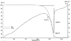
Figure 2 displays the TG-DTA curves of the marble waste. It was found that the thermal behavior of marble waste is characterized by the occurrence of three thermic events at different temperatures in DTA curve, which can be described as: i) endothermic event at approximately 150 ºC, which indicates release of water absorbed on the particle surface; ii) small endothermic event at 771.6 °C that is related to dolomite decomposition; and iii) intense endothermic event at 864.9 °C that is related to the calcite decomposition. The TG curve showed that the decomposition of carbonates (dolomite and calcite) was accompanied by a high mass loss (42.26%) due to the release of CO2. The total mass loss obtained corroborates the high value of the loss on ignition (Table 1).
Morphological aspects of the marble waste powder particles can be observed in the SEM image in Figure 3a. It is noted that the marble waste particles have an irregular morphology and a smooth surface. The marble waste powder has a wide particle size distribution, but the most of the particles are concentrated in the size range < 10 µm. The EDS spectrum in Figure 3b shows characteristic peaks of calcium (Ca), magnesium (Mg), carbon (C) and oxygen (O), reinforcing the results of chemical analysis (Table 1) and XRD analysis (Figure 1). The detected platinum peaks are associated with the sample metallization process.

3.2. Structural characterization of the synthesized powders
The structural characterization via X-ray diffraction of the synthesized powders are presented in Figure 4. XRD results revealed that all the synthesized powders were composed of β-calcium pyrophosphate (β-CPP, β-Ca2P2O7) and magnesium whitlockite (Mg-WH, (CaMg)3(PO4)2). These obtained phosphate phases are in accordance with the literature37-39. In fact, the marble waste used in this work contains 52.43 wt.% CaO and 9.62 wt.% MgO (Table 1), resulting in a Mg/Ca ratio of 0.18. On the other hand, it is known that an Mg/Ca ratio > 0.05 favors the replacement of calcium by magnesium in the crystalline lattice of calcium phosphates, leading to the formation of magnesium whitlockite40,41. It is also worth mentioning that the diffractograms in Figure 1 did not show any diffraction peaks of calcite and dolomite from the marble waste. This result suggests that the digestion process of carbonates (calcite and dolomite) present in the marble waste was fully efficient17,36,39.

XRD patterns of the synthesized powders: a) Ca/P (0.5:1); b) Ca/P (1:1); c) Ca/P (1.5:1); d) Ca/P (2:1); e) Ca/P (2.5:1) and f) Ca/P (3:1).
As found in Figure 4, all the synthesized powders corresponded to a biphasic phosphate bioceramic of the β-CPP/Mg-WH type, regardless of the Ca/P ratio. However, small but important differences can be observed in the new synthesized biphasic phosphate bioceramics. For the biphasic bioceramics obtained with Ca:P (1:1) and Ca:P (2:1) molar ratios, it was observed that the main peak at ~ 29.4º corresponded to the calcium β-CPP phase. In this case, the formation of β-CPP was facilitated by the molar concentration used in these samples associated with calcination at 900 ºC42. It is also noted that these biphasic bioceramics presented low intensity magnesium whitlockite diffraction peaks. For the other molar ratios (Ca:P), it was observed that the main peak at ~ 31.1° corresponds to the magnesium whitlockite phase. In addition, the diffraction peaks related to the β-CPP phase had their intensities decreased. This fact may be related to the increase in magnesium concentration in these samples, since by varying the proportion of calcium, a greater quantity of marble waste was supplied to the digestion solution. In fact, as shown in Table 1, marble waste is rich in calcium and magnesium.
The synthesized biphasic phosphate bioceramic powders of the β-CPP/Mg-WH type presented the following average crystallite size values determined by the Scherrer equation: i) Ca:P (0.5:1) = 55.94 nm; ii) Ca:P (1:1) = 42.56 nm; iii) Ca:P (1.5:1) = 43.16 nm; iv) Ca:P (2:1) = 57.55 nm; v) Ca:P (2.5:1) = 45.56 nm; and vi) Ca:P (3:1) = 47.84 nm. Therefore, all β-CPP/Mg-WH biphasic bioceramics present mean crystallite sizes within the nano-sized range. This result is very relevant, since calcium phosphate-based bioceramic nanoparticles have unique properties and functions that are highly attractive for medical applications2,11.
3.3. FTIR analysis
Figure 5 displays typical FTIR spectra of the biphasic calcium-magnesium phosphate bioceramic powders synthesized using marble waste. The FTIR spectrum obtained for the biphasic bioceramic synthesized using a Ca:P (1:1) ratio is shown in Figure 5a. This biphasic phosphate bioceramic (β-CPP/Mg-WH) mainly presented a main phase corresponding to the β-CPP phase (Figure 4b). The identification of the vibrational groups of calcium-magnesium phosphate materials are described as follow17,23,43-45. Vibrational bands were found at 3438.74 cm-1 and 1628.58 cm-1, indicating the presence of water molecules and OH- groups on the particle surface. The vibrational band at 2356.86 cm-1 corresponds to the CO2 group, which is related to the atmosphere of the environment where the equipment is installed. The vibrational bands at 1219.31 cm-1 and 969.58 cm-1 demonstrate the presence of the PO43- group. The vibrational band detected at 740.81 cm-1 represents the bonds corresponding to the Ca2+ ion. The vibrational bands detected at 553.70 cm-1 and 484.26 cm-1 are associated with the Ca-O and Mg-O groups, respectively. The FTIR spectrum of the biphasic bioceramic synthesized using a Ca:P (2.5:1) ratio was very similar, as shown in Figure 5b. More precisely, only small differences can be observed between the FTIR spectra, which are probably related to the different amounts of the β-CPP and Mg-WH phases present in these biphasic bioceramics.

3.4. Morphological analysis
The morphological aspects of the synthesized calcium phosphate-magnesium phosphate powders can be observed in Figure 6. SEM micrographs revealed that the nano-sized biphasic powders are composed of highly agglomerated plates with non-uniform sizes. Such observed agglomerated plates are due to the calcination step at 900 ºC, which caused the initial solid sintering between the synthesized nanoparticles. However, apparently, the morphology of the nanoparticles presents equiaxed shapes. This result is relevant, since nanometric powders tend to favor the wettability and capillarity properties of phosphate-based bioceramics, and, thus, can aid in the repair and reconstruction of damaged bone tissue11,13.

SEM images of the synthesized powders: a) Ca/P (0.5:1); b) Ca:P (1:1) c) Ca/P (1.5:1); and d) Ca/P (2.5:1).
3.5. Thermal stability
The thermal behavior described in terms of TG-DTA curves of the biphasic calcium phosphate-magnesium phosphate bioceramics synthesized using marble waste is shown in Figure 7. The results indicated that the synthesized biphasic bioceramics showed high thermal stability when heated to ~1000 ºC, independently of the molar ratio (Ca:P) used. In fact, the biphasic bioceramic obtained with a Ca:P (1:1) molar ratio presented a total mass loss of 0.60% (Figure 7a), while that obtained with Ca:P (2.5:1) molar ratio had a total mass loss was of 0.87% (Figure 7b). In the DTA curves, endothermic events are observed at 192.2 °C (Figure 7a) and 186.3 ºC (Figure 7b) due to the loss of adsorbed water from the biphasic bioceramics (β-CPP/Mg-WH). These results are quite relevant, since bioceramics, when implanted in a biological environment, must present high thermal stability without weight loss, in case of temperature changes.

4. Conclusions
The experimental results indicated that it is feasible to synthesize calcium phsophate-magnesium phosphate bioceramics using marble waste composed essentially of carbonates (calcite and dolomite). The valorization of marble waste to produce bioceramics for medical applications can be an attractive technological alternative for the final disposal of this polluting solid waste. It was found by XRD analysis that all the synthesized powders were composed of a biphasic mixture of β-CPP (β-calcium pyrophosphate) and Mg-WH (magnesium whitlockite). However, the predominance of β-CPP or Mg-WH in biphasic bioceramics is dependent on the molar ratio (Ca:P). The synthesized powders presented an average crystallite size in the 42.56 -57.55 nm range, indicating a nanostructured nature. FTIR results confirmed the presence of vibrational bands related to the β-CPP and Mg-WH phases identified in the biphasic bioceramics. SEM images revealed that the powder particles are highly agglomerates with equiaxed morphology. TG-DTA analyses indicated low mass loss and high thermal stability during heating. Therefore, these findings suggest that the marble waste offers a promising strategy for use as a cheap carbonate source in the manufacture of biphasic calcium phosphate-magnesium phosphate bioceramics.
5. Acknowledgements
The authors acknowledge the Foundation for Research Support of the State of Rio de Janeiro – Brazil (FAPERJ) - Process No. E-26/201.137/2022 and National Council for Scientific and Technological Development – Brazil (CNPq) - Process No. 306147/2023-8.
6. References
- 1 Dorozhkin SV. A history of calcium orthophosphates (CaPO4) and their biomedical applications. Morphologie. 2017;101(334):143-53.
- 2 Holanda JNF. Nanostructured calcium phosphate-based bioceramics from waste materials. In: Martínez LMT, Kharissova OV, Kharisov BI, editors. Handbook of ecomaterials. New York: Springer International Publishing; 2017. p. 1-18.
- 3 Wang J, Tang Y, Cao Q, Wu Y, Wang Y, Yuan B, et al. Fabrication and biological evaluation of 3D-printed calcium phosphate ceramic scaffolds with distinct macroporous geometries through digital light processing technology. Regen Biomater. 2022;9:rbac005.
- 4 Rujiraprasert P, Suriyasangpetch S, Srijunbarl A, Singthong T, Makornpan C, Nampuksa K, et al. Calcium phosphate ceramic as a model for enamel substitute material in dental applications. BDJ Open. 2023;9:25.
- 5 Skliarenko Y, Kolomiiets VV, Balatskyi VV, Galuza Y, Koryak OS, Macewicz LL, et al. The impact of the physicochemical properties of calcium phosphate ceramics on biocompatibility and osteogenic differentiation of mesenchymal stem cells. BMC Res Notes. 2024;17:295.
- 6 Goldberg MA, Krohicheva PA, Fomin AS, Khairutdinova DR, Antonova OS, Baikin AS, et al. In situ magnesium calcium phosphate cements formation: from one pot powders precursors synthesis to in vitro investigations. Bioact Mater. 2020;5(3):644-58.
- 7 Afonina A, Kizalaite A, Zarkov A, Drabavicius A, Goto T, Sekino T, et al. Synthesis of whitlockite nanopowders with different magnesium content. Ceram Int. 2022;48(21):32125-30.
- 8 Heng C, Liu W, Zheng X, Ma X, Hui J, Fan D. Dopamine and DNA functionalized manganese whitlockite nanocrystals for magnetic resonance imaging and chemo-photothermal therapy of tumors. Colloids Surf B Biointerfaces. 2023;222:113120.
- 9 Kizalaite A, Klimavicius V, Balevicius V, Niaura G, Salak AN, Yang JC, et al. Dissolution–precipitation synthesis and thermal stability of magnesium whitlockite. CrystEngComm. 2023;25:4370-9.
- 10 Gharsallah S, Alsawi A, Hammami B, Khitouni M, Charnay C, Chemingui M. Synthesis and characterization of new composite materials based on magnesium phosphate cement for fluoride retention. Materials. 2023;16:718.
- 11 Dorozhkin SV. Functionalized calcium orthophosphates (CaPO4) and their biomedical applications. J Mater Chem B Mater Biol Med. 2019;7:7471-89.
- 12 Sun JS, Tsuang YH, Liao CJ, Liu HC, Hang YS, Lin FH. The effects of calcium phosphate particles on the growth of osteoblasts. J Biomed Mater Res. 1997;37:324-34.
- 13 Dorozhkin SV. Multiphasic calcium orthophosphate (CaPO4) bioceramics and their biomedical applications. Ceram Int. 2016;42(6):6529-54.
- 14 Bouler JM, Pilet P, Gauthier O, Verron E. Biphasic calcium phosphate ceramics for bone reconstruction: A review of biological response. Acta Biomater. 2017;53:1-12.
- 15 Butler DH, Shahack-Gross R. Formation of biphasic hydroxylapatite-Beta magnesium tricalcium phosphate in heat treated salmonid vertebrae. Sci Rep. 2017;7:3610.
- 16 Basu S, Basu B. Doped biphasic calcium phosphate: synthesis and structure. J Asian Ceram Soc. 2019;7(3):265-83.
- 17 Corrêa THA, Toledo R, Silva NS, Holanda JNF. Novel nano-sized biphasic calcium phosphate bioceramics (β-CPP/β-TCP) derived of lime mud waste. Mater Lett. 2019;243:17-20.
- 18 Windarti T, Nulandaya L, Widjijono W, Nuryono N. Synthesis and characterization of biphasic calcium phosphate substituted cerium as a potential osteoporotic bone filler. Period Polytech Chem Eng. 2023;67(2):242-55.
- 19 Corrêa THA, Holanda JNF. Recycling of tilapia carcass waste to obtain nanostructured biphasic calcium phosphate powder. Ceramica. 2023;69(362):261-5.
-
20 ABIROCHAS: Associação Brasileira da Indústria de Rochas Ornamentais. Produção brasileira de Lavra [Internet]. Brasília: ABIROCHAS; 2021 [cited 2024 July 11]. Available from: https://abirochas.com.br/wp-content/uploads/2022/01/Producao-Brasileira-Lavra-2021.pdf
» https://abirochas.com.br/wp-content/uploads/2022/01/Producao-Brasileira-Lavra-2021.pdf - 21 Mulk S, Korai AL, Azizullah A, Shahi L, Khattak MNK. Marble industry effluents cause an increased bioaccumulation of heavy metals in Mahaseer (Tor putitora) in Barandu River, district Buner, Pakistan. Environ Sci Pollut Res Int. 2017;24:23039-56.
- 22 Iqbal Q, Musarat MA, Ullah N, Alaloul WS, Rabbani MBA, Al Madhoun W, et al. Marble dust effect on the air quality: an environmental assessment approach. Sustainability. 2022;14:3831.
- 23 Thakur AK, Pappu A, Thakur VK. Resource efficiency impact on marble waste recycling towards sustainable green construction materials. Curr Opin Green Sustain Chem. 2018;13:91-101.
- 24 Santos T, Gonçalves JP, Andrade HMC. Partial replacement of cement with granular marble residue: effects on the properties of cement pastes and reduction of CO2emission. SN Applied Sciences. 2020;2:e1605.
- 25 Munir MJ, Kazmi SMS, Wu Y, Hanif A, Arif Khan MU. Thermally efficient fired clay bricks incorporating waste marble sludge: an industrial-scale study. J Clean Prod. 2018;174:1122-35.
- 26 Khan Z, Gul A, Shah SAA, Qazi S, Wahab N, Badshah E, et al. Utilization of marble waste in clay bricks: a step towards lightweight energy efficient construction materials. Civ Eng J. 2021;7(9):1488-500.
- 27 Ahmad S, Shah MUH, Ullah A, Shah SN, Rehan MS, Khan IA, et al. Sustainable use of marble waste in industrial production of fired clay bricks and its employment for treatment of flue gases. ACS Omega. 2021;6(35):22559-69.
- 28 Castellano J, Sanz V, Canas E, Sánchez E. Industry-scalable wall tile composition based on circular economy. Bol Soc Esp Ceram Vidr. 2022;61:374-82.
- 29 Ramos JCR, Passalini PGS, Holanda JNF. Utilization of marble waste as a sustainable replacement for calcareous in the manufacture of red-firing wall tiles. Constr Build Mater. 2023;377:131115.
- 30 Buyuksagis IS, Uygunoglu T, Tatar E. Investigation on the usage of waste marble powder in cement-based adhesive mortar. Constr Build Mater. 2017;154:734-42.
- 31 Shukla A, Gupta N, Gupta A. Development of green concrete using waste marble dust. Mater Today Proc. 2020;26:2590-4.
- 32 Hou W, Zhang Q, Zhuang Z, Zhang Y. Sustainable reusing marble powder and granite powder in cement-based materials: a Review. ACS Sustain Chem& Eng. 2024;12(7):2484-510.
- 33 Miculescu F, Mocanu AC, Maidaniuc A, Dascălu CA, Miculescu M, Voicu SI, et al. Biomimetic calcium phosphates derived from marine and land bioresources. In: Thirumalai J, editor. Hydroxyapatite: advances in composite nanomaterials, biomedical applications and its technological facets. London: IntechOpen; 2018. p. 89-108.
- 34 Mocanu AC, Stan GE, Maidaniuc A, Miculescu M, Antoniac IV, Ciocoiu RC, et al. Naturally-derived biphasic calcium phosphates through increased phosphorus-based reagent amounts for biomedical applications. Materials. 2019;12(3):381.
- 35 Algamala Y, Khalila NM, Saddiqd A, Baghdadi AM. Antimicrobial activity of hydroxyapatite nanoparticles prepared from marble wastes. Main Group Chem. 2022;21:865-73.
- 36 Gomes LC, Di Lello BC, Campos JB, Sampaio M. Synthesis and characterization of calcium phosphates produced from chicken eggshell. Ceramica. 2012;59(348):448-52.
- 37 Landi E, Logroscino G, Proietti L, Tampieri A, Sandri M, Sprio S. Biomimetic Mg-substituted hydroxyapatite: from synthesis to in vivo behaviour. J Mater Sci Mater Med. 2008;19(1):239-47.
- 38 Kim HD, Jang HL, Ahn HY, Lee HK, Park J, Lee ES, et al. Biomimetic whitlockite inorganic nanoparticles-mediated in situ remodeling and rapid bone regeneration. Biomaterials. 2017;112:31-43.
- 39 Kim DW, An JS, Cho IS. Effects of Mg and Sr co-addition on the densification and biocompatible properties of calcium pyrophosphate. Ceram Int. 2018;44(8):9689-95.
- 40 Le Geros RZ. Calcium Phosphates. In: Myers HM, editor. Monographs in oral biology and medicine. San Francisco: Karger; 1991.
- 41 Driessens FC, Boltong MG, Maeyer EA, Wenz R, Nies B, Planell JA. The Ca/P range of nanoapatitic calcium phosphate cements. Biomaterials. 2002;23(19):4011-7.
- 42 Zhao S, Jiang Q, Peel S, Wang X, He F. Effects of magnesium-substituted nanohydroxyapatite coating on implant osseointegration. Clin Oral Implants Res. 2013;24(suppl. A100):34-41.
- 43 Gunasekaran S, Anbalagan G. Thermal decomposition of natural dolomite. Bull Mater Sci. 2007;30:339-44.
- 44 Vasant SR, Joshi MJ. Synthesis and characterization of nanoparticles of calcium pyrophosphate. Mod Phys Lett B. 2011;25(1):53-62.
- 45 Ebrahimi M, Botelho MG, Dorozhkin SV. Biphasic calcium phosphates bioceramics (HA/TCP): Concept, physicochemical properties and the impact of standardization of study protocols in biomaterials research. Mater Sci Eng C. 2017;71:1293-312.


