 Congenital Zika virus infection in laboratory animals: a comparative review highlights translational studies on the maternal-foetal interface
Abstract
The 2015-16 Zika virus (ZIKV) epidemic has posed unprecedented concern for maternal-infant health, mainly due to the substantial risk of microcephaly and other neurological birth abnormalities associated with congenital ZIKV syndrome (CZS). As licenced vaccines and effective antivirals are still unavailable, attention has been focused on post-delivery in vitro or translational in vivo studies to understand the impact of maternal ZIKV infection on placentation and neurodevelopmental consequences for the foetus. Here, we review clinical and translational studies highlighting ZIKV-induced maternal-foetal interface dysfunction, adding to our previous observations of experimental ZIKV vertical transmission to pregnant rhesus monkeys and newly published post-epidemic findings about the theme. This comparative review focuses on the mechanisms by which the virus has a cytopathic effect on trophoblasts and macrophages during placentation in humans, nonhuman primates, and rodent transgenic models, crosses the placental barrier, replicates, and establishes a persistent uteroplacental infection. When considering the mechanism of ZIKV-induced birth defects in humans and other susceptible hosts, it becomes apparent how the various stages of the ZIKV cycle in the host (both the parent and offspring) unfold. This understanding presents specific opportunities for pharmacological intervention and the development of preventative vaccines.
Key words:
Zika virus infection; vertical transmission; teratogenesis; haemochorial placenta; maternal-foetal interface; nonhuman primates; AG129 mice; sofosbuvir; antiviral drugs
Historical and epidemiologic aspects of ZIKV infection
Zika virus (ZIKV), a mosquito-borne flavivirus whose vectors are Aedes aegypti and Aedes albopictus, is widely disseminated in Brazil1 and can infect humans and nonhuman primates. Indeed, ZIKV was first isolated from a rhesus monkey in 1947 and Aedes africanus in 1948.2 In humans, the infection was first described in Nigeria, Africa.3 Ordinarily, ZIKV infections have been reported to be sporadic and commonly associated with mild disease. The first well-known outbreak of the ZIKV occurred in the Yap Islands of Micronesia in 2007. According to local public health reports, the Zika infection rate ranged from 73%,4 the outbreak of French Polynesia in 2013-14 followed by general prevalence rates of 49% with an estimated 32,000 infected patients between October 2013 and April 2014.5,6 In the Brazilian outbreak, neurological disorders such as Guillain-Barré syndrome in adults reported an incidence between April-July 2015 among those ≥ 12 years of age was 5.6 cases/100,000 population/year and increased markedly with increasing age to 14.7 cases/100,000 among those ≥ 60 years of age.7 An effective link between the vertical transmission of the ZIKV and microcephaly emerged from the epidemic in Brazil in 2015-2016, with infection rates estimated to range from 10% to 80%.8,9,10 Earlier, during the 2013-2014 Zika outbreak in French Polynesia, the risk of microcephaly related to ZIKV infection was 0.95%, as estimated from eight cases identified retrospectively in the population of 270,000 inhabitants, with a 66% ZIKV infection rate.11,12 In contrast, the Brazilian Live Births Information System reported thousands of microcephaly or other neurodevelopmental-associated anomalies between July 2015 and February 2016.13,14 The Brazilian ZIKV outbreak, two distinct waves of infection, extended across all Brazilian regions in 2015 and 2016. 1,673 272 cases were notified, of which 41,473 (2-5%) were in pregnant women. During the most severe first wave, 1950 cases of microcephaly were confirmed [1373 (70.4%0] occurred in the Northeast region, with a peak monthly estimated at 49.9 cases per 10,000 live births.15 In the Brazilian outbreak, approximately one-third of liveborn children with prenatal ZIKV exposure presented with at least one abnormality compatible with congenital infection, while the risk of presenting with at least two abnormalities in combination was less than 1.0%.16
The spectrum of sequelae of congenital ZIKV infection, broadly referred to as congenital ZIKV syndrome (CZS), in which microcephaly is a major manifestation, includes (but is not limited to) parenchymal or cerebellar calcification, ventriculomegaly, hydrocephalus, arthrogryposis, and visual and hearing disorders.17 ZIKV infection during pregnancy can be associated with neurodevelopmental abnormalities, even in normocephalic infants or those born to asymptomatic mothers. Neurodevelopmental outcomes possibly associated with congenital ZIKV infections include shorter attention spans, longer processing times of visual stimuli, postnatal microcephaly, lissencephaly, hypotonia, hypertonia, arthrogryposis, hearing tissues (inner ear), transient developmental delay, delayed myelination, persistent intracranial calcifications, ventriculomegaly, cerebellar hypoplasia, and cortical abnormalities, as revised by Caldwell and colleagues.18 Early identification and intervention for neurodevelopmental disorders can improve cognitive, social, and behavioural functioning.19 The objectives of this manuscript were to compare ZIKV-induced maternal-foetal interface changes in different laboratory animal species, highlight the differences among ZIKV vertical transmission in the light of anatomical differences in placentation, and add some unpublished results of our previous study evolving ZIKV infection at the early third of prepregnancy of rhesus monkeys and its vertical viral transmission.
Relevance of the maternal-foetal interface for ZIKV vertical transmission
Physiologically, during early human pregnancy, the uterine mucosa transforms into the decidua, into which the foetal placenta implants and where placental trophoblast cells and maternal cells intermingle and communicate.20 In discoid placentation, maternal and foetal circulations are interwoven, with trophoblast and endothelial cell layers differently organised depending on the species: haemomonochorial in humans and haemotrichorial in mice, which represents an additional endothelial barrier to protect the foetus from invading pathogens. Another similarity between the human and NHP species that may contribute to foetal infection is the long gestational period of Old-World monkeys (150 to 210 days) and human beings (280 days), in contrast with rodents (20 days).21,22 As estimated from translational studies with rhesus monkeys, the viraemia period of ZIKV-infected (at the first gestational trimester) dams lasts 7 to 10 days;23 such prolonged maternal viraemia can trigger transplacental viral transposition, virus crossing into the amniotic fluid (AF), and in utero foetal death.24 The ZIKV strain infects and replicates in primary human placental macrophages (Hofbauer cells) and poorly in cytotrophoblasts. Viral replication induces type I interferon (IFN), proinflammatory cytokines, and antiviral gene expression but causes minimal cell death. In addition to suggesting a mechanism for intrauterine transmission in which ZIKV gains access to the foetal compartment by directly infecting placental cells and disrupting the placental barrier,25 other authors have suggested that ZIKV can open the paracellular pathway of STB cells.26 In human ZIKV vertical transmission, monocytes/macrophages (Hofbauer cells) have been referred to as “Trojan horses” since they may carry infectious virus particles to immune-privileged sites such as the placental, blood-testis, and blood-brain barriers.27 Another hypothesis is that low-affinity circulating maternal antibodies could enhance ZIKV replication by binding to Fc receptor (FcR)-bearing cells via antibody-dependent infection enhancement (ADE), i.e., cross-reactive antibodies (against other flaviviruses) transported transplacentally via neonatal FcRs, which could improve ZIKV replication in pregnancy-associated progenitor cells (PAPCs).28 The prolonged period of ZIKV viremia observed in pregnant macaques and human beings has been justified by the hypothesis that the infected foetus and/or placenta role as a ZIKV reservoir that cannot be efficiently cleared by the dam immune system however, this hypothesis needs to be best investigated.22
Together, observations in NHPs and humans indicate that foetal abnormalities caused by ZIKV infection can occur regardless of maternal signs. A Brazilian epidemiological report reinforced that previous dengue fever epidemics may be related to microcephaly incidence and the idea of a window of cross-protection and a window of increased risk.29 This is a controversial issue, as an experimental study using a pregnant rhesus monkey did not reveal foetal abnormalities at delivery; however, more ZIKV RNA was detected in the placenta of macaques immunised to DENV, suggesting that DENV immunity could enhance ZIKV infection of the placenta.30
ZIKV infection in the circulation of pregnant women and macaques crosses the placental barrier (endothelial cells and trophoblasts), and trophoblasts represent the first viral replication site in the foetus, since primary trophoblasts express ZIKV cell entry receptors. There are controversies about whether ZIKV-infected trophoblast function may be preserved and become a compartment of foetal viral dissemination.31 As demonstrated in dizygotic twins, which present different outcomes after infection with a Brazilian isolate of ZIKV, trophoblasts from the ZIKV-nonaffected twin secreted increased levels of inflammatory chemokines (RANTES/CCL5 and IP10) because the nonaffected twin more efficiently activated genes (induced by IFN-g) involved in placental ZIKV immune protection, thus preventing ZIKV dissemination into developing foetus tissues.32 Another report about discordant outcomes in dizygotic twins in Brazil is justified by gene mutations in the MTOR and Wnt pathways, which regulate foetal neurodevelopment.33,34 Indeed, foetal protection requires the induction of a robust placental antiviral response, with type I IFN binding to the IFN alpha receptor (IFNAR) expressed in many cell types and upregulating IFN-stimulated genes (ISGs) in the placenta.35 ZIKV single-stranded RNA elicits an antiviral response via receptor acid-inducible gene I (RIG-I), resulting in robust type I and III IFN responses.36 The decidua microenvironment is physiologically tolerogenic.37 During ZIKV infection, dNK cells (the main immune cell population of the first-trimester decidua) produce proinflammatory cytokines that may regulate ZIKV infection during pregnancy.38 On the other hand, the immune system recruit neutrophils, natural killer cells (dNKs), macrophages, T cells and dendritic cells (DCs) in the stromal decidua microenvironment, which alters the decidua, possibly leading to placenta immune-mediated injuries. The expression of IFN type I diminished extravillous trophoblast invasion by the spiral artery (EVT-mediated spiral artery remodelling), elevating the risk of hypertension and preeclampsia in pregnant women39 and pregnant rhesus monkeys.23 Additionally, in a type I IFN receptor-deficient (IFNAR-/-) mouse model, placental ZIKV infection can cause indirect damage to the foetus due to reduced uteroplacental perfusion, leading to intrauterine foetal growth delay and early embryonic death.40
Foetal neuropathologic findings associated with congenital ZIKV infection in animal models
Congenital ZIKV syndrome, described in the early Brazilian epidemic,41 has been extensively investigated through translational studies using immunodeficient mice (AG129)42 and immunocompetent pregnant nonhuman primates.23,43,44 Preclinical studies have confirmed that foetal neural progenitor cells (NPCs) are the principal target cells contributing to abnormal brain embryo development, as observed in ZIKV-infected pregnant mice and rhesus monkeys. In addition to pathogenic mechanisms, animal models are valuable tools for investigating the efficacy of repositioned or new antiviral drugs.45In vitro studies have also been performed to understand the mechanisms underlying neuropathologic findings and the cytopathic effects of ZIKV on the death of NPCs and optic progenitor cells (OPCs).46,47 Other in vitro studies confirmed that the ZIKV E (envelope) protein causes cell cycle arrest, a decrease in cell proliferation, and an increase in the mitotic length of dividing human NPCs through dysregulating the cyclinD1-p21-p53 pathway and changing intercellular calcium levels and ATP stimulation in ZIKV E protein-expressing hNPCs.48 The direct neurotoxicity of the ZIKV envelope protein may be explained by the overexpression of polyadenosine diphosphate-ribose polymerase 1 in hNPCs.49 Another hypothesis is that ZIKV replication in the central nervous system (CNS) leads to direct cellular injury by increasing cellular glycolysis, which may cause intracellular stress and endoplasmic reticulum dysfunction50 associated with neuroinflammationm.51 AXL, a phosphatidylserine (PS) receptor, has been proposed to play a key role as an attachment factor for ZIKV infection in the host cell.52 However, genetic ablation of AXL does not affect ZIKV entry or ZIKV-mediated cell death in human induced pluripotent stem cell (iPSC)-derived NPCs or cerebral organoids.53 ZIKV tropism for glial cells is facilitated by the expression of the receptor tyrosine kinase (RTK) AXL, which impacts the transcriptional induction of type I IFN, proinflammatory cytokines and chemokines in infected NPCs, thus stimulating ZIKV replication.54 Consequently, this process results in the downregulation of foetal neurogenesis and the upregulation of NPC apoptosis, disrupting NPC cell cycle proliferation/self-renewal, which is essential for normal mammalian brain development.55 Other flaviviruses may also enter the CNS, causing peripheral neuropathy, including West Nile virus (WNV), Japanese encephalitis virus (JEV), and tick-borne encephalitis virus (TBEV).18 However, the neurotropic flaviviruses, which have direct cytolytic/oncolytic effects on NPCs, have shown beneficial effects on preclinical GBM models by killing glioma stem cells (GSCs).56 A 43-year-old woman was diagnosed with glioblastoma after the tumour resection was infected with the ZIKV outbreak in Brazil. Following the infection resolution, the glioblastoma regressed, and no recurrence was observed.57
ZIKV-induced inflammatory and vascular placental injuries alter the “immune clock” of pregnancy
The “immune clock of pregnancy” refers to dynamic changes in gestational immune status, which are chronologically necessary to maintain maternal immune homeostasis, thus supporting foetal growth while protecting the mother and foetus against infectious agents. Healthy-term pregnancy outcomes require synchronization of implantation competency by the blastocyst with endometrial receptivity. This short-term period of crosstalk between the blastocyst and the uterus, the so-called “window of implantation”, is controlled not only by ovarian oestrogen and progesterone hormones58 but by signalling molecules, including cytokines, growth factors, homeobox transcription factors, lipid mediators and morphogen genes, which function through autocrine, paracrine and juxtracrine interactions to support the effectiveness of blastocyst implantation.59 The interference of ZIKV and other congenital infections at the foetal-maternal interface in terms of the local cellular mechanisms that control maternal immune tolerance may help understand the teratogenesis mechanism.60
In a preclinical study of Asian-lineage ZIKV vertical transmission in pregnant rhesus monkeys, we confirmed the exacerbated placental anergic/suppressive mechanism of myeloid-derived suppressor cells (MDSCs),23 as suggested by Asian-lineage ZIKV infection of pregnant women’s blood leading to exacerbated M2-skewed immunosuppression of nonclassical monocytes in conjunction with global suppression of the type I interferon signalling pathway and aberrant expression of host genes associated with pregnancy complications.61 The phenotype of MDSCs was more frequent in placental cells from ZIKV-infected-Sofosbuvir(SOF)-treated dams, as well as in those from the negative control group, than in those from the ZIKV-infected-nontreated group. Not by chance, placentas from non-SOF-treated dams contained more of the viral replicative marker, anti-double-stranded RNA (dsRNA)-marked cells than those from SOF-treated dams. In both the untreated and SOF-treated groups, Hofbauer cells (CD3- HLADR- CD4+ CD14+) and T lymphocytes (CD3+) were detected in the placenta,23 confirming that the placental anergic/suppressive mechanism works by controlling viral-induced placental inflammation and preventing foetal death, as previously described by others. In two pregnant women with ZIKV infection, amniotic fluid collected by amniocentesis at 20 weeks gestation was RNA negative, and ZIKV RNA remained positive in the dam’s plasma until delivery, but the virus was not transmitted to the newborns. Similarly, two cases of maternal immunocompromised status were associated with a prolonged ZIKV viremia period.62
Non-invasive in vivo imaging studies have shown robust maternal-placental-foetal inflammation (chronic decidualitis, lymphoplasmacytic infiltrates, and neutrophilic leukocytoclastic vasculitis associated with spiral artery placenta) and calcification during ZIKV infection. Such uterine vasculitis affects the oxygen permeability of the placental villus, resulting in a significant decrease in oxygen delivery to the foetus.63 Similarly, in immunocompromised pregnant IFNAR-/- mice infected with ZIKV, placental inflammation was associated with impaired blood flow circulation in uteroplacental and foetal brain vessels, as revealed by colour Doppler ultrasound examination as early as 12.5 embryonic days, leading to intrauterine growth delay.40 Semmes and Coyne attributed severe ZIKV-induced placental inflammation in mice to a direct innate antiviral response64 and consequent foetal growth restriction or death due to blood supply restriction caused by microvascular disruption.65 Similarly, Pomar and colleagues described reduced umbilical artery blood flow and placentomegaly (placental thickness > 40 mm) in pregnant women with congenital ZIKV infection.66
The placental microenvironmental inflammation induced by ZIKV infection may also involve pericyte activation. Despite the poor description of ZKV infection, placental pericytes are essential for endothelial cell proliferation and placental microvasculature development. They surround the abluminal surface of capillary blood vessels through contractile cytoplasmic extensions that wrap around the endothelial cells lining the capillaries and venules. These antigen-presenting cells (APCs) play an essential role through contact-mediated and paracrine crosstalk with endothelial cells, and virus-infected pericytes may enhance viral penetration of the placenta and foetal blood-brain barrier, leading to neuroinflammatory and developmental sequelae in the foetus.67 Mouse models confirmed that ZIKV replication in pericytes from the choroid plexus impaired the permeability of the endothelial barrier and tissue perfusion.68 Similarly, human cytomegalovirus (HCMV) infection of the brain and retinal pericytes in vitro induces HCMV-associated neuroinflammation, reduced perfusion, and retinal dysfunction in neonates.69 In addition, the interaction of HCMV with pericytes in the placental microvasculature results in lytic infection. However, further studies investigating the infectivity of ZIKV in human placental pericytes and its potential role in ZIKV dissemination or the mechanisms of placental and foetal inflammation are needed.
Propositions to prevent congenital Zika syndrome
Translational studies using the rodent model interferon-α/β knockout mice (AG129) have highlighted the protective role of endogenous type I IFN against ZIKV infection,42,70 while other studies have reinforced the importance of secreted IFN-I by trophoblastic cells, which represents a possible mediator of pregnancy complications, including spontaneous abortions and growth restriction, in the context of congenital ZIKV infection.71 Classically associated with viral infections, type I IFNs activate downstream signalling pathways that increase the transcription of IFN-stimulated genes in the microenvironment, inhibiting viral replication and limiting viral spread.72 For example, ZIKV RNA activates Toll-like receptor-3 (TLR-3),73 dependent on Toll/interleukin-1 receptor/resistance protein (TIR).74 In addition, retinoic acid-inducible gene-I (RIG-I) mediates the innate immune response, triggering IFN production and antiviral actions to control ZIKV infection and placental inflammation.75
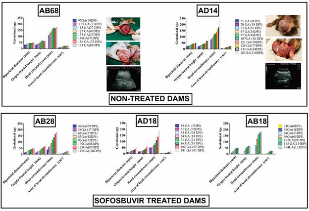
Sofosbuvir is a nucleotide analogue prodrug successfully used to treat chronic hepatitis caused by the hepatitis C virus (HCV), a Flaviviridae family member like the ZIKV. Its intracellular active triphosphate form inhibits HCV RNA polymerase and replication.76,77 The HCV and ZIKV RdRp-encoding genes are in the NS5B and NS5 domains. These genes are highly conserved among Flaviviridae members.78 The antiviral activity of SOF against ZIKV replication was demonstrated in vitro.79,80 Additionally, preclinical studies have confirmed its protective effect against ZIKV-induced teratogenesis in macaques.23,81 In addition, for the prevention of ZIKV vertical transmission and associated CZS, SOFs had less severe RNAaemia than did non-SOFs. The offspring of nontreated dams presented ZIKV neurotropism, whereas those of SOF-treated dams did not, suggesting an antiviral protective effect. At delivery, the offspring of nontreated dams exhibited macroscopic neural abnormalities such as lissencephaly, ventriculomegaly, and subdural haematoma (Figure, Table). Histological analysis revealed marked disorganisation of the cerebellar white matter folium, fold layer oedema, and ischaemic damage to the Purkinje and granular layers.23 The antiviral efficacy of SOF was confirmed to reduce ZIKV replication and prevent long-term sequelae in AG129 mice.82 In another SOF-treated rodent model, NOD/SCID mice, intravenous ZIKV infection prevented vertical transmission. Ouabain (an inhibitor of Na+, K+-ATPase) has been shown to act as an anti-ZIKV replication drug by binding to the ZIKV NS5-RdRp and NS3-helicase proteins83 and in a mouse model of CZS.84 The macrolide antibiotic azithromycin (AZM) reduces ZIKV proliferation and has cytoprotective effects on glial cell lines and human astrocytes.85 AZM upregulates the expression of host type I and III interferons and several downstream interferon-stimulated genes (ISGs) in response to ZIKV infection.86 No new clinical studies have been published to date, and other in vitro studies on anti-flavivirus agents have been published.87

Gestational ultrasonographic follow-up showing foetal protection in pregnant rhesus monkeys inoculated with Zika virus (ZIKV) and treated with or without sofosbuvir (SOF). The parameters adopted included the biparietal diameter, occiput-frontal length, head circumference, area of head circumference and thorax constriction. AB68 offspring showing postnatal congenital ZIKV-induced injury. AD14 offspring exhibiting normal development.23
Summary of clinical and virological findings of offspring from Zika virus (ZIKV)-infected macaque dams treated or not with sofosbuvir (SOF) as previously reported by Gardinali and colleagues23
More than 50 ZIKV vaccine candidates are now in different preclinical and clinical phases.88,89,90 Proof-of-concept studies in mice and marmosets91 demonstrated a protective effect against ZIKV-induced teratogenesis during pregnancy.
Promising phase-1 clinical trials have evaluated the safety and efficacy of candidate anti-Zika vaccines. One of them was based on inactivated, viral vectors, and DNA vaccines. Immunogenicity ranged from 10% to 100% in geometric mean titre (GMT) (6.3; 95% confidence interval (CI): 3.7-10.8) observed among recipients of single-dose inactivated anti-Zika vaccine. For DNA vaccines, the seroconversion rate ranged from 60% to 100%, with the highest seroconversion rate (100%) and GMT (2871; 95% CI: 705.3-11688). For the viral vector vaccine (Ad26.ZIKV.001), the seroconversion rate was 100%, and GMT peaked after two shots with both low and high-dose vaccines.92
Despite worldwide efforts, no effective vaccine or monoclonal antibodies against ZIKV were licensed for human use until May 2024. Unfortunately, ZIKV continues to circulate low in some areas and can re-emerge in naïve populations.90,93 Public education initiatives targeting insecticide applications and innovative approaches, for example, manipulating vector bacterial symbionts such as Wolbachia, to combat mosquito transmission arboviruses need an approach integrating antiviral research, vaccination, and vector control.85 From 2015 to 2020, 3,591 cases of CZS were confirmed in Brazil, with an incidence of 44.03 cases per 1,000 live births and a specific mortality of 12.35 deaths per 1,000 live births.94 The World Health Organisation is alert to continued ZIKV vaccine development efforts. The initiative for Vaccine Research and the National Institutes of Health National Institute of Allergy and Infectious Diseases co-hosted a meeting of experts in March 2018 to identify strategies to demonstrate vaccine effectiveness given the worsening incidence of ZIKV disease.95 A wide variety of formulations are being studied, including live virus vaccines, inactivated vaccines, whole-virus vaccines, subunit vaccines, mRNA, DNA, protein, and vector-based platforms.88 In a novel live-attenuated ZIKV strain (titled Z7) generated by inserting 50 RNA nucleotides (nt) into the 5′ untranslated region (UTR) of the ZIKV Cambodian strain (FSS13025), the neurovirulence, immune antagonism, and mosquito infectivity of Z7 were attenuated compared with those of other isolates. Z7 induced robust humoral and cellular immune responses that completely prevented viremia after infection with the ZIKV strain PRVABC59 in type I IFN-deficient (Ifnar1−/−) mice; however, additional studies confirming the prevention of ZIKV-induced teratogenesis need to be performed in new preclinical studies.96
In conclusion
The results of the present study indicate that ZIKV replicates in trophoblast cells, transposes the placental barrier, and induces inflammation. Subsequently, ZIKV quickly reaches foetal blood, crosses the foetal haematoencephalic barrier (neuroinvasion), replicates in foetal NPCs, and induces neuroinflammation, causing CZS, foetal death and death. New preventive and therapeutic approaches to prevent foetal ZIKV infections remain necessary since no licenced product is available until we finish this review.
ACKNOWLEDGEMENTS
To Igo Vieira de Sousa, Valdeci de Souza Peçanha, and Marco Aurelio Correa (Instituto de Ciência e Tecnologia em Biomodelos - ICTB - Fiocruz) for their support and technical help with our NHP study, and Departamento de Primatologia/ICTB-Fiocruz for providing the pregnant rhesus monkeys for this study.
REFERENCES
- 1 Chouin-Carneiro T, David MR, Nogueira FB, dos Santos FB, Lourenço-de-Oliveira R. Zika virus transmission by Brazilian Aedes aegypti and Aedes albopictus is virus dose and temperature-dependent. PLoS Negl Trop Dis. 2020; 14(9): e0008527.
- 2 Dick GWA, Haddow AJ. Uganda S virus. A hitherto unrecorded virus isolated from mosquitoes in Uganda. (I). Isolation and pathogenicity. Trans R Soc Trop Med Hyg. 1952; 46(6): 600-18.
- 3 Macnamara FN. Zika virus: a report on three cases of human infection during an epidemic of jaundice in Nigeria. Trans R Soc Trop Med Hyg. 1954; 48(2): 139-45.
- 4 Duffy MR, Chen TH, Hancock WT, Powers AM, Kool JL, Lanciotti RS, et al. Zika virus outbreak on Yap Island, Federated States of Micronesia. N Engl J Med. 2009; 360(24): 2536-43.
- 5 de Laval F, Leparc-Goffart I, Meynard JB, Daubigny H, Simon F, Briolant S. Zika virus infections. Med Sante Trop. 2016; 26(2): 145-50.
- 6 Aubry M, Teissier A, Huart M, Merceron S, Vanhomwegen J, Roche C, et al. Zika virus seroprevalence, French Polynesia, 2014-2015. Emerg Infect Dis. 2017; n23(4): 669-72.
- 7 Styczynski AR, Malta JMAS, Krow-Lucal ER, Percio J, Nóbrega ME, Vargas A, et al. Increased rates of Guillain-Barré syndrome associated with Zika virus outbreak in the Salvador metropolitan area, Brazil. PLoS Negl Trop Dis. 2017; 11(8): e0005869.
- 8 Zanluca C, de Melo VCA, Mosimann ALP, dos Santos GIV, dos Santos CND, Luz K. First report of autochthonous transmission of Zika virus in Brazil. Mem Inst Oswaldo Cruz. 2015; 110(4): 569-72.
- 9 Calvet GA, Santos FBD, Sequeira PC. Zika virus infection: epidemiology, clinical manifestations and diagnosis. Curr Opin Infect Dis. 2016; 29(5): 459-66.
- 10 Teixeira MG, Costa MCN, de Oliveira WK, Nunes ML, Rodrigues LC. The epidemic of Zika virus-related microcephaly in Brazil: detection, control, etiology, and future scenarios. Am J Public Health. 2016; 106(4): 601-5.
- 11 Cauchemez S, Besnard M, Bompard P, Dub T, Guillemette-Artur P, Eyrolle-Guignot D, et al. Association between Zika virus and microcephaly in French Polynesia, 2013-15: a retrospective study. Lancet. 2016; 387(10033): 2125-32.
- 12 Johansson MA, Mier-y-Teran-Romero L, Reefhuis J, Gilboa SM, Hills SL. Zika and the risk of microcephaly. N Engl J Med. 2016; 375(1): 1-4.
- 13 Jaenisch T, Rosenberger KD, Brito C, Brady O, Brasil P, Marques ET. Risk of microcephaly after Zika virus infection in Brazil, 2015 to 2016. Bull World Health Organ. 2017; 95(3): 191-8.
- 14 de Araújo TVB, Ximenes RAA, Miranda-Filho DB, Souza WV, Montarroyos UR, de Melo APL, et al. Association between microcephaly, Zika virus infection, and other risk factors in Brazil: final report of a case-control study. Lancet Infect Dis. 2018; 18(3): 328-36.
- 15 de Oliveira WK, de França GVA, Carmo EH, Duncan BB, Kuchenbecker RS, Schmidt MI. Infection-related microcephaly after the 2015 and 2016 Zika virus outbreaks in Brazil: a surveillance-based analysis. Lancet. 2017; 390(10097): 861-70.
- 16 Ximenes RAA, Miranda-Filho DB, Brickley EB, Araújo TVB, Montarroyos UR, Abtibol-Bernardino MR, et al. Risk of adverse outcomes in offspring with RT-PCR confirmed prenatal Zika virus exposure: an individual participant data meta-analysis of 13 cohorts in the Zika Brazilian Cohorts Consortium. Lancet Reg Health Am. 2023; 17: 100395.
- 17 Freitas DA, Souza-Santos R, Carvalho LMA, Barros WB, Neves LM, Brasil P, et al. Congenital Zika syndrome: a systematic review. PLoS One. 2020; 15(12): e0242367.
- 18 Caldwell M, Boruah AP, Thakur KT. Acute neurologic emerging flaviviruses. Ther Adv Infect Dis. 2022; 9: 20499361221102664.
- 19 Rice ME, Galang RR, Roth NM, Ellington SR, Moore CA, Valencia-Prado M, et al. Vital signs: Zika-associated birth defects and neurodevelopmental abnormalities possibly associated with congenital Zika virus infection - U.S. Territories and Freely Associated States, 2018. MMWR Morb Mortal Wkly Rep. 2018; 67(31): 858-67.
- 20 Vento-Tormo R, Efremova M, Botting RA, Turco MY, Vento-Tormo M, Meyer KB, et al. Single-cell reconstruction of the early maternal-fetal interface in humans. Nature. 2018; 563(7731): 347-53.
- 21 Buse E, Markert UR. The immunology of the macaque placenta: a detailed analysis and critical comparison with the human placenta. Crit Rev Clin Lab Sci. 2019; 56(2): 118-45.
- 22 Caine EA, Jagger BW, Diamond MS. Animal models of Zika virus infection during pregnancy. Viruses. 2018; 10(11): 598.
- 23 Gardinali NR, Marchevsky RS, Oliveira JM, Pelajo-Machado M, Kugelmeier T, Castro MP, et al. Sofosbuvir shows a protective effect against vertical transmission of Zika virus and the associated congenital syndrome in rhesus monkeys. Antiviral Res. 2020; 182: 104859.
- 24 Magnani DM, Rogers TF, Maness NJ, Grubaugh ND, Beutler N, Bailey VK, et al. Fetal demise and failed antibody therapy during Zika virus infection of pregnant macaques. Nat Commun. 2018; 9(1): 1624.
- 25 Quicke KM, Bowen JR, Johnson EL, McDonald CE, Ma H, O'Neal JT, et al. Zika virus infects human placental macrophages. Cell Host Microbe. 2016; 20(1): 83-90.
- 26 Miranda J, Martín-Tapia D, Valdespino-Vázquez Y, Alarcón L, Espejel-Nuñez A, Guzmán-Huerta M, et al. Syncytiotrophoblast of placentae from women with Zika virus infection has altered tight junction protein expression and increased paracellular permeability. Cells. 2019; 8(10): 1174.
- 27 McDonald EM, Anderson J, Wilusz J, Ebel GD, Brault AC. Zika virus replication in myeloid cells during acute infection is vital to viral dissemination and pathogenesis in a mouse model. J Virol. 2020; 94(21): e00838-20.
- 28 Zulu MZ, Martinez FO, Gordon S, Gray CM. The elusive role of placental macrophages: the hofbauer Cell. J Innate Immun. 2019; 11(6): 447-56.
- 29 Carvalho MS, Freitas LP, Cruz OG, Brasil P, Bastos LS. Association of past dengue fever epidemics with the risk of Zika microcephaly at the population level in Brazil. Sci Rep. 2020; 10: 1752.
- 30 Crooks CM, Weiler AM, Rybarczyk SL, Bliss MI, Jaeger AS, Murphy ME, et al. Previous exposure to dengue virus is associated with increased Zika virus burden at the maternal-fetal interface in rhesus macaques. PLoS Negl Trop Dis. 2021; 15(7): e0009641.
- 31 Aagaard KM, Lahon A, Suter MA, Arya RP, Seferovic MD, Vogt MB, et al. Primary human placental trophoblasts are permissive for Zika virus (ZIKV) replication. Sci Rep. 2017; 27(7): 41389.
- 32 Amaral MS, Goulart E, Caires-Júnior LC, Morales-Vicente DA, Soares-Schanoski A, Gomes RP, et al. Differential gene expression elicited by ZIKV infection in trophoblasts from congenital Zika syndrome discordant twins. PLoS Negl Trop Dis. 2020; 14(8): e0008424.
- 33 Caires-Júnior LC, Goulart E, Melo US, Araujo BHS, Alvizi L, Soares-Schanoski A, et al. Discordant congenital Zika syndrome twins show differential in vitro viral susceptibility of neural progenitor cells. Nat Commun. 2018; 9(1): 475.
- 34 da Silva LRO, Oliveira P, Sardi S, Soares G, Bandeira AC, Costa RS, et al. Zika virus congenital syndrome and MTOR gene variants: insights from a family of dizygotic twins. Heliyon. 2021; 7(4): e06878.
- 35 Casazza RL, Lazear HM, Miner JJ. Protective and pathogenic effects of interferon signaling during pregnancy. Viral Immunol. 2020; 33(1): 3-11.
- 36 Kell AM, Gale M. RIG-I in RNA virus recognition. Virology. 2015; 479-480: 110-21.
- 37 Fu B, Wei H. Decidual natural killer cells and the immune microenvironment at the maternal-fetal interface. Sci China Life Sci. 2016; 59(12): 1224-31.
- 38 Espino A, Gouilly J, Chen Q, Colin P, Guerby P, Izopet J, et al. The mechanisms underlying the immune control of Zika virus infection at the maternal-fetal interface. Front Immunol. 2022; 13: 1000861.
-
39 Simoni MK, Negatu SG, Park JY, Mani S, Arreguin MC, Amses K, et al. Type I interferon alters invasive extravillous trophoblast function. bioRxiv [Preprint]. 2024. Available from: https://pubmed.ncbi.nlm.nih.gov/38559122/
» https://pubmed.ncbi.nlm.nih.gov/38559122/ - 40 Forster D, Schwarz JH, Brosinski K, Kalinke U, Sutter G, Volz A. Obstetric ultrasonography to detect fetal abnormalities in a mouse model for Zika virus infection. Viruses. 2020; 12(1): 72.
- 41 Brasil P, Pereira JP, Moreira ME, Nogueira RMR, Damasceno L, Wakimoto M, et al. Zika virus infection in pregnant women in Rio de Janeiro. N Engl J Med. 2016; 375(24): 2321-34.
- 42 Sumathy K, Kulkarni B, Gondu RK, Ponnuru SK, Bonguram N, Eligeti R, et al. Protective efficacy of Zika vaccine in AG129 mouse model. Sci Rep. 2017; 7: 46375.
- 43 Coffey LL, Keesler RI, Pesavento PA, Woolard K, Singapuri A, Watanabe J, et al. Intraamniotic Zika virus inoculation of pregnant rhesus macaques produce fetal neurologic disease. Nat Commun. 2018; 9(1): 2414.
- 44 Dudley DM, Van Rompay KK, Coffey LL, Ardeshir A, Keesler RI, Bliss-Moreau E, et al. Miscarriage and stillbirth following maternal Zika virus infection in nonhuman primates. Nat Med. 2018; 24(8): 1104-7.
- 45 Elfiky AA, Azzam EB, Shafaa MW. The anti-HCV, Sofosbuvir, versus the anti-EBOV Remdesivir against SARS-CoV-2 RNA dependent RNA polymerase in silico. Mol Divers. 2022; 26(1): 171-81.
- 46 Salick MR, Wells MF, Eggan K, Kaykas A. Modelling Zika virus infection of the developing human brain in vitro using stem cell derived cerebral organoids. J Vis Exp JoVE. 2017; (127): 56404.
- 47 Harding AT, Ocwieja K, Jeong M, Zhang Y, Leger V, Jhala N, et al. Human otic progenitor cell models of congenital hearing loss reveal potential pathophysiologic mechanisms of Zika virus and cytomegalovirus infections. mBio. 2024; 15(4): e0019924.
- 48 Arora H, Prajapati B, Seth P. Potential role of lncRNA in impairing cellular properties of human neural progenitor cells following exposure to Zika virus E protein. Exp Neurol. 2023; 368: 114493.
- 49 Steiner JP, Bachani M, Malik N, Li W, Tyagi R, Sampson K, et al. Neurotoxic properties of the Zika virus envelope protein. Exp Neurol. 2023; 367: 114469.
- 50 Gladwyn-Ng I, Cordón-Barris L, Alfano C, Creppe C, Couderc T, Morelli G, et al. Stress-induced unfolded protein response contributes to Zika virus-associated microcephaly. Nat Neurosci. 2018; 21(1): 63-71.
- 51 Gilbert-Jaramillo J, Garcez P, James W, Molnár Z, Clarke K. The potential contribution of impaired brain glucose metabolism to congenital Zika syndrome. J Anat. 2019; 235(3): 468-80.
- 52 Xie S, Zhang H, Liang Z, Yang X, Cao R. AXL, an important host factor for DENV and ZIKV replication. Front Cell Infect Microbiol. 2021; 11: 575346.
- 53 Wells MF, Salick MR, Wiskow O, Ho DJ, Worringer KA, Ihry RJ, et al. Genetic ablation of AXL does not protect human neural progenitor cells and cerebral organoids from Zika virus infection. Cell Stem Cell. 2016; 19(6): 703-8.
- 54 Meertens L, Labeau A, Dejarnac O, Cipriani S, Sinigaglia L, Bonnet-Madin L, et al. Axl mediates ZIKA virus entry in human glial cells and modulates innate immune responses. Cell Rep. 2017; 18(2): 324-33.
- 55 Faizan MI, Abdullah M, Ali S, Naqvi IH, Ahmed A, Parveen S. Zika virus-induced microcephaly and its possible molecular mechanism. Intervirology. 2016; 59(3): 152-8.
- 56 Calderón-Peláez MA, Anaya SJM, Bedoya-Rodríguez IJ, González-Ipuz KG, Vera-Palacios D, Buitrago IV, et al. Zika virus: a neurotropic warrior against high-grade gliomas-unveiling its potential for oncolytic virotherapy. Viruses. 2024; 16(4): 561.
- 57 Garcez PP, Guasti A, Ventura N, Higa LM, Andreiuolo F, de Freitas GPA, et al. Case report: regression of glioblastoma after flavivirus infection. Front Med. 2023; 10: 1192070.
- 58 Conneely OM, Mulac-Jericevic B, DeMayo F, Lydon JP, O'Malley BW. Reproductive functions of progesterone receptors. Recent Prog Horm Res. 2002; 57: 339-55.
- 59 Dey SK, Lim H, Das SK, Reese J, Paria BC, Daikoku T, et al. Molecular cues to implantation. Endocr Rev. 2004; 25(3): 341-73.
- 60 Aghaeepour N, Ganio EA, Mcilwain D, Tsai AS, Tingle M, Van Gassen S, et al. An immune clock of human pregnancy. Sci Immunol. 2017; 2(15): eaan2946.
- 61 Foo SS, Chen W, Chan Y, Bowman JW, Chang LC, Choi Y, et al. Asian Zika virus strains target CD14+ blood monocytes and induce M2-skewed immunosuppression during pregnancy. Nat Microbiol. 2017; 2(11): 1558-70.
- 62 Barzon L, Percivalle E, Pacenti M, Rovida F, Zavattoni M, Del Bravo P, et al. Virus and antibody dynamics in travelers with acute Zika virus infection. Clin Infect Dis. 2018; 66(8): 1173-80.
- 63 Hirsch AJ, Roberts VHJ, Grigsby PL, Haese N, Schabel MC, Wang X, et al. Zika virus infection in pregnant rhesus macaques causes placental dysfunction and immunopathology. Nat Commun. 2018; 9(1): 263.
- 64 Semmes EC, Coyne CB. Innate immune defenses at the maternal-fetal interface. Curr Opin Immunol. 2022; 74: 60-7.
- 65 Cohen J. Animals show how Zika harms fetuses. Science. 2016; 352(6287): 752-3.
- 66 Pomar L, Lambert V, Madec Y, Vouga M, Pomar C, Matheus S, et al. Placental infection by Zika virus in French Guiana. Ultrasound Obstet Gynecol. 2020; 56(5): 740-8.
- 67 Butsabong T, Felippe M, Campagnolo P, Maringer K. The emerging role of perivascular cells (pericytes) in viral pathogenesis. J Gen Virol. 2022; 102(8): 001634.
- 68 Kim J, Alejandro B, Hetman M, Hattab EM, Joiner J, Schroten H, et al. Zika virus infects pericytes in the choroid plexus and enters the central nervous system through the blood-cerebrospinal fluid barrier. PLoS Pathog. 2020; 16(5): e1008204.
- 69 Alcendor DJ. Human vascular pericytes and cytomegalovirus pathobiology. Int J Mol Sci. 2019; 20(6): 1456.
- 70 Miner JJ, Cao B, Govero J, Smith AM, Fernandez E, Cabrera OH, et al. Zika virus infection during pregnancy in mice causes placental damage and fetal demise. Cell. 2016; 165(5): 1081-91.
- 71 Yockey LJ, Jurado KA, Arora N, Millet A, Rakib T, Milano KM, et al. Type I interferons instigate fetal demise after Zika virus infection. Sci Immunol. 2018; 3(19): eaao1680.
- 72 Mor G, Aldo P, Alvero AB. The unique immunological and microbial aspects of pregnancy. Nat Rev Immunol. 2017; 17(8): 469-82.
- 73 Luo H, Winkelmann ER, Fernandez-Salas I, Li L, Mayer SV, Danis-Lozano R, et al. Zika, dengue and yellow fever viruses induce differential antiviral immune responses in human monocytic and first trimester trophoblast cells. Antiviral Res. 2018; 151: 55-62.
- 74 Chen Y, Lin J, Zhao Y, Ma X, Yi H. Toll-like receptor 3 (TLR3) regulation mechanisms and roles in antiviral innate immune responses. J Zhejiang Univ Sci B. 2021; 22(8): 609-32.
- 75 Lu AY, Gustin A, Newhouse D, Gale M. Viral protein accumulation of Zika virus variants links with regulation of innate immunity for differential control of viral replication, spread, and response to interferon. J Virol. 2023; 97(5): e0198222.
- 76 Xie X, Zou J, Shan C, Shi PY. Small molecules and antibodies for Zika therapy. J Infect Dis. 2017; 216(Suppl. 10): S945-50.
- 77 Bullard-Feibelman KM, Govero J, Zhu Z, Salazar V, Veselinovic M, Diamond MS, et al. The FDA-approved drug sofosbuvir inhibits Zika virus infection. Antiviral Res. 2017; 137: 134-40.
- 78 Mesci P, Macia A, Moore SM, Shiryaev SA, Pinto A, Huang CT, et al. Blocking Zika virus vertical transmission. Sci Rep. 2018; 8(1): 1218.
- 79 Mumtaz N, Jimmerson LC, Bushman LR, Kiser JJ, Aron G, Reusken CBEM, et al. Cell-line dependent antiviral activity of sofosbuvir against Zika virus. Antiviral Res. 2017; 146: 161-3.
- 80 Sacramento CQ, de Melo GR, de Freitas CS, Rocha N, Hoelz LVB, Miranda M, et al. The clinically approved antiviral drug sofosbuvir inhibits Zika virus replication. Sci Rep. 2017; 7(1): 40920.
- 81 Watanabe S, Tan NWW, Chan KWK, Vasudevan SG. Assessing the utility of antivirals for preventing maternal-fetal transmission of Zika virus in pregnant mice. Antiviral Res. 2019; 167: 104-9.
- 82 Ferreira AC, Zaverucha-do-Valle C, Reis PA, Barbosa-Lima G, Vieira YR, Mattos M, et al. Sofosbuvir protects Zika virus-infected mice from mortality, preventing short- and long-term sequelae. Sci Rep. 2017; 7(1): 9409.
- 83 Carvalho DCM, da Silva PG, Dantas WM, da Silva SJR, da Silva CTA, Chaves EJF, et al. Antiviral activity of ouabain against a Brazilian Zika virus strain. Sci Rep. 2022; 12(1): 12598.
- 84 Guo J, Jia X, Liu Y, Wang S, Cao J, Zhang B, et al. Inhibition of Na+/K+ ATPase blocks Zika virus infection in mice. Commun Biol. 2020; 3(1): 380.
- 85 Côrtes N, Lira A, Prates-Syed W, Dinis Silva J, Vuitika L, Cabral-Miranda W, et al. Integrated control strategies for dengue, Zika, and Chikungunya virus infections. Front Immunol. 2023; 14: 1281667.
- 86 Li C, Zu S, Deng YQ, Li D, Parvatiyar K, Quanquin N, et al. Azithromycin protects against Zika virus infection by upregulating virus-induced Type I and III interferon responses. Antimicrob Agents Chemother. 2019; 63(12): e00394-19.
- 87 Lu JW, Huang CK, Chen YC, Lee GC, Ho YJ. Virucidal activity of trehalose 6-monolaurate against dengue virus in vitro. Drug Dev Res. 2023; 84(8): 1699-708.
- 88 Poland GA, Ovsyannikova IG, Kennedy RB. Zika vaccine development: current status. Mayo Clin Proc. 2019; 94(12): 2572-86.
- 89 Wang Y, Ling L, Zhang Z, Marin-Lopez A. Current advances in Zika vaccine development. Vaccines. 2022; 10(11): 1816.
- 90 Woodson SE, Morabito KM. Continuing development of vaccines and monoclonal antibodies against Zika virus. NPJ Vaccines. 2024; 9(1): 91.
- 91 Kim IJ, Lanthier PA, Clark MJ, De La Barrera RA, Tighe MP, Szaba FM, et al. Efficacy of an inactivated Zika vaccine against virus infection during pregnancy in mice and marmosets. NPJ Vaccines. 2022; 7(1): 9.
- 92 Yeasmin M, Molla MMA, Masud HMAA, Saif-Ur-Rahman KM. Safety and immunogenicity of Zika virus vaccine: a systematic review of clinical trials. Rev Med Virol. 2023; 33(1): e2385.
- 93 Mugabe VA, Borja LS, Cardoso CW, Weaver SC, Reis MG, Kitron U, et al. Changes in the dynamics of dengue incidence in South and Central America are possibly due to cross-population immunity after Zika virus epidemics. Trop Med Int Health. 2021; 26(3): 272-80.
- 94 Vilharba BLA, Yamamura M, de Azevedo MV, Fernandes WS, Santos-Pinto CDB, de Oliveira EF. Disease burden of congenital Zika virus syndrome in Brazil and its association with socioeconomic data. Sci Rep. 2023; 13(1): 11882.
- 95 Vannice KS, Cassetti MC, Eisinger RW, Hombach J, Knezevic I, Marston HD, et al. Demonstrating vaccine effectiveness during a waning epidemic: a WHO/NIH meeting report on approaches to development and licensure of Zika vaccine candidates. Vaccine. 2019; 37(6): 863-8.
- 96 Nazneen F, Thompson EA, Blackwell C, Bai JS, Huang F, Bai F. An effective live-attenuated Zika vaccine candidate with a modified 5' untranslated region. NPJ Vaccines. 2023; 8(1): 50.
-
Financial support: YCV was supported by a CNPq fellowship, MAP is supported by FAPERJ [“Cientista do Nosso Estado” E-26/200.882/2021(259728)] and CNPq (Nº 09/2022 - Bolsas de Produtividade em Pesquisa - PQ 304797/2022-7), MPM is supported by FAPERJ (“Cientista do Nosso Estado” E-CNE: E-26/200.528/2023) and CNPq (Bolsas de Produtividade em Pesquisa - PQ 317510-2021-5).