 Evaluation of the in vitro activity of synthetic derivatives of N-cyclohexyl-3-(3-methylphenyl)-1,2,4-oxadiazole-5-amine on the strain y of Trypanosoma cruzi and an in vivo toxicity study
Abstract
BACKGROUND Chagas disease (CD) is caused by Trypanosoma cruzi. Treatment is based on benznidazole (Bz), although it has significant limitations, such as low efficacy in the chronic phase. Therefore, the search for new therapies with greater selectivity and antiparasitic activity is necessary. In this context, 1,2,4-oxadiazole stands out for its biological properties, including antiparasitic activities.
OBJECTIVE To evaluate the in vitro activity of N-cyclohexyl 3-(3-methylphenyl)-1,2,4-oxadiazol-5-amine derivatives against the Y strain of T. cruzi and an in vivo toxicity study.
METHODS Cytotoxicity was evaluated in LLC-MK2 cells by the MTT assay, while the antiparasitic effect on the three T. cruzi life forms was determined by counting. Flow cytometry analyses were then conducted to investigate possible death pathway mechanisms and antioxidant and antiacetylcholinesterase activities. Scanning electron microscopy (SEM) was also performed to observe morphological changes caused by the compounds. Finally, in vivo acute toxicity tests were performed on ZebraFish embryos.
RESULTS The results presented show distinct cellular toxicity profiles in LLC-MK2 cells, in addition to demonstrating antiparasitic activity at different concentrations. In amastigotes, cytotoxic effects were stimulated. The molecules also induced an increase in reactive oxygen species and membrane damage, in addition to loss of integrity and morphological changes. The antioxidant activity revealed a high capacity for scavenging free radicals, suggesting an alteration of the redox balance of the parasite, in addition to showing inhibition of acetylcholinesterase, an important enzyme present in the formation of parasites, which choline is a constituent. In the ZebraFish model, molecule 2a showed dose-dependent embryonic toxicity, with an LC50 of 14-15 µM.
MAIN CONCLUSIONS The calculated conclusions appear to indicate an antiparasitic effect associated with cell death mechanisms. However, further studies are needed to reduce toxicity in the animal model and increase delivery to the site of action.
Key words:
Trypanosoma cruzi
; Chagas disease; oxadiazoles; antiproliferative effect
Chagas disease (CD) is an infection caused by Trypanosoma cruzi, affecting approximately 6 million people in the Americas, including Bolivia, Argentina, Brazil, and Colombia, as well as the United States and Europe.1,2 In most cases, the disease is asymptomatic or presents with mild, nonspecific symptoms.
The acute phase ends when the parasitaemia disappears and the remaining parasites reside, in most cases, in the deep tissues.3 Finally, the chronic phase manifests as a multisystemic disease that primarily affects the cardiovascular and digestive systems.4 There are only two medications that demonstrate proven efficacy against T. cruzi infection, which include benznidazole (Bz) and Nifurtimox (Nfx), both used since 1970 and administered as monotherapy for 60 days.
Nifurtimox treatment has frequent adverse effects, including anorexia, nausea, vomiting, and polyneuropathy, and is therefore discontinued in up to 75% of cases.5 While Bz remains the first-choice medication option, it may present adverse effects such as contact dermatitis, skin rash or photosensitisation.
Although Bz has guaranteed efficacy in the acute phase of the disease, it has a poor long-term tolerability profile, in addition to ineffectiveness in the chronic phase and lack of cure in chronically infected adults.6 This highlights a clear and substantial priority for new, better, and safer antiparasitic drugs for CD that demonstrate trypanocidal action and greater selectivity against parasites.
The compound oxadiazole is a five-membered heterocyclic compound containing two nitrogen atoms and one oxygen atom. It is part of an aromatic linking group capable of connecting to a variety of substituents and exhibits the same biological activity as esters, amides and carbamates, and therefore, it behaves as a bioisostere, with better hydrolytic and metabolic properties.7
1,2,4-Oxadiazole has been frequently studied due to its important biological activities, such as antibacterial, anti-inflammatory, antituberculous, antifungal, antioxidant and antiparasitic.8
The aim of this study was to evaluate the effects of three synthetic 1,2,4-oxadiazole derivatives on the life cycle forms of the parasite, as well as its cytotoxicity, cell killing activities, morphological alterations, and antioxidant activities.
Therefore, the target product must be at least as effective and better than the current treatment in terms of safety and clinical outcomes.
MATERIALS AND METHODS
Chemicals - The N-cyclohexyl-3-(3-methylphenyl)-1,2,4-oxadiazol-5-amine derivative was synthesised from a solution of arylamidoximes and dicyclohexylcarbodiimide (DCC 1) in DMF for 10 min under microwave irradiation by the Laboratório de Síntese de Compostos Bioativos of the Universidade Federal de Pernambuco.8 It was screened the 1,2,4-oxadiazole derivatives, which were designated as molecules 2a, 2f, and 2i (Fig. 1). Furthermore, these molecules were diluted in sterile dimethyl sulfoxide (DMSO) to obtain stock solutions at 0.2 M concentrations.

chemical structures of molecules 2a, 2f and 2i. Molecules derived from oxadiazole. In (A) molecule 2a, in (B) molecule 2f, and in (C) molecule 2i.
Benznidazole, used as a reference drug and positive control in our assays, was donated by the Laboratório Farmacêutico de Pernambuco (LAFEPE). For the assays, serial dilutions were performed using a sterile phosphate buffer solution (PBS). All substances were diluted in sterile DMSO to provide stock solutions at a concentration of 0.2 M, in order to obtain working solutions, such that the DMSO concentration did not exceed 0.5%.
Cytotoxicity assay - The cytotoxicity of the oxadiazole derivatives (2a, 2f, and 2i) was evaluated to assess their selectivity for T. cruzi using the 3-(4,5-Dimethyl-2-thiazolyl)-2,5-diphenyl-2H-tetrazolium bromide (MTT) in LLC-MK2 cell line (monkey renal epithelial cells), obtained from the Rio de Janeiro Cell Bank, as previously described.9 Briefly, the cells were cultured in Dulbecco's Modified Eagle Medium (DMEM).
After this period, the experimental groups were incubated with the MTT solution (2.5 mg/mL), and after 3 h, the formazan salt was solubilised by the addition of DMSO.
The plates were incubated for 17 h, and their absorbance was read at 570 nm to obtain the percentage of viable cells compared to the control group. Cell viability percentages were estimated using the concentration required to reduce 50% of cell viability (CC50 µM).
Antiparasitic effect - The epimastigote forms (Y strain) of T. cruzi were provided by the Trypanosomatid Collection of the Instituto Oswaldo Cruz, Fiocruz, Rio de Janeiro, Brazil. The parasites were cultured in liver infusion tryptose (LIT) medium, with the addition of antibiotics and supplemented with 10% foetal bovine serum (FBS).
At a concentration of 1 x 10⁶ cells/mL, the cultures were seeded in 96-well plates and treated with the derivatives (2a, 2f, and 2i) and Bz at different concentrations (100-1.5 µM).
Untreated cells were used as a negative control. The antiproliferative effect was quantified by counting parasites using the Neubauer chamber and the percentage of viable cells when compared with untreated cells was used to estimate the concentration capable of inhibiting the proliferation of epimastigote forms by 50% (IC50).
Evaluation of the effect on trypomastigote forms - After host cell infection, trypomastigote forms were obtained at a concentration of 1 x 10⁶ trypomastigotes/mL and maintained in DMEM medium with 10% foetal calf serum (FCS).
At this concentration, the parasites were incubated for 24 h with the study molecules and Bz in 96-well plates. Untreated parasites were used as a negative control and considered 100% viable, and 0.5% DMSO was used as a vehicle.
The percentage of cell viability was calculated to estimate the lethal concentration for 50% of the trypomastigotes (LC50). Furthermore, the selectivity index (SI) of the trypomastigote forms relative to the host cells (CC50/LC50) was calculated.
The counting of trypomastigote forms in the Neubauer chamber was performed after 24 h, considering that these forms do not multiply in vitro without host cells and, therefore, begin to die naturally after this period.10
Evaluation of the effect on amastigote forms - To evaluate the effect of the derived molecules on intracellular amastigote forms, 1 x 10⁵ LLC-MK2/mL were cultured in 24-well plates and incubated for 24 h at 37ºC.
Soon after, the trypomastigote forms (1 x 10⁶) were added to LLC-MK2 cells in DMEM medium with 2% FBS and incubated again for 48 h. After this period, the medium was changed and the wells were treated at concentrations of 30 and 60 µM with the study molecules (2a, 2f, and 2i) and Bz for 24 h.
Subsequently, the slides were fixed with Bouin's solution and stained with Giemsa and after 24 h, the amastigote forms internalised in each infected and uninfected cell were counted, as well as the number of amastigotes/100 infected cells and the survival index, which evaluates the capacity of a molecule to eliminate parasites from a host cell.11
Flow cytometry assays - Flow cytometry was used to evaluate the profile and mechanisms of cell death caused by the study molecules. Epimastigotes were used at a concentration of 1 x 10⁶ and treated (100 and 50 µM) for 24 h.12
Evaluation of cell death mechanisms - The mechanisms of cell death using the fluorescence markers Annexin V-phycoerythrin (V-PE) and 7-ActinomycinD (7-AAD) were evaluated with characteristics indicative of cell death by apoptosis and necrosis, respectively, using epimastigote forms (1 x 10⁶) induced by the study molecules (100 and 50 µM) for 24 h.
After this period, the cultures were washed with PBS (1x) and with binding buffer (2x), containing 10 mM Hepes, 140 mM NaCl, 2.5 mM CaCl2 and pH 7.4.
Then, 100 µL of the binding buffer and 10 µL of the markers were added, according to the manufacturer's instructions (Annexin V/PE Apoptosis Detection Kit I, BD Bioscienses). The experimental groups were analysed using FL2 and FL3 detectors, orange and red fluorescence, for AvPE and 7-AAD, respectively.
At least 10,000 events were performed and divided into four quadrants: viable cells, with low staining for both dyes; necrotic cells, with staining for 7-AAD; apoptosis, with high staining for AvPE; and double-labelled cells. If loss of membrane integrity or externalisation of phosphatidylserine occurs, it is due to staining with 7-AAD or AvPE, respectively.12
Assessment of cytoplasmic reactive oxygen species (ROS) production - This assay is based on the investigation of cytoplasmic oxidative stress using the fluorescence marker 2'7'-dichlorofluorescein diacetate (DCFH-DA).
Gating strategy: unstained cells were used as a negative control, while cells treated with 50 µM of Lupeox were used as a positive control to induce cytoplasmic oxidative stress.
Epimastigote forms (1 x 10⁶) were plated in 24-well plates, using the study molecules (100 and 50 µM) for 24 h. Then, 10 µL of the DCFH-DA solution was added and incubated for 3 h.
The plate was kept in the dark until the end of the treatment and after 24 h of incubation, the cells were centrifuged and finally analysed by flow cytometry (10,000 events per sample) in the FACSCalibur equipment.12
Assessment of mitochondrial transmembrane potential (ΔΨm) determination - Finally, to assess changes in mitochondrial transmembrane potential induced by the study molecules, a lipophilic and cationic dye called Rhodamine 123 (Rho123), a marker that emits red fluorescence, was used.13
Gating strategy: unstained cells were used as a negative control, while cells treated with 50 µM of rotenone were used as a positive control to induce changes in mitochondrial transmembrane potential.
The epimastigote forms (1 x 10⁶) were incubated with the study molecules (100 and 50 µM) for 24 h. After this incubation period, 10 µL of Rho123 was added for 30 min. The samples were centrifuged, washed twice with PBS, and resuspended in PBS (500 µL/tube). Finally, they were analysed by flow cytometry.
The results were expressed according to the fluorescence intensity, using the FL2 detector.12
Antioxidant and antiacetylcholinesterase activities in vitro - Antioxidant activity was evaluated by the DPPH (2,2-diphenyl-1-picrylhydrazyl) method following the methodology described by Becker et al.14 with modifications, and by the ABTS (2,2'-azino-bis(3-ethylbenzothiazoline-6-sulfonic acid)) method as described by Re et al.15
Both tests were conducted in a 96-well flat-bottom microplate using a BioTek ELISA reader, model ELX 800.
The inhibitory activity of recombinant acetylcholinesterase (AChE) enzyme was measured in 96-well flat-bottom plates using a BioTek ELISA reader, model ELX 800, with the "Gen5 V2.04.11" software, based on the methodology described by Rhee et al.16 and Trevisan et al.17
All solutions were used as negative standards, except for the sample. All samples were analysed in triplicate.
Scanning electron microscopy (SEM) analysis - To analyse the morphological changes induced by the study molecules, the epimastigote forms were incubated (1 x 10⁶) for 24 h with the study molecules and Bz (50 and 25 µM). Glutaraldehyde (2.5%) was then added to each sample.
The fixed samples were exposed to increasing amounts of 30, 50, 70, 90, and 100% ethanol for dehydration, followed by centrifugation at 800g for 5 min. After this process, the slides were coated with gold layers (20 nm thick) using a QT150-ES-Quorum Metallizer and finally analysed using a Quanta 450 FEG-FEI SEM.12
Acute toxicity analysis in ZebraFish embryos - To explore the in vivo testing, acute toxicity analyses were performed on ZebraFish embryos, according to the Organisation for Economic Co-operation and Development (OECD) FET236, conducted in accordance with study plan ZS-TOX-06/25-1.
WT ZebraFish (Danio rerio) embryos were obtained through natural spawning of breeding stock, according to the breeding protocol for the progenitors. The animals were kept in a recirculating water system (density of 2 fish/L), with water quality maintained by mechanical, biological, and chemical filtration, and passed through a UV light sterilisation unit.
Water chemical parameters were maintained at pH ~7, temperature 27ºC, oxygen ~6 mg/L, total ammonia < 0.01 mg/L, and conductivity of 600 μS/cm. The photoperiod was set at 14 h/12 h, with the light phase beginning at 7:00 am (190 lux). Fish (juveniles and adults) were fed brine shrimp (Artemia sp., Artemia salina do RN®, Brazil) and commercial flake food (Alcon Basic®, Brazil; 60% protein and 15% fat). Food was offered three times a day.
Adult fish were placed in breeding tanks (three males: two females) containing a removable, transparent partition with holes separating males and females, but allowing chemical and visual contact between individuals.
The partition was removed in the first hour of light (07:00 h) and spawning occurred naturally. One hour after spawning, embryos were collected, checked for fertilisation (under a stereomicroscope at 40x magnification; MOTIC), and counted.
The percentage of fertilised eggs (> 70%) was verified by visual identification of the blastula stage at 3 hpf. The coagulation percentage of the negative control group (< 10%) was also verified. Each item was weighed to prepare 50 mL of the highest concentration. After preparing the stock solution, dilutions were performed to obtain the concentrations of the study molecule (2a).
Water from the recirculation system was used as a negative control. Fertilised eggs were separated, washed with system water, and transferred to 24-well polystyrene plates (1 embryo/well).
Statistical analysis - All statistical analyses and graphical representations were performed using R software. Data were expressed as mean ± standard error of the mean (SEM). Dose-response curves and survival analyses were generated using nonlinear regression models using the drc and ggplot2 packages. The significance level adopted was p < 0.05.
RESULTS
Cytotoxicity in host cells (LLC-MK2) - The in vitro antiparasitic activity of the oxadiazole-derived molecules was evaluated in three parasite stages. In epimastigotes, after 24 and 48 h of treatment, the IC50 values for 2a, 2f, and 2i were 31.3 µM and 7.6 µM, 44.4 µM and 2.6 µM, 7.0 µM and 3.7 µM and for Bz (17.3 µM and 3.5 µM). In trypomastigotes, after 24 h of treatment, the LC50 values for 2a, 2f, and 2i were 14.2 µM, 2.4 µM, and 2.6 µM, and for Bz (59.1 µM), as shown in Table I.
Estimated values of CC50, IC50 and LC50 of molecules 2a, 2f and 2i and of the reference drug benznidazole (Bz)
The SI was calculated from the CC50/LC50 (LLC-MK2 cells/trypomastigotes), and it was observed that molecule 2a appeared to be the most promising when compared to the other molecules (and even the reference drug).
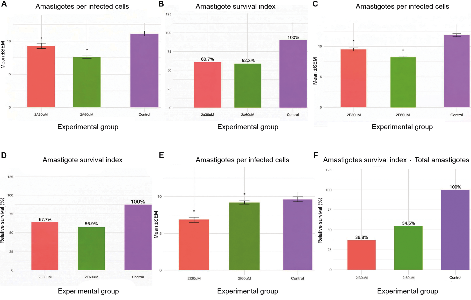
The antiamastigote effect is shown in Fig. 2. In items A and B, it was possible to identify a significant and expressive antiparasitic reduction in the intracellular load of amastigotes of molecule 2a compared to the control group and Bz (data not shown). The survival index was 60.7% and 52.3% for the two concentrations tested (30 and 60 μM), respectively.
In items C and D of Fig. 2, we observed that the 2f molecule showed dose-dependent activity against amastigote forms, with greater reduction of parasites at the concentration of 60 μM, suggesting a good antiparasitic effect in vitro. Furthermore, the survival index indicated moderate toxicity at 30 μM (67.7%) and increased toxicity with the dose (56.9% at 60 μM), evidencing a dose-dependent relationship for both efficacy and toxicity.
In the subsequent items (E and F), still in Fig. 2, the effect of molecule 2i is observed, which also significantly attracted its effect; however, it presented an even more important effect on the survival index results at the lowest concentration (30 μM).
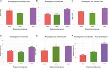
antiamastigote effect (24 h) of molecules 2a, 2f and 2i. Antiamastigote assay showing the number of amastigotes per infected cell (A, C, E) and the survival index (B, D, F) for the studied molecules 2a, 2f, and 2i, respectively. (A-B) correspond to molecule 2a, (C-D) to 2f, and (E-F) to 2i.
Flow cytometry assay - The killing mechanisms identified by the flow cytometry assay were important for evaluating the killing profile of the substances involved in this study. Epimastigote forms and death markers, such as 7AAD and AxV PE, were used to evaluate the apoptotic and necrotic death pathways, respectively.
The control group (n = 10) was evaluated by quadrant analysis (UL/UR/LL/LR), with mean percentage distribution in the gated population (%) of UL 0.55 ± 0.16%, UR 3.48 ± 0.84%, LL 94.99 ± 0.90% and LR 0.98 ± 0.12% [mean ± standard deviation (SD)].
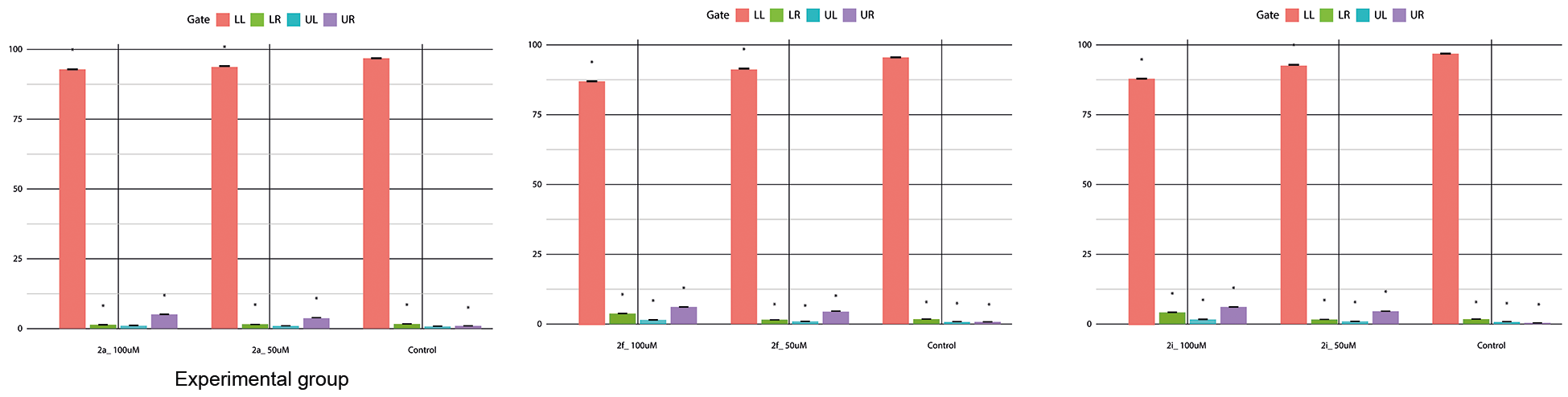
In Fig. 3, in A, molecule 2a can be seen, in the dotplot and bar quadrants, which, at the concentrations tested (50 and 100 μM), did not induce cell death by apoptosis or necrosis. The molecule's profile is virtually identical to that of the control group, with only a slight increase in late apoptosis at the higher dose.
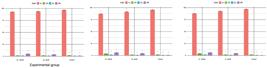
flow cytometry analysis of cell death in epimastigotes. Epimastigote forms of the Trypanosoma cruzi Y strain were incubated for 24 h and analysed by flow cytometry using 7-AAD and Annexin V-PE staining after treatment with molecules 2a, 2f, and 2i. Representative dot plots show the distribution of viable cells (LL), early apoptotic cells (LR), necrotic cells (UL), and late apoptotic cells (UR).
Also in Fig. 3, in B, the graphs of the 2f molecule at a concentration of 100 μM, it is possible to see a significant increase in late apoptosis/necrosis (evidenced by the UR quadrant - purple) and a small increase in early apoptosis (LR - green), compared to the control group. However, at the lower concentration, a profile close to the control was obtained, but with a slight increase in UR.
Molecule 2i, still in Fig. 3, according to the dotplot graph in C, showed greater dispersion in the early apoptosis (LR) quadrants and more evident in late apoptosis (LR), in relation to the control.
Furthermore, the DCFH-DA marker was used to monitor the increased production of ROS in order to assess the cellular integrity profile using oxidised DCF (2'7'-dichlorofluorescein).
For ROS control with DCF, fluorescence in FL1-DCF was expressed as a geometric mean. In the controls (n = 10), the geometric mean was 11.61 ± 0.55 (min-max: 10.37-12.10), demonstrating consistency of the control.
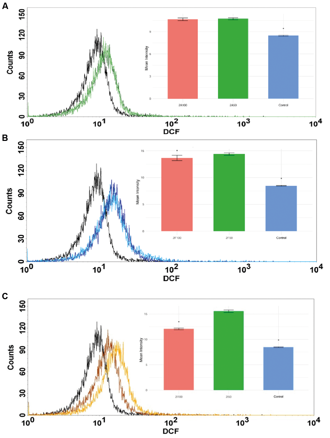
According to Fig. 4, in A, flow cytometry analysis showed that treatment with molecule 2a, at concentrations of 50 and 100 μM, promoted a significant increase in the production of cytoplasmic ROS in relation to the control (p < 0.05).
According to the histogram in Fig. 4, in B, it is possible to observe a shift to the right of the fluorescence peaks; however, no significant differences are observed between the two concentrations, suggesting saturation of the pro-oxidant effect from 50 μM of molecule 2f.
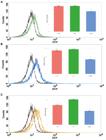
cytoplasmic reactive oxygen species (ROS) production in epimastigotes. Flow cytometry analysis of cytoplasmic ROS production in Trypanosoma cruzi epimastigotes treated with molecules 2a, 2f, and 2i for 24 h. Histograms show the fluorescence intensity profiles of ROS production compared to the control group (CT, black curve). For molecule 2a, dark green and light green curves represent treatments at 100 µM and 50 µM, respectively. For molecule 2f, dark blue and light blue curves represent treatments at 100 µM and 50 µM, respectively. For molecule 2i, orange and brown curves represent treatments at 100 µM and 50 µM, respectively.
The 2i molecule was also represented by flow cytometry, as shown in Fig. 4, in C, which revealed that there was a significant increase in the production of cytoplasmic ROS (in relation to the control). The largest increase was observed at 50 μM followed by 100 μM, both with a rightward shift in the DCF fluorescence histogram.
The relative reduction of ROS at 100 μM compared to 50 μM may be associated with cytotoxic effects, leading to a lower cellular capacity to generate ROS.
Furthermore, the dye Rhodamine 123 (Rho123) was also used to assess the mitochondrial transmembrane potential (ΔΨm). Mitochondrial potential monitored by Rho123 (FL1-Rho123), with fluorescence expressed as geometric mean (Geo Mean). In the controls analysed (n = 6), the average Geo Mean was 91.23 ± 0.22 (min-max: 90.90-91.48), indicating control stability.
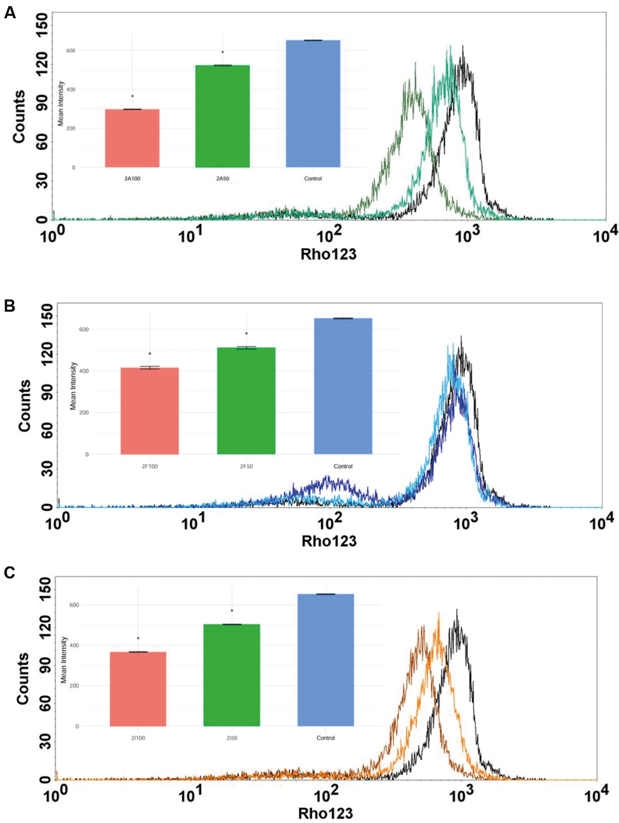
Staining with the Rho123 dye showed, according to the graph above, in Fig. 5, in A, a significant and dose-dependent reduction in mitochondrial fluorescence after treatment with molecule 2a, as observed at the lowest concentration (50 μM) in relation to the highest concentration (100 μM).
The histogram shifts to the left as the dose increases, indicating depolarisation of the mitochondrial membrane potential (relative to the control group).
Meanwhile, the 2f molecule promoted a slight depolarisation of its mitochondrial transmembrane potential using Rho123, at both concentrations in a dose-dependent manner (p < 0.05 vs control). The histograms indicate a slight shift to the left with increasing concentration, shown in Fig. 5, in B.
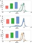
mitochondrial membrane potential (ΔΨm) evaluation in epimastigotes. Flow cytometry analysis of mitochondrial membrane potential (ΔΨm) in Trypanosoma cruzi epimastigotes treated with molecules 2a, 2f, and 2i for 24 h. The mitochondrial membrane potential was evaluated based on relative fluorescence intensity. In all histograms, the black curve represents the control group (CT). For molecule 2a, dark green and light green curves represent treatments at 100 µM and 50 µM, respectively; for molecule 2f, dark blue and light blue curves represent treatments at 100 µM and 50 µM, respectively; and for molecule 2i, brown and orange curves represent treatments at 100 µM and 50 µM, respectively.
The Rho123 assay demonstrated that the 2i molecule promoted a significant dose-dependent depolarisation of the membrane potential in cells treated with 50 μM and 100 μM, compared to the control group. Furthermore, the histogram shows a progressive shift to the left with increasing dose, reflecting lower fluorescence and loss of ΔΨm (Fig. 5, in C).
In antioxidant assays, all samples demonstrated the ability to inhibit free radicals. This potential for inhibiting oxidising radicals was evaluated by measuring the inhibition of DPPH and ABTS radicals, which are widely recognised models for assessing a substance's ability to neutralise free radicals (Table II).
The results obtained indicate that the samples exhibited a high capacity for radical inhibition, both for the ABTS radical and for the DPPH radical, with the latter being more prominent in sample 2a, with average inhibitory concentration (IC50) values of 8.56 ± 0.99 μg.mL-1, being more active than commercial antioxidants BHT and Trolox.
For acetylcholinesterase inhibition, only sample 2a showed enzyme inhibition activity, being classified as having high inhibition, with an IC50 of 18.87 ± 0.30 μg.mL-1, showing it to be a promising molecule in the studies of inhibition of this enzyme (Table II).
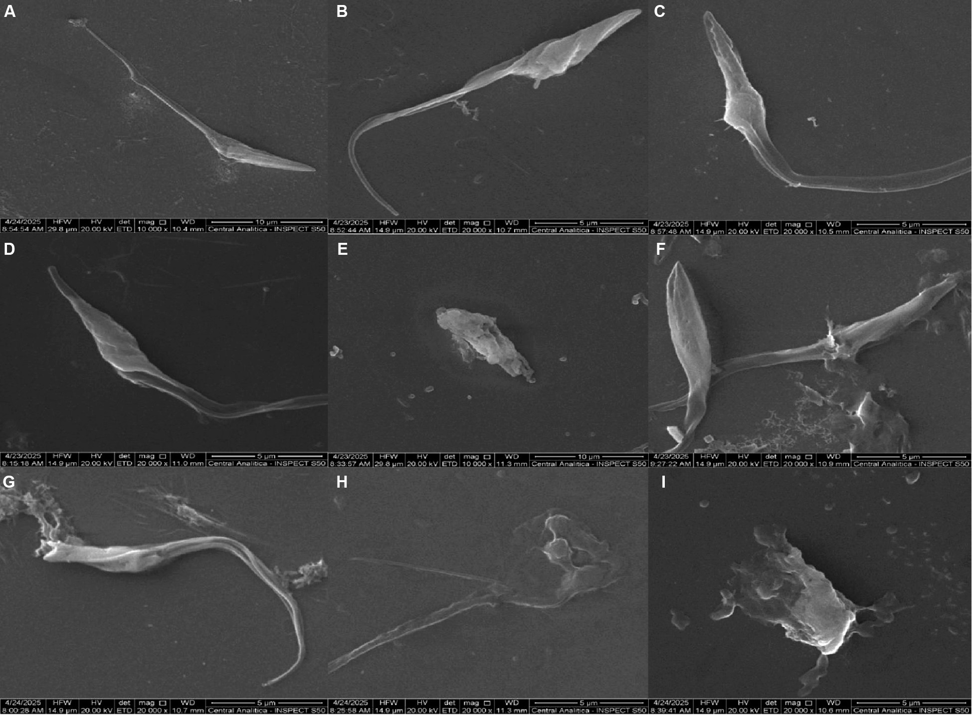
Evaluation of structural changes in the parasite - In order to analyse epimastigote forms microscopically, based on the observation and analysis of the microstructural characteristics of materials, SEM tests were performed at two different concentrations (25 µM and 50 µM) for 24 h, along with the study molecules and the reference drug (Bz: 25 µM and 50 µM).
SEM microscopy revealed different morphologies depending on the concentration and molecule used.
In Fig. 6, in A, the control group can be seen, presenting normal morphology and typical shape, with no apparent morphological alteration. In B and C, the cells treated with Bz are observed, showing irregularity in the body and tail and membrane roughness. In D and E (molecule 2a), changes in thickened, irregular body and membrane swelling, in addition to collapsed shape, loss of shape and absence of flagellum, at the two reported concentrations, respectively.
Images F and G of Fig. 6 show cells treated with molecule 2f, in which the epimastigote forms present membrane pores, a deformed central region, and an expanded and collapsed shape with loss of morphology. Molecule 2i (items H and I) presented membrane rupture with extravasated content and total loss of morphology with the presence of vesicles/fragmented structures around it.
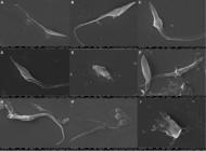
scanning electron microscopy (SEM) of epimastigotes. Representative images showing morphological alterations in Trypanosoma cruzi Y strain epimastigotes. In (A), the control group displays normal morphology and typical elongated shape. In (B) and (C), cells treated with benznidazole (Bz) exhibit body and tail irregularities. In (D) and (E), corresponding to treatments with molecule 2a, cells show collapsed shape and loss of typical morphology. In (F) and (G) show cells treated with molecule 2f, characterised by the presence of membrane pores. Finally, in (H) and (I), corresponding to treatments with molecule 2i, exhibit severe membrane rupture with extravasated cellular content and total loss of morphology.
Evaluation of the in vivo toxicity - After in vitro assays using molecule screening (2a, 2f, and 2i), it was observed that the killing profile and SI were higher for molecule 2a. Therefore, new assays were performed with wild-type (WT) ZebraFish embryos/larvae (code: ZS-TOX-06/25-1) at concentrations of 100, 50, 25, 12.5, and 6.25 µM. The mortality rate was measured at 24, 48, 72, and 96 h.
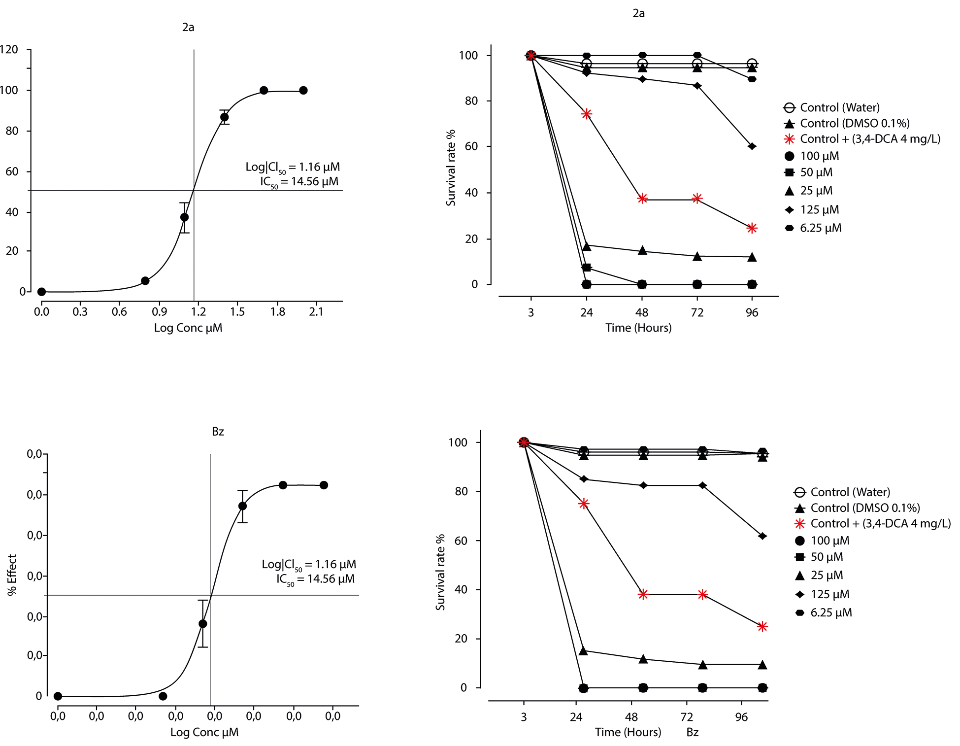
In the dose-response assay (Fig. 7A-1), the sigmoid curve demonstrated a direct relationship between concentration and mortality, with an LC50 calculated at 14.56 µM. This value indicates the concentration required to induce 50% lethality in the tested population, serving as a toxicological reference parameter. The curve also revealed a behaviour with a steep transition range, suggesting that small variations in dose result in significant changes in mortality.
In the time-related survival assay (Fig. 7A-2), it was evident that the toxicity of molecule 2a is time-dependent. At high concentrations, above 50 µM, mortality occurred acutely, with almost complete disappearance of survival in less than 24 h.

concentration effect and mortality/survival rate analysis. Analysis of the concentration-dependent effects of molecule 2a and benznidazole (Bz) on embryonic mortality and survival. Graphs (1) represent the effect of increasing concentrations of each compound on embryonic mortality, while graphs (2) show the corresponding survival rates. Treatments with molecule 2a and Bz demonstrate dose-dependent variations in embryonic viability.
However, at doses of 12.5-25 µM, mortality was progressive, increasing gradually until 96 h, which indicates a cumulative effect and possible involvement of metabolic or bioaccumulation mechanisms in the embryo. On the other hand, at concentrations below 6.25 µM, no significant change in survival rate was observed over 96 h, suggesting a safety range for sublethal assays.
The effect of Bz concentration on embryo mortality was also determined. The LC50 value was 14.7 µM, corresponding to the median lethal concentration, with a 95% confidence interval (CI) between 13.7 µM and 15.8 µM (graph B and A).
Furthermore, the time-related effect on the survival rate of ZebraFish embryos at different concentrations was also evaluated. It was observed that the higher the concentration, the lower the survival rate over time, indicating a dose- and time-dependent effect.
DISCUSSION
Recently, our research group described the activity of 1,2,4-oxadiazole derivatives against tumour cell lines and their possible immune-mediated mechanisms through a systematic review.18 Furthermore, it evaluated their antitumour and immunomodulatory effects in melanoma cells,19 and explored their leishmanicidal20 and trypanocidal activities in vitro.12
Oxadiazoles are five-membered heterocyclic compounds that have been widely explored in various scientific areas, such as the pharmaceutical industry, in drug discovery, for example.
Among the known isomers, the one of greatest interest is 1,2,4-oxadiazole due to its diverse biological activities, such as anti-inflammatory,21 anticancer,22 anticonvulsant23 and antiparasitic.24
The three oxadiazole derivatives (2a, 2f and 2i) prepared here were evaluated in vitro against the epimastigote, trypomastigote and replicative amastigote forms of T. cruzi and in vivo (2a) to evaluate their cytotoxic effect. First, we used the Y strain of T. cruzi, classified for its high in vitro infectivity and partial resistance to Bz, in addition to constituting an important research model.25,26
Initially, the cytotoxic effect of the study molecules was evaluated in host cells, which revealed differences in the cellular toxicity profile when compared. The LLC-MK2 cell line (monkey renal epithelial cells) is widely used in in vitro studies of T. cruzi precisely because of its high susceptibility to infection, viability in culture and ease of laboratory manipulation.27
The oxadiazole derivatives caused a statistically significant reduction in cellular function compared to the control groups. Furthermore, host cell toxicity was also measured by estimating the concentration required to reduce host cell viability by 50% (CC50).
According to the results, Bz presented an estimated CC50 of 59.1 µM, at which it is possible to observe moderate toxicity, probably due to its structure, with a nitroimidazole group (-NO2), known in the literature for generating ROS after its bioactivation, affecting host cells.
The compounds 2f (CC50 14 µM) and 2i (CC50 25.7 µM) are lipophilic and electronegative molecules, containing trifluoromethyl (-CF3) and fluorine (-F) groups, respectively.28 Both structural behaviours are found in the literature, which reveal that one favours penetration into the cell membrane, but can also affect sensitive organelles and generate cell death (2f) and can affect pharmacokinetic properties and unwanted interactions (2i).29
Molecule 2a (CC50 µM) has an unsubstituted phenolic ring linked to a 1,2,4-oxadiazole system. The presence of a cyclohexyl substituent confers a hydrophobic character and increased mass and lipophilicity, characteristics that can reduce cell coverage and the layer by cytotoxic targets, and consequently, contribute to an increase in CC50.30
This result confirms the importance of considering the therapeutic window when comparing compounds. Recent data in the literature indicate that a group of pyrazol-imidazoline derivatives also exhibited low toxicity, with CC50 > 100 µM.31
Furthermore, there is evidence in the literature that explores the structural effects on the bioactivity and toxicity of the evaluated compounds.
The SI is the ability of a molecule to interact effectively with a desired target compared to an undesired one. SI metrics use the ratio of the IC50s obtained between the targets. The higher the SI, the more selective the molecule of interest.32,33
Our data show that, with the exception of molecule 2f, the other molecules 2a and 2i exhibited high SIs compared to the control group, demonstrating that our molecules have promising potential against T. cruzi strains.
Literature data have already demonstrated such activity of molecule 2a in breast cancer and cervical cancer cell lines, which exhibited compound activity at an IC50 of 19.5 μM in MCF-7 cells and 78.7 μM in HeLA cells.34 Nevertheless, molecule 2i has also demonstrated activity in tumour lines of lung carcinoma (A549), PC-3 prostate adenocarcinoma, and breast adenocarcinoma (MDA-MB-231), although.35
The oxadiazole derivatives presented have as a chemical nucleus a modified hydrazide group (-NH-NH-R) attached to the oxadiazole ring, with structural variations in the substituted phenyl ring.29
This structural configuration suggests that these molecules may act as protonophores, that is, weak lipophilic acids capable of transporting protons from the mitochondrial intermembrane space to the matrix, inducing mitochondrial uncoupling.36
Furthermore, when analysing the histograms, it is also possible to notice a shift to the right of the fluorescence peaks for both doses, indicating greater DCF intensity. However, no statistically relevant difference was observed between the two concentrations evaluated, suggesting saturation of the pro-oxidant effect.
Performing the same analysis with the 2f molecule, it can be inferred that both concentrations evaluated (50 and 100 μM) promoted a significant increase in cytoplasmic ROS production.
This can be clearly observed in the results obtained by cytometry with depolarisation of the mitochondrial membrane channel. Protonophores utilise the mitochondrial pH gradient to transport protons from the inner membrane space to the mitochondrial matrix.37
As a consequence of the electrochemical influx of protons across the inner membrane, there will be a disruption in the supply of the ATP synthase enzyme responsible for ATP synthesis, resulting in a reduction in nutrient metabolism.
The mitochondria will need to increase their metabolic rate to compensate for the gradient leakage, potentially disrupting the parasite's energy-gathering mechanisms and resulting cell death.38
In our study, we evaluated the activity of oxadiazole derivatives against the amastigote forms of T. cruzi. All molecules demonstrated significant cytotoxic activity, especially when compared to the control treatment with Bz.
One of the main defence mechanisms of amastigote forms is the ability to invade host cells, replicating in the cytoplasm without being recognised, which makes therapies targeting extracellular forms difficult.
Furthermore, transiently dormant and metabolically inactive amastigote forms are resistant to Bz treatment.39 As previously mentioned, molecules 2a, 2f, and 2i can undergo changes in their structural conformation, and such changes in their radicals, acting as protonophores, can increase the molecule's lipophilicity.
This property facilitates intracellular penetration and enhances the efficacy of treatment against intracellular parasites.40 As already discussed, changes in mitochondrial membrane potential and ROS production were observed in cells treated with molecules 2a, 2f, and 2i.
This effect may be related to the structural nature of the molecules, which can act directly on the mitochondrial membrane; modulation of proton flux may thus influence ROS production.40 Excessive ROS production can cause lipid peroxidation of cell membranes, promoting apoptosis and/or late apoptosis, as evidenced in our flow cytometry results.41
Simultaneously, in addition to the activities mentioned above, the ability of the molecules to neutralise free radicals in solution was evaluated through the analysis of antioxidant activity using methodologies such as DPPH and ABTS.
Research with new 1,3,4-oxadiazole derivatives also showed antioxidant activity, with an IC50 value of 2.2 µg/mL. From a biological perspective, this observed activity is especially relevant because T. cruzi requires a redox system, based on trypanothione reductase and related enzymes, to withstand the oxidative stress imposed by the host.42
In this study, molecule 2a showed the highest efficiency, being suggested as a hydrogen donor in the DPPH test, probably due to aromatic conjugation and the presence of NH groups.
However, its activity was limited in the ABTS test, where it showed lower solubility or reduced efficiency in electron donation in aqueous medium.14
On the other hand, molecule 2f exhibited a stable antioxidant profile, being more effective in the ABTS test due to the presence of the trifluoromethyl group in its structure, which increases its polarity.
Similarly, molecule 2i showed better performance in the ABTS system, indicating that the structural substituents in these compounds alter their antioxidant capacity according to the type of radical and the physicochemical properties.43
These changes are in line with the literature that reports the antioxidant activity of these heterocyclic compounds, which can vary according to the nature of their radical and the physicochemical properties of the molecules. Furthermore, the compounds studied demonstrated a high capacity for scavenging the ABTS radical and reducing the ferric ion.44
Furthermore, in the results demonstrated in the evaluation of acetylcholinesterase inhibition, molecule 2a was also greater than the controls, including Bz and the other molecules.
Although T. cruzi does not have a functional cholinergic system like mammals, they possess the presence of choline and functional membrane acetylcholine.45
Therefore, the inhibition of choline formation could be evaluated using acetylcholinesterase (AChE) enzyme inhibitors. Regarding the ultrastructure of T. cruzi, research indicates that the parasite requires specific sterols at all stages of its cell cycle, which ensure cell viability and proliferation.
These sterols, specifically the ergosterol biosynthesis pathway, are essential for the parasite, since they are unable to survive solely on host cholesterol.46
Therefore, T. cruzi is vulnerable to inhibitors of the 14α-demethylase (CYP51) enzyme, an enzyme that catalyses the conversion of lanosterol to ergosterol, resulting in structural alterations of various organelles and cytotoxic consequences.
Additionally, we employed SEM to evaluate the molecules under study (2a, 2f, and 2i) at concentrations of 25 and 50 µM.
In SEM assay, the molecules (2a, 2f and 2i), at concentrations of 25 and 50 μM, caused different changes in the parasitic forms (epimastigotes), including membrane irregularity, roughness, swelling, cell collapse, and loss of the flagellum. This is comparable to what was observed,47 who reported ultrastructural changes in treated trypomastigote forms.
After the in vitro assays performed and the results analysed, molecule 2a was an excellent candidate for initiating in vivo assays with ZebraFish. The results show that molecule 2a exhibited dose-dependent embryonic toxicity (LC50 14-15 μM).
The use of ZebraFish embryos as an in vivo model is validated by their high sensitivity to toxic compounds and their applicability in drug screening. The LC50 obtained (14.56 µM) suggests that molecule 2a exhibits moderate toxicity, with a relatively narrow concentration range between no effect (below 6.25 µM) and significant lethality (above 12.5 µM).
Results in ZebraFish using Bz and molecule 2a were also performed. Molecule 2a demonstrated marked toxicity in Danio rerio embryos compared to Bz.
While Bz exhibited the expected dose-dependent profile, with an IC50 of 14.7 µM and mortality at the highest concentrations, molecule 2a caused early mortality at intermediate concentrations, suggesting significant cytotoxic potential.
Even when tested in combination with Bz, the LC50 remained very similar. The chemical nucleus of 2a may contribute to the observed toxicity, while Bz, in this same arrangement, appears to have only a secondary role in acute lethality.
This is consistent with findings in the literature, which report that 1,2,4-oxadiazole derivatives almost frequently exhibit embryonic toxicity in ZebraFish.30
Many drug interaction effects only emerge when metabolism is active. However, the embryo has limited metabolic capacity and differences in biotransformation pathways, which may explain the absence of observed interactions.48
Further studies are needed to assess how the covariance of toxicity in ZebraFish is influenced by factors such as absorption, metabolism, and toxicity mechanisms. The next step may be to reduce exposure in the organism and increase delivery to the site of action through controlled release, such as loaded nanoparticles, to limit the free availability of the compound.49
In conclusion, the molecules studied showed different activities against T. cruzi. The mechanism of action was largely related to mitochondrial dysfunction, redox imbalance, and apoptotic processes. Molecule 2a stood out for its great antiparasitic activity in vitro, justifying its evaluation in an in vivo model, which showed dose-dependent embryonic toxicity. These results provide important information on the therapeutic window and the need for optimisation strategies such as controlled release systems. Further in vivo assays in murine infection models are needed, as well as the need for complementary molecular targets.
ACKNOWLEDGEMENTS
To the Laboratório de Bioprospecção Farmacêutica e Bioquímica Clínica (LBFBC), Central Analítica, Universidade Federal do Ceará (UFC), Fundação Oswaldo Cruz (FIOCRUZ-CE), and Z-Safe Biotecnologia LTDA.
Funding
How to cite:
DATA AVAILABILITY
The datasets generated and/or analysed during the current study are not publicly available but are available from the corresponding author upon reasonable request.
REFERENCES
-
1 Bern C, Messenger LA, Whitman JD, Maguire JH. Chagas disease in the United States: a public health approach. Clin Microbiol Rev. 2019; 33: 10.1128/cmr.00023-19. doi: 10.1128/CMR.00023-19.
» https://doi.org/10.1128/CMR.00023-19 -
2 Antinori S, Galimberti L, Bianco R, Grande R, Galli M, Corbellino M, et al. Chagas disease in Europe: a review for the internist in the globalized world. Eur J Intern Med. 2017; 43: 6-15. doi: 10.1016/j.ejim.2017.05.001.
» https://doi.org/10.1016/j.ejim.2017.05.001 -
3 Echavarría NG, Echeverría LE, Stewart M, Gallego C, Saldarriaga C. Chagas disease: chronic Chagas cardiomyopathy. Curr Probl Cardiol. 2021; 46: 100507. doi: 10.1016/j.cpcardiol.2019.100507.
» https://doi.org/10.1016/j.cpcardiol.2019.100507 -
4 Clark EH, Messenger LA, Whitman JD, Bern C. Chagas disease in immunocompromised patients. Clin Microbiol Rev. 2024; 37: e00099-23. doi: 10.1128/cmr.00099-23.
» https://doi.org/10.1128/cmr.00099-23 -
5 Forsyth CJ, Hernandez S, Olmedo W, Abuhamidah A, Traina MI, Sanchez DR, et al. Safety profile of nifurtimox for treatment of Chagas disease in the United States. Clin Infect Dis. 2016; 63: 1056-62. doi: 10.1093/cid/ciw477.
» https://doi.org/10.1093/cid/ciw477 -
6 Crespillo-Andújar C, Venanzi-Rullo E, López-Vélez R, Monge-Maillo B, Norman F, López-Polín A, et al. Safety profile of benznidazole in the treatment of chronic Chagas disease: experience of a referral centre and systematic literature review with meta-analysis. Drug Saf. 2018; 41: 1035-48. doi: 10.1007/s40264-018-0696-5.
» https://doi.org/10.1007/s40264-018-0696-5 -
7 Wang JJ, Sun W, Jia WD, Bian M, Yu LJ. Research progress on the synthesis and pharmacology of 1,3,4-oxadiazole and 1,2,4-oxadiazole derivatives: a mini review. J Enzyme Inhib Med Chem. 2022; 37: 2304-19. doi: 10.1080/14756366.2022.2115036.
» https://doi.org/10.1080/14756366.2022.2115036 -
8 Melo de Oliveira VN, Dos Santos FG, Ferreira VPG, Araújo HM, Do Ó Pessoa C, Nicolete R, et al. Focused microwave irradiation-assisted synthesis of N-cyclohexyl-1,2,4-oxadiazole derivatives with antitumor activity. Synth Commun. 2018; 48(19): 2522-32. doi: 10.1080/00397911.2018.1509350.
» https://doi.org/10.1080/00397911.2018.1509350 -
9 Mosmann T. Rapid colorimetric assay for cellular growth and survival: application to proliferation and cytotoxicity assays. J Immunol Methods. 1983; 65(1-2): 55-63. doi: 10.1016/0022-1759(83)90303-4.
» https://doi.org/10.1016/0022-1759(83)90303-4 -
10 Duran RJ, Muñoz-Calderon A, Gómez KA, Potenza M. In vitro differentiation of Trypanosoma cruzi epimastigotes into metacyclic trypomastigotes using a biphasic medium. STAR Protoc. 2021; 2(3): 100703. doi: 10.1016/j.xpro.2021.100703.
» https://doi.org/10.1016/j.xpro.2021.100703 -
11 Marín C, Ramírez-Macías I, Rosales MJ, Muro B, Reviriego F, Navarro P, et al. In vitro leishmanicidal activity of 1,3-disubstituted 5-nitroindazoles. Acta Trop. 2015; 148: 170-8. https://doi.org/10.1016/j.actatropica.2015.04.028.
» https://doi.org/10.1016/j.actatropica.2015.04.028 -
12 Rocha YM, Magalhães EP, Chaves MM, Marinho MM, de Oliveira VNM, De Oliveira RN, et al. Antiparasitary and antiproliferative activities in vitro of a 1,2,4-oxadiazole derivative on Trypanosoma cruzi Parasitol Res. 2022; 121(7): 2141-56. doi: 10.1007/s00436-022-07554-z.
» https://doi.org/10.1007/s00436-022-07554-z -
13 Barraca A, Sgarbi G, Solaini G, Lenaz G. Rhodamine 123 as a probe of mitochondrial membrane potential: evaluation of proton flux through F0 during ATP synthesis. Biochim Biophys Acta Bioenerg. 2003; 1606(1-3): 137-46. doi: https://doi.org/10.1016/S0005-2728(03)00110-5.
» https://doi.org/10.1016/S0005-2728(03)00110-5 -
14 Becker MM, Nunes GS, Ribeiro DB, Silva FEPS, Catanante G, Marty JL. Determination of the antioxidant capacity of red fruits by miniaturized spectrophotometry assays. J Braz Chem Soc. 2019; 30: 1108-14. doi: https://doi.org/10.21577/0103-5053.20190003.
» https://doi.org/10.21577/0103-5053.20190003 -
15 Re R, Pellegrini N, Proteggente A, Pannala A, Yang M, Rice-Vans C. Antioxidant activity applying an improved ABTS radical cation decolorization assay. Free Radic Biol Med. 1999; 26(9-10): 1231-7. doi: 10.1016/s0891-5849(98)00315-3.
» https://doi.org/10.1016/s0891-5849(98)00315-3 -
16 Rhee IK, Van de Meent M, Ingkanina K, Verpoorte R. Screening for acetylcholinesterase inhibitors from Amaryllidaceae using silica gel thin-layer chromatography in combination with bioactivity staining. J Chromatogr A. 2001; 915(1-2): 217-23. doi: 10.1016/s0021-9673(01)00624-0.
» https://doi.org/10.1016/s0021-9673(01)00624-0 -
17 Trevisan MTS, Macedo FVV. Seleção de plantas com atividade anticolinasterase para tratamento da doença de Alzheimer. Quim Nova. 2003; 26: 301-4. doi: https://doi.org/10.1590/S010040422003000300002.
» https://doi.org/10.1590/S010040422003000300002 -
18 Araújo HM, de Moura GA, Rocha YM, Rodrigues JPV, Nicolete R. Oxadiazole derivatives as anticancer and immunomodulatory agents: a systematic review. Curr Med Chem. 2023; 30: 3472-85. doi: 10.2174/0929867329666220929145619.
» https://doi.org/10.2174/0929867329666220929145619 -
19 Araújo HM, de Moura GA, Rocha YM, Gomes CVP, de Oliveira RN, Nicolete LDF, et al. In vitro antitumor and immunomodulatory activities of 1, 2, 4-oxadiazole derivatives. Biochem Biophys Rep. 2025; 41: 10195. doi: https://doi.org/10.1016/j.bbrep.2025.101950.
» https://doi.org/10.1016/j.bbrep.2025.101950 -
20 Pinheiro CVG, Rocha YM, Rodrigues JPV, de Moura GA, de Oliveira JR, Lourenço FD, et al. In silico and in vitro assessment of anti-Leishmania infantum activity of a novel cyclohexyl-1, 2, 4-oxadiazole derivative. Mol Biochem Parasitol. 2025; 262: 111674. doi: 10.1016/j.molbiopara.2025.111674.
» https://doi.org/10.1016/j.molbiopara.2025.111674 -
21 Mohamed MFA, Marzouk AA, Nafady A, El-Gamal DA, Allam RM, Abuo-Rahma GE, et al. Design, synthesis and molecular modeling of novel aryl carboximidamides and 3-aryl-1, 2, 4-oxadiazoles derived from indomethacin as potent anti-inflammatory iNOS/PGE2 inhibitors. Bioorg Chem. 2020; 105: 104439. doi: 10.1016/j.bioorg.2020.104439.
» https://doi.org/10.1016/j.bioorg.2020.104439 -
22 Kumar D, Patel G, Chavers AK, Chang K-H, Shah K. Synthesis of novel 1, 2, 4-oxadiazoles and analogues as potential anticancer agents. Eur J Med Chem. 2011; 46: 3085-3092. doi: https://doi.org/10.1016/j.ejmech.2011.03.031.
» https://doi.org/10.1016/j.ejmech.2011.03.031 -
23 Mohammadi-Khanaposhtani M, Shabani M, Faizi M, Aghaei I, Jahani R, Shafari Z, et al. Design, synthesis, pharmacological evaluation, and docking study of new acridone-based 1, 2, 4-oxadiazoles as potential anticonvulsant agents. Eur J Med Chem. 2016; 112: 91-8. doi: 10.1016/j.ejmech.2016.01.054.
» https://doi.org/10.1016/j.ejmech.2016.01.054 -
24 Pitasse-Santos P, Sueth-Santiago V, Lima MEF. 1, 2, 4-and 1, 3, 4-Oxadiazoles as scaffolds in the development of antiparasitic agents. J Braz Chem Soc. 2018; 29(3): 435-56. doi: https://doi.org/10.21577/0103-5053.20170208.
» https://doi.org/10.21577/0103-5053.20170208 -
25 Sykes ML, Kennedy EK, Avery VM. Impact of laboratory-adapted intracellular Trypanosoma cruzi strains on the activity profiles of compounds with anti-T. cruzi activity. Microorganisms. 2023; 11: 476. doi: https://doi.org/10.3390/microorganisms11020476.
» https://doi.org/10.3390/microorganisms11020476 -
26 Franco CH, Alcantara LM, Chatelain E, Freitas-Junior LH, Moraes CB. Drug discovery for Chagas disease impact of different host cell lines on assay performance and hit compound selection. Trop Med Infect Dis. 2019; 4: 82. doi: 10.3390/tropicalmed4020082.
» https://doi.org/10.3390/tropicalmed4020082 -
27 Duran-Rehbein GA, Vargas-Zambrano JC, Cuellar A, Puerta CJ, Gonzalez JM. Mammalian cellular culture models of Trypanosoma cruzi infection a review of the published literature. Parasite. 2014; 21: 38. doi: 10.1051/parasite/2014040.
» https://doi.org/10.1051/parasite/2014040 -
28 Liu X-H, Wen Y-H, Cheng L, Xu T-M, Wu N-J. Design, synthesis, and pesticidal activities of pyrimidin-4-amine derivatives bearing a 5-(trifluoromethyl)-1, 2, 4-oxadiazole moiety. J Agric Food Chem. 2021; 69: 6968-80. doi: 10.1016/j.ejmech.2011.03.031.
» https://doi.org/10.1016/j.ejmech.2011.03.031 -
29 Zainab SR, Khan JZ, Nadeem H, Tipu MK, Irshad N. Safety assessment of novel oxadiazole derivatives in acute and sub-acute toxicity studies. Naunyn Schmiedebergs Arch Pharmacol. 2025; 398: 3631-53. doi: 10.1007/s00210-024-03474-0.
» https://doi.org/10.1007/s00210-024-03474-0 -
30 Yang S, Ren C-L, Ma T-Y, Zou W-Q, Dai L, Tian X-Y, et al. 1, 2, 4-oxadiazole-based bio-isosteres of benzamides synthesis, biological activity and toxicity to zebrafish embryo. Int J Mol Sci. 2021; 22: 2367. doi: 10.3390/ijms22052367.
» https://doi.org/10.3390/ijms22052367 -
31 De Oliveira EC, Lara LS, Orlando LMR, Lanera SC, De Souza TP, Figueiredo NS, et al. Anti-Trypanosoma cruzi potential of new pyrazole-imidazoline derivatives. Molecules. 2025; 30: 3082. doi: https://doi.org/10.3390/molecules30153082.
» https://doi.org/10.3390/molecules30153082 -
32 Huggins DJ, Sherman W, Tidor B. Rational approaches to improving selectivity in drug design. J Med Chem. 2012; 55: 1424-44. doi: https://doi.org/10.1021/jm2010332.
» https://doi.org/10.1021/jm2010332 -
33 Lica JJ, Wieczór M, Grabe GJ, Heldt M, Jancz M, Misiak M, et al. Effective drug concentration and selectivity depends on fraction of primitive cells. Int J Mol Sci. 2021; 22: 4931. doi: https://doi.org/10.3390/ijms22094931.
» https://doi.org/10.3390/ijms22094931 -
34 Hassanzadeh F, Elham J, Mohammadreza Z, Ghadamali K, Golnaz V. Synthesis, cytotoxic evaluation, and molecular docking studies of some new 1, 3, 4-oxadiazole-based compounds. Res Pharm Sci. 2020; 15: 454-62. doi: 10.4103/1735-5362.297848.
» https://doi.org/10.4103/1735-5362.297848 -
35 Aydin E, Senturk AM, Kucuk HB, Guzel M. Cytotoxic activity and docking studies of 2-arenoxybenzaldehyde n-acyl hydrazone and 1, 3, 4-oxadiazole derivatives against various cancer cell lines. Molecules. 2022; 27: 7309. doi: 10.3390/molecules27217309.
» https://doi.org/10.3390/molecules27217309 -
36 Childress ES, Salamoun JM, Hargett SR, Alexopoulos SJ, Chen S-H, Shah DP, et al. [1, 2, 5] Oxadiazolo [3, 4-b] pyrazine-5, 6-diamine derivatives as mitochondrial uncouplers for the potential treatment of nonalcoholic steatohepatitis. J Med Chem. 2020; 63: 2511-26. doi: 10.1021/acs.jmedchem.9b01440.
» https://doi.org/10.1021/acs.jmedchem.9b01440 -
37 Kenwood BM, Calderone JA, Taddeo EP, Hohen KL, Santos WL. Structure–activity relationships of furazano [3, 4-b] pyrazines as mitochondrial uncouplers. Bioorg Med Chem Lett. 2015; 25: 4858-61. doi: 10.1016/j.bmcl.2015.06.040.
» https://doi.org/10.1016/j.bmcl.2015.06.040 -
38 Murray JH, Burgio AL, Beretta M, Hargett SR, Harris TE, Olzomer E, et al. Oxadiazolopyridine derivatives as efficacious mitochondrial uncouplers in the prevention of diet-induced obesity. J Med Chem. 2023; 66: 3876-95. doi: 10.1021/acs.jmedchem.2c01573.
» https://doi.org/10.1021/acs.jmedchem.2c01573 -
39 Bustamente JM, Sanchez-Valdez F, Padilla AM, White B, Wang W, Tarleton RL. A modified drug regimen clears active and dormant trypanosomes in mouse models of Chagas disease. Sci Transl Med. 2020; 12: eabb7656. doi: 10.1126/scitranslmed.abb7656.
» https://doi.org/10.1126/scitranslmed.abb7656 -
40 Khailova LS, Firsov AM, Kotova EA, Antonenko YN. Interaction of potent mitochondrial uncouplers with thiol-containing antioxidants. Antioxidants. 2019; 8: 194. doi: 10.3390/antiox8060194.
» https://doi.org/10.3390/antiox8060194 -
41 Su LJ, Zhang JH, Gomez H, Murugan R, Hong X, Xu D, et al. Reactive oxygen species-induced lipid peroxidation in apoptosis, autophagy, and ferroptosis. Oxid Med Cell Longev. 2019; 2019: 5080843. doi: 10.1155/2019/5080843.
» https://doi.org/10.1155/2019/5080843 -
42 Nazarbahjat N, Ariffin A, Abdullah Z, Abdulla MA, Shia JKS, Leong KH. Synthesis, characterization, drug-likeness properties and determination of the in vitro antioxidant and cytotoxic activities of new 1,3,4-oxadiazole derivatives. Med Chem Res. 2016; 25: 2015-29. doi: 10.1007/s00044-016-1660-5.
» https://doi.org/10.1007/s00044-016-1660-5 -
43 Khalaf NA, Said GE, Abdel-Latif E, Metwally HM. Exploring 1,3,4-oxadiazolyl sulfide derivatives as antidiabetic candidates: synthesis, antioxidant activity, SAR-study, molecular docking, and DFT-insights. BMC Chem. 2025; 19: 316. doi: 10.1186/s13065-025-01678-w.
» https://doi.org/10.1186/s13065-025-01678-w -
44 Mihailovic N, Markovic V, Matic IZ, Stanisavljevic NS, Jovanovic ZS, Trifunovic S, et al. Synthesis and antioxidant activity of 1,3,4-oxadiazoles and their diacylhydrazine precursors derived from phenolic acids. RSC Adv. 2017; 7: 8550-60. doi: 10.1039/c6ra28787e.
» https://doi.org/10.1039/c6ra28787e -
45 Barnadas-Carceller B, Martinez-Peinado N, Gómez LC, Ros-Lucas A, Gabaldón-Figueira JC, Diaz-Mochon JJ, et al. Identification of compounds with activity against Trypanosoma cruzi within a collection of synthetic nucleoside analogs. Front Cell Infect Microbiol. 2023; 12: 1067461. doi: 10.3389/fcimb.2022.1067461.
» https://doi.org/10.3389/fcimb.2022.1067461 -
46 Chen CK, Leung SSF, Guilbert C, Jacobson MP, McKerrow JH, Podust LM. Structural characterization of CYP51 from Trypanosoma cruzi and Trypanosoma brucei bound to the antifungal drugs posaconazole and fluconazole. PLoS Negl Trop Dis. 2010; 4: e651. doi: https://doi.org/10.1371/journal.pntd.0000651.
» https://doi.org/10.1371/journal.pntd.0000651 -
47 Dos Santos Filho JM, Moreira DRM, De Simone CA, Ferreira RS, McKerrow JH, Meira CS, et al. Optimization of anti-Trypanosoma cruzi oxadiazoles leads to identification of compounds with efficacy in infected mice. Bioorg Med Chem. 2012; 20: 6423-33. doi: 10.1016/j.bmc.2012.08.047.
» https://doi.org/10.1016/j.bmc.2012.08.047 -
48 Ali S, van Mil HGJ, Richardson MK. Large-scale assessment of the zebrafish embryo as a possible predictive model in toxicity testing. PLoS One. 2011; 6: e21076. doi: 10.1371/journal.pone.0021076.
» https://doi.org/10.1371/journal.pone.0021076 -
49 Gomes DC, Medeiros TS, Pereira ELA, Da Silva JFO, Oliveira JWF, Fernandes-Pedrosa MF, et al. From benznidazole to new drugs: nanotechnology contribution in Chagas disease. Int J Mol Sci. 2023; 24(18): 13778. doi: 10.3390/ijms241813778.
» https://doi.org/10.3390/ijms241813778
Correspondence
+ Corresponding author: roberto.nicolete@fiocruz.br | https://orcid.org/0000-0002-6101-1155Conflict of interest
Handling editor:
FIRST REVIEW ROUND - REVIEWERS COMMENTS
REVIEWER #1
Reviewing this manuscript was a pleasure. The work is well-structured and presents interesting findings that may contribute to future alternative therapies for Chagas disease. I have a few minor suggestions for the authors, primarily concerning clarification of terms and improvements to the clarity and flow of the manuscript:
1. The stock concentration values are not described in the Methods section.
2. The acronym MTT should be defined at its first occurrence in the text.
3. The acronym DMEM should be defined at its first occurrence in the text.
4. The origin of the LLC-MK2 cell line is mentioned only in the Discussion; this information should already be provided in the Methods section.
5. Regarding the figure titles, I assumed they followed the order in which they appear. However, in the text you refer to Figures 1, 2, and 3, but the figures themselves are not labeled as such.
6. Please improve the quality of the figures and increase the font size, as even with zooming it was difficult to visualize the data and compare it with what is described in the text.
REVIEWER #2
This paper investigates the in vitro anti-Trypanosoma cruzi activity of three N-cyclohexyl-3-(3-methylphenyl)-1,2,4-oxadiazole-5-amine derivatives (2a, 2f, 2i) across epimastigote, trypomastigote, and intracellular amastigote stages of the Y strain, and explores putative death mechanisms via flow cytometry (Annexin V/7-AAD, DCFH-DA, Rh123) and SEM. It also assesses in vivo acute embryotoxicity of the most selective compound (2a) in zebrafish embryos under OECD FET236. The work addresses an important unmet need in Chagas disease drug discovery, where current therapies are limited, particularly regarding chronic infection. It provides starting points for structure–activity–toxicity (SAT) considerations within the oxadiazole scaffold class and flags embryotoxicity risks early in the process.
Please replace the "Conclusion" section with "Results" in the abstract.
Introduction: When mentioning the biological activities of the 1,2,4-oxadiazole class in the final paragraph of the introduction, the authors should include its antiparasitic activity (as stated in the abstract). The specific objective of the study must also be clearly stated in this final paragraph.
Results and Discussion: The study identifies compound 2a as the most selective and 2i as the most potent. Consider to perform functional assays (ATP quantification/mitochondrial respiration) and include positive controls to calibrate the flow cytometry readings and gating to validate the proposed mechanism of action.
Efficacy against amastigotes is critical for translational relevance. Provide a complete dose-response curve (at least 6 concentrations), IC50 values and percentage of infected cells.
Consider to expand host cell cytotoxicity assays to include human-relevant cells (derived cardiomyocytes or human fibroblasts) to better estimate the therapeutic index in disease-relevant tissues.
The Conclusion section must be integrated as part of the main text.
The manuscript needs revision for English.
Verify and ensure correct units, labels, and legends for all tables and graphs.
Avoid using abbreviations when referring to Figures and Tables within the text. Standardize the format using title case (e.g., Figure 1, Table 2).
Titles and Resolution: All figures are missing titles. Figures 2, 3, 4, and 5 are in low resolution, making it impossible to read the graphs and legends.
Table 1: Change the numbering from "Table I" to "Table 1".
Units: Include the concentration unit for the CC50 (LLC-MK2) values.
AUTHORS' RESPONSE TO THE REVIEWERS
February, 14th, 2026.
Dear Editor,
I am sending the revised form of the manuscript entitled "EVALUATION OF THE IN VITRO ACTIVITY OF SYNTHETIC DERIVATIVES OF N-CYCLOHEXYL-3(3-METHYLPHENYL)-1,2,4-OXADIAZOLE-5AMINE ON THE STRAIN Y OF Trypanosoma cruzi AND AN IN VIVO TOXICITY STUDY" to be considered for publication in the "MIOC".
The suggestions made for this revision have been incorporated in the text. We trust that corrections and additions made to the manuscript have improved it considerably and we hope that you will now find it suitable for publication.
We would like to thank you for the interest by our study.
The specific criticisms and suggestions made to this study will be pointed below and changes in the manuscript were also highlighted, as indicated:
In response to the reviewers' comments:
Reviewer #1
Reviewing this manuscript was a pleasure. The work is well-structured and presents interesting findings that may contribute to future alternative therapies for Chagas disease. I have a few minor suggestions for the authors, primarily concerning clarification of terms and improvements to the clarity and flow of the manuscript:
R: Thanks for your interest and suggestions made for improving the manuscript!
1. The stock concentration values are not described in the Methods section.
It was inserted, as required (page 2, section Chemicals).
2. The acronym MTT should be defined at its first occurrence in the text.
It was inserted, as required (page 2, section Cytotoxicity Assay).
3. The acronym DMEM should be defined at its first occurrence in the text.
It was inserted, as required (page 2, section Cytotoxicity Assay).
4. The origin of the LLC-MK2 cell line is mentioned only in the Discussion; this information should already be provided in the Methods section.
It was inserted, as required (page 2, section Cytotoxicity Assay).
5. Regarding the figure titles, I assumed they followed the order in which they appear. However, in the text you refer to Figures 1, 2, and 3, but the figures themselves are not labeled as such.
It was corrected, as required for the entire text.
6. Please improve the quality of the figures and increase the font size, as even with zooming it was difficult to visualize the data and compare it with what is described in the text.
Thanks! All figures were improved, as required.
Reviewer #2
This paper investigates the in vitro anti-Trypanosoma cruzi activity of three N-cyclohexyl-3-(3-methylphenyl)-1,2,4-oxadiazole-5-amine derivatives (2a, 2f, 2i) across epimastigote, trypomastigote, and intracellular amastigote stages of the Y strain, and explores putative death mechanisms via flow cytometry (Annexin V/7-AAD, DCFH-DA, Rh123) and SEM. It also assesses in vivo acute embryotoxicity of the most selective compound (2a) in zebrafish embryos under OECD FET236. The work addresses an important unmet need in Chagas disease drug discovery, where current therapies are limited, particularly regarding chronic infection. It provides starting points for structure–activity–toxicity (SAT) considerations within the oxadiazole scaffold class and flags embryotoxicity risks early in the process.
Please replace the "Conclusion" section with "Results" in the abstract.
Thanks! It was corrected, as required (page 1, highlighted in the text).
Introduction: When mentioning the biological activities of the 1,2,4-oxadiazole class in the final paragraph of the introduction, the authors should include its antiparasitic activity (as stated in the abstract). The specific objective of the study must also be clearly stated in this final paragraph.
It was added in the text, as required (page 2, highlighted in the Introduction section).
Results and Discussion: The study identifies compound 2a as the most selective and 2i as the most potent. Consider to perform functional assays (ATP quantification/mitochondrial respiration) and include positive controls to calibrate the flow cytometry readings and gating to validate the proposed mechanism of action.
Thanks for the suggestion to expand our assays. In this study was conducted an antioxidant assay and also was included now the acetylcholinesterase inhibition assay to reinforce the in vitro effects of the compounds tested and better speculate the differences between each one derivative regarding distinct targets on the parasite. We added the methodology and also better discussed information regarding these mechanisms, especially for 2a molecule, in the revised text highlighted in the pages 4, 6 and 8 and also in the results from Table 2. Moreover, a discussed text correlating ROS inhibition and mitochondrial activity was already pointed in the Discussion section (pages 7 and 8): "Performing the same analysis with the 2f molecule, it can be inferred that both concentrations evaluated (50 and 100 μM) promoted a significant increase in cytoplasmic ROS production.
This can be clearly observed in the results obtained by cytometry with depolarization of the mitochondrial membrane channel. Protonophores utilize the mitochondrial pH gradient to transport protons from the inner membrane space to the mitochondrial matrix (37).
As a consequence of the electrochemical influx of protons across the inner membrane, there will be a disruption in the supply of the ATP synthase enzyme responsible for ATP synthesis, resulting in a reduction in nutrient metabolism.
The mitochondria will need to increase their metabolic rate to compensate for the gradient leakage, potentially disrupting the parasite's energy-gathering mechanisms and resulting cell death (38)"
Regarding the flow cytometry assay, the positive control of the gating strategy in the 7aad and annexin v experiment is justified by the robustness of the marker, as you observe in the graph. Some articles also do not use this positive control. Although they do not use a known agent to induce apoptosis as a positive control, they compare biological states (apoptotic vs. non-apoptotic) in real populations. For Ros production and ΔΨm determination the positive controls were described in the text (highlighted in pages 3 and 4). We have also performed our assay in line with this similar approach, e.g by the manuscripts listed below and highlighted in the References section:
-ARAÚJO, Héverton Mendes et al. In vitro antitumor and immunomodulatory activities of 1, 2, 4-oxadiazole derivatives. Biochemistry and Biophysics Reports, v. 41, p. 101950, 2025.
-DE, Pradip et al. Triple fluorescence staining to evaluate mechanism-based apoptosis following chemotherapeutic and targeted anti-cancer drugs in live tumor cells. Scientific reports, v. 8, n. 1, p. 13192, 2018.
-ANTHONY, R. S. et al. Flow cytometry using annexin V can detect early apoptosis in peripheral blood stem cell harvests from patients with leukaemia and lymphoma. Bone marrow transplantation, v. 21, n. 5, p. 441-446, 1998.
Efficacy against amastigotes is critical for translational relevance. Provide a complete dose-response curve (at least 6 concentrations), IC50 values and percentage of infected cells.
Thank you for your suggestion! We agree that an IC50 determination requires a full dose-response curve with multiple concentrations spanning the dynamic range. In the intracellular amastigote assay conducted here, only two concentrations (30 and 60 µM) were tested due to mainly previous standardization of these concentrations employed in previous assays from our research group and also to experimental limitations at the time of the study, which prevents a robust logistic fit of 4 more parameters. This is in line with published guidelines indicating that a true IC50 prediction requires ≥5 spanning the most response range and multiple concentrations above and below 50% effect.
To meet your request properly, we report (i) the percentage of infected cells at each concentration and (ii) the normalized parasite load (amastigotes per 100 cells) expressed as % survival relative to the untreated control (Figure 2). In addition, we provide an IC50 estimated by logarithmic interpolation between 30 and 60 µM (IC50 ≈ 72 µM), clearly labeled. This is an approximation due to the limited number of concentrations. The complete dose-response curve (≥6 concentrations) will be developed for further studies.
Consider to expand host cell cytotoxicity assays to include human-relevant cells (derived cardiomyocytes or human fibroblasts) to better estimate the therapeutic index in disease-relevant tissues.
We appreciate the reviewer's suggestion regarding the inclusion of human-relevant host cells (e.g., human fibroblasts or cardiomyocytes) to better estimate the tissue-related therapeutic index. In this study, cytotoxicity was evaluated in LLC-MK2 cells because (i) LLC-MK2 is a highly permissive, non-phagocytic epithelial host cell widely used for T. cruzi propagation and intracellular amastigote assays, allowing for robust and reproducible infection readings, and (ii) using the same host cell line as the intracellular assay allows for a consistent in vitro selectivity index (CC50/IC50), minimizing interference from host cell death. It is important to emphasize that the choice of host cell influences T. cruzi susceptibility profiles and, therefore, any in vitro model represents a balance between productivity/reproducibility and tissue specificity. We would like to clarify that in future work we will expand the toxicity analysis to relevant human cells in order to refine the estimates of the translational therapeutic index. Please, see some articles employing the LLC-MK2 cells, as in our study (highlighted in the References section):
-BETTIOL, Esther et al. Identification of three classes of heteroaromatic compounds with activity against intracellular Trypanosoma cruzi by chemical library screening. PLoS neglected tropical diseases, v. 3, n. 2, p. e384, 2009.
-DE MENEZES, Ramon RPPB et al. Antiparasitic effect of (−)-α-bisabolol against Trypanosoma cruzi Y strain forms. Diagnostic microbiology and infectious disease, v. 95, n. 3, p. 114860, 2019.
-PITASSE-SANTOS, Paulo et al. A novel protocol for the synthesis of 1, 2, 4-oxadiazoles active against trypanosomatids and drug-resistant leukemia cell lines. Tropical Medicine and Infectious Disease, v. 7, n. 12, p. 403, 2022.
The Conclusion section must be integrated as part of the main text.
It was adjusted, as pointed it (highlighted in page 9). Thanks!
The manuscript needs revision for English.
It was checked and improved it.
Verify and ensure correct units, labels, and legends for all tables and graphs.
It was checked for the entire manuscript.
Avoid using abbreviations when referring to Figures and Tables within the text. Standardize the format using title case (e.g., Figure 1, Table 2).
It was corrected in the entire manuscript (highlighted).
Titles and Resolution: All figures are missing titles. Figures 2, 3, 4, and 5 are in low resolution, making it impossible to read the graphs and legends.
It was corrected for all figures. Thanks!
Table 1: Change the numbering from "Table I" to "Table 1".
It was corrected (highlighted in page 5).
Units: Include the concentration unit for the CC50 (LLC-MK2) values.
It was corrected (highlighted in page 2, section Cytotoxicity Assay).
Thank you for your attention and valuable contributions!
Roberto Nicolete, PhD
- peer review recommendation: accept
REVIEWERS COMMENTS
REVIEWER #1
No comments
REVIEWER #2
No comments
- peer review recommendation: accept