Use of point-of-care ultrasound in the analysis of arterial abnormalities in people with chronic kidney disease on hemodialysis: a cross-sectional study
Abstract
Background Individuals with chronic kidney disease (CKD) have twice the risk of peripheral arterial disease (PAD) and are more susceptible to arterial calcification. Diagnosing arterial abnormalities involves a combination of clinical assessment and noninvasive tests. The ankle-brachial index (ABI) is a cost-effective method for assessing arterial abnormalities and predicting mortality. Continuous Doppler ultrasound is the gold standard for ABI assessment due to its greater accuracy, and point-of-care ultrasound (POCUS) enables bedside measurement of this index.
Objectives To analyze the prevalence of arterial abnormalities in individuals undergoing hemodialysis using POCUS.
Methods We conducted an observational, analytical, cross-sectional study of 85 patients on hemodialysis, using a clinical and sociodemographic questionnaire and calculating the ABI. The primary outcome was the analysis of the presence of arterial abnormalities through the measurement of ABI using the POCUS method..
Results The prevalence of ABI results suggestive of PAD and arterial calcification was 24.7% and 22.4%, respectively. Diabetes mellitus (p = 0.02) and advanced age (p = 0.01) were identified as the main risk factors for PAD.
Conclusions The POCUS-assisted assessment identified a prevalence of 24.7% for PAD and 22.4% for arterial calcification among patients with CKD. Advanced age and diabetes mellitus were the main risk factors associated with PAD. The data suggest that POCUS is an innovative approach for the bedside diagnosis of arterial abnormalities in patients on hemodialysis.
Keywords:
peripheral arterial disease; ultrasonography; chronic kidney disease; ankle brachial index; hemodialysis
Resumo
Contexto Indivíduos com doença renal crônica (DRC) apresentam risco duas vezes maior de doença arterial periférica (DAP) e são mais suscetíveis a calcificação arterial. O diagnóstico das alterações arteriais envolve a combinação de avaliação clínica e testes não invasivos. O índice tornozelo-braquial (ITB) é um método de avaliação de alterações arteriais, preditivo de mortalidade, com bom custo-benefício. O método Doppler contínuo é considerado padrão-ouro para análise de ITB, pois proporciona maior precisão, e a ultrassonografia point-of-care (POCUS) permite obter essa medida à beira leito.
Objetivos Analisar a prevalência de alterações arteriais em indivíduos em hemodiálise por meio da POCUS.
Métodos Estudo transversal, analítico e observacional realizado com 85 pacientes em hemodiálise, por meio da aplicação de questionário clínico e sociodemográfico e cálculo do ITB. O desfecho do estudo consistiu na análise da presença de alterações arteriais por meio da mensuração do ITB por meio do método POCUS.
Resultados Evidenciou-se prevalência de 24,7% e 22,4% de pacientes com ITB sugestivo de DAP e de calcificação arterial, respectivamente. Diabetes melito (p = 0,02) e idade avançada (p = 0,01) foram identificados como os principais fatores de risco para DAP.
Conclusões A avaliação com auxílio da POCUS identificou prevalência de 24,7% para DAP e 22,4% para calcificação arterial entre os pacientes com DRC. Idade avançada e diagnóstico de diabetes melito foram os principais fatores de risco associados à DAP. Os dados indicam que a POCUS é uma abordagem inovadora para diagnosticar alterações arteriais à beira leito em pacientes em hemodiálise.
Palavras-chave:
doença arterial periférica; ultrassonografia; doença renal crônica; índice tornozelo-braço; hemodiálise
INTRODUCTION
Peripheral arterial disease (PAD) is characterized by arterial involvement of the extremities, especially the lower extremities. PAD results from the obstruction of arterial blood flow due to atherosclerosis, culminating in intermittent claudication, pain, muscle atrophy, hair loss, and thickening of the toenails.1,2
PAD affects more than 230 million people, including 10% to 25% of the population over 55 years of age, a risk that increases with age. Smoking and diabetes mellitus (DM) are the main risk factors, followed by hypertension, dyslipidemia, obesity, sedentary lifestyle, history of cardiovascular disease (CVD), male sex, and Black race.3-6 PAD significantly impairs quality of life and presents high morbidity and mortality, being considered an independent marker of increased risk of death from CVD.6
The risk of PAD is twice as high in individuals with chronic kidney disease (CKD) compared to the general population.5 Patients with CKD have a high cardiovascular risk and frequently present with most cardiovascular and PAD risk factors concomitantly.7,8 In addition, patients with CKD who undergo hemodialysis are more susceptible to arterial calcification, which is widely distributed throughout the body, especially the lower extremities. The process of arterial calcification is associated with major cardiovascular events and increased mortality in these patients.9
A combination of clinical evaluation and noninvasive tests is required to diagnose arterial abnormalities. The initial evaluation should include a detailed medical history, looking for symptoms such as intermittent claudication or leg fatigue. The physical examination should include assessment of the lower extremities, including pallor or capillary refill time.10 The ankle-brachial index (ABI) is one of the most cost-effective methods for diagnosing arterial abnormalities, as well as an important predictor of mortality in this at-risk population.11,12 This index is considered a primary screening tool, with sensitivity and specificity values > 80%.13,14
Approximately 70% of individuals with arterial abnormalities are asymptomatic and, hence, underdiagnosed. Thus, routinely calculating the ABI during physical examinations in general practice becomes even more important.15,16 Measuring the ABI in patients at high risk of developing arterial abnormalities is advisable, even in the absence of symptoms.16
The ABI is defined as the ratio between the highest systolic blood pressure (SBP) of a lower limb (dorsalis pedis or posterior tibial artery) and the highest SBP of an upper limb (brachial artery). The normal ABI range is considered 0.9 to 1.3. Values ≤ 0.9 are associated with PAD, whereas values ≥ 1.3 indicate arterial calcification and warrant investigation.13,17,18 There are different methods for measuring the ABI. The continuous-wave Doppler method is considered the gold standard because it allows for greater accuracy19,20 and is easier to use than stethoscopy for measuring lower limb SBP.21
A Doppler-based modality, point-of-care ultrasound (POCUS), is becoming increasingly common in clinical evaluation. This procedure allows for bedside patient assessment and provides a faster, simpler, cheaper, and safer diagnosis than other imaging tests.22 POCUS enables visualization of blood vessels and qualitative assessment of arterial flow, which facilitates diagnosis of arterial abnormalities, especially in patients with diabetes who typically have non-compressible arteries due to the calcification process.23 POCUS has numerous applications, such as assisting in ABI calculation, which can increase measurement accuracy in various populations. One such population is people undergoing hemodialysis, who generally have mobility restrictions and can benefit from this portable technology. POCUS combines portability, direct anatomical visualization, and dynamic assessment capabilities.9
Early detection of arterial abnormalities is essential for preventing ischemic complications. In a recent meta-analysis of 15 studies (9067 arteries), the overall sensitivity and specificity of bedside ultrasound were 0.86 (95% CI 0.81-0.90) and 0.95 (95% CI 0.78-0.97), respectively. Although arteriography is the gold standard for diagnosing PAD, it is an invasive procedure that requires iodinated contrast and exposes patients to ionizing radiation, which limits its routine use in patients with CKD due to the risk of contrast-induced nephropathy.24
POCUS has high diagnostic performance for vascular system assessment, with sensitivity ranging from 90% to 97% and specificity up to 93%.25 Comparison with traditional screening methods, such as the ABI (69%-79% sensitivity) and the finger-brachial index (83%-85% sensitivity), highlights the technical and diagnostic advantage of POCUS,11,20,26 especially in clinical contexts that demand immediate evaluation, such as hemodialysis. Thus, the technique represents an important advance in the early detection and assessment of the severity of vascular changes, favoring more precise and timely clinical interventions.
In the present study, POCUS was used to analyze the prevalence of arterial abnormalities in individuals undergoing hemodialysis.
METHODS
Study design
This observational, analytical, cross-sectional study was conducted in accordance with the Strengthening the Reporting of Observational Studies in Epidemiology (STROBE) guidelines. Eligible participants were patients undergoing hemodialysis at the Clínica de Doenças Renais do Vale do São Francisco (CLIRENAL), located in the municipality of Paulo Afonso, Bahia, Brazil.
Because the sample size was calculated based on the finite population of patients undergoing hemodialysis at the clinic (n = 311), the finite population formula was used.27 Assuming a 95% confidence level, an expected population prevalence of 28%,28 and a maximum tolerated sampling error of 8%, the ideal sample size was calculated at 88 participants. The final sample included 85 individuals, representing virtually all eligible patients at the institution after application of the exclusion criteria.
Context
In 2022, the city of Paulo Afonso, in the northeastern region of Brazil, had a population of 112,870 inhabitants, of whom 43.6% had a per capita income of ≤ 0.5 times the minimum wage, with 96.4% of all children between 6 and 14 years of age were enrolled in school. The city also had a structured health service network, including all levels of care.29
CLIRENAL is the leading provider of hemodialysis and nephrology services in the Paulo Afonso region. As a clinic affiliated with the Brazilian Unified Health System, it receives patients from Paulo Afonso and surrounding municipalities, encompassing the states of Bahia, Alagoas, and Pernambuco.
During the study period, a total of 311 patients underwent hemodialysis at the clinic. These patients, distributed across 3 daily shifts, were subdivided into groups that were treated on alternate days, Monday through Saturday.
Participants
Patients were invited to participate during hemodialysis treatment, after the study’s objectives had been explained. There was no sampling process (probabilistic or non-probabilistic). Those who met all of the inclusion criteria and none of the exclusion criteria were assessed. However, a minimum sample size was determined to estimate the relationship between the main outcomes, minimizing type II error (false negative).
The participants were selected based on the following inclusion criteria: a confirmed diagnosis of stage 5 CKD (glomerular filtration rate below 15 mL/min/1.73 m2) and current conventional hemodialysis. The exclusion criteria were the presence of an arteriovenous fistula and/or lower limb amputation.
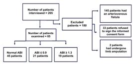
Clinic visits were conducted during all 3 hemodialysis shifts, from Monday to Saturday, and a clinical and sociodemographic questionnaire was administered to 265 patients on hemodialysis of both sexes, regardless of age or concomitant medical conditions. During subsequent bedside visits with the included patients, POCUS and a physical examination were performed, including palpation of arterial pulses in the lower limbs (pedal and posterior tibial) and blood pressure measurement, which was prioritized during the 20-minute dialysis session to minimize interference with patients’ blood pressure levels. A total of 180 patients were excluded from the study due to the presence of an arteriovenous fistula, lower limb amputation, refusal to participate, or refusal to provide written informed consent (Figure 1).
Variables
The independent variables were sex, age, BMI, race, duration of dialysis, risk factors (sedentary lifestyle, alcohol use, smoking), comorbidities (hypertension, family history of CVD, DM, CVD, dyslipidemia), and signs and symptoms compatible with PAD.
Data source
The patients’ clinical and demographic data were collected through medical record review and a semi-structured questionnaire. The questionnaire provided information on age, sex, race, duration of dialysis, presence of vascular access for dialysis, BMI, smoking history, sedentary lifestyle, and family history of CVD.
The following comorbidities were investigated: hypertension, DM, heart disease, stroke, and previous amputations. The presence of signs and symptoms compatible with PAD was characterized by intermittent claudication (muscle pain, mainly in the calves, that appears with exertion and worsens at rest), trophic changes in the extremities (dry and/or cold skin, reduced hair and thickened nails), and non-healing wounds or ulcers.
ABI calculation
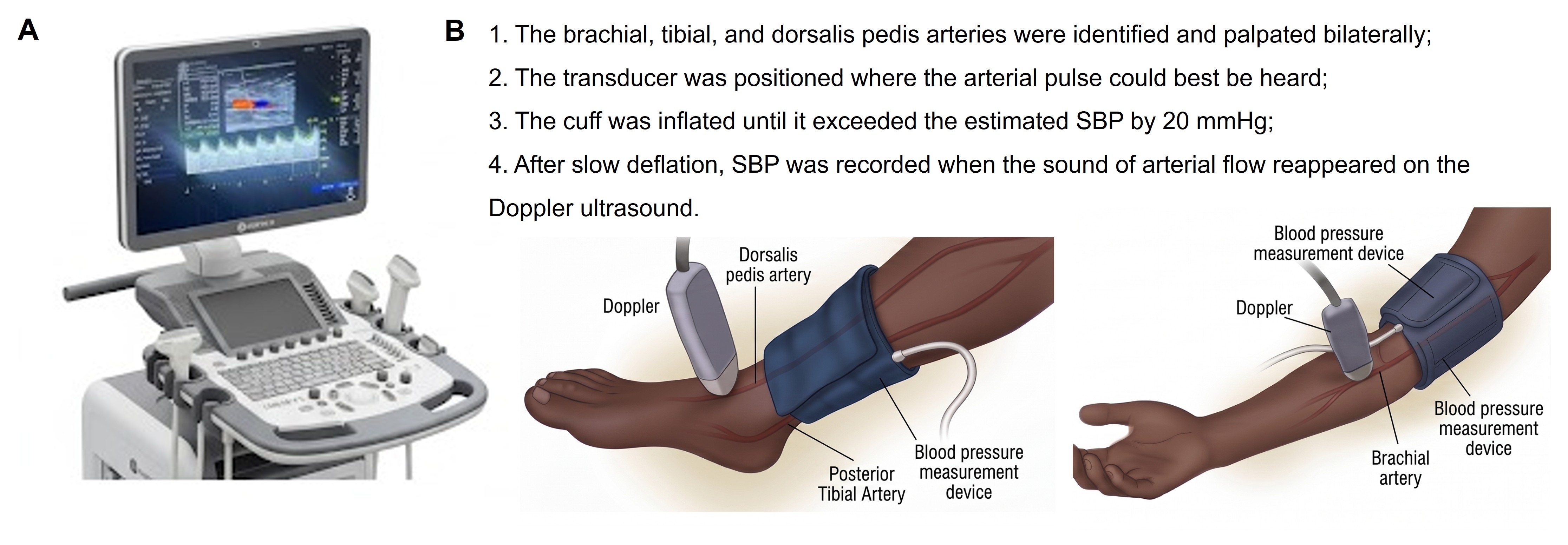
The data for ABI calculation were obtained using a LOGIQ V2 portable ultrasound device in pulsed Doppler mode, with a linear transducer (Figure 2A). The researchers underwent specific prior training to standardize their technique. In cases of dubious results or changes in measurements, the examination was repeated by a more experienced professional to ensure the overall reliability of the data.

A. Image of the ultrasound device used in the study. B. Steps for measuring systolic blood pressure and obtaining the ankle-brachial index at the bedside. Caption: SBP = systolic blood pressure.
SBP values were recorded during the first 20 minutes of the dialysis session, after 5 minutes of rest in a semi-supine position in the hemodialysis chair. The cuff, whose size was appropriate for the arm and ankle circumference, was selected and positioned approximately 2 to 3 cm above the cubital fossa and malleoli, respectively (Figure 2B).
The ABI of the patient’s right and left limbs was then obtained by dividing the highest SBP of the right ankle by the highest SBP of the upper limbs and by dividing the highest SBP of the left ankle by the highest SBP of the upper limbs, respectively.17,21 ABI results between 0.9 and 1.3 were considered normal, indicating normal blood flow in the lower limb; results ≥ 1.3 were considered suggestive of arterial calcification; and results ≤ 0.9 indicated arterial stenosis and were considered PAD-related.13,17
Statistical analysis
The data were organized using descriptive statistics and presented in table format. The chi-square test was used to assess the relationship between arterial abnormalities (PAD or arterial calcification) and clinical and sociodemographic variables. Results were considered significant when p < 0.05.
Ethical aspects
This study was approved by the institutional research ethics committee (certificate: 58588522.3.0000.5666) and was conducted in accordance with the Brazilian National Health Council Resolution 510/2016. CLIRENAL’s authorization and collaboration were also requested.
RESULTS
A total of 85 patients on hemodialysis were assessed. Of these, 21 (24.7%) had an ABI ≤ 0.9, consistent with PAD (mean: 0.7), and 19 (22.4%) had an ABI ≥ 1.3, consistent with arterial calcification (mean: 1.5).
The prevalence of PAD was higher among women (66.7%, n = 14), those > 60 years of age (76.2%, n = 16), those with a normal BMI (52.4%, n = 11), those who had been on hemodialysis < 12 months (61.9%, n = 13), and those who were sedentary (85.7%, n = 18). The distribution of cases was homogeneous in relation to race (mixed race 38.1% [n = 8], Black 33.3% [n = 7], and White 28.6% [n = 6]).
Arterial calcification was more prevalent among men (63.2%, n = 12), those > 60 years of age (52.6%, n = 10), those with a normal BMI (68.4%, n = 13), those who had been on hemodialysis < 12 months (57.9%, n = 11), and those who were sedentary (78.9%, n = 15). The distribution of cases was homogeneous in relation to race (mixed race 26.3% [n = 5], Black 36.8% [n = 7], and White 36.8% [n = 7]).
Among patients with arterial abnormalities (PAD or arterial calcification), alcohol consumption was more prevalent among those with arterial calcification (63.2%, n = 12), while smoking was more prevalent among those with PAD (57.1%, n = 12). The most prevalent comorbidities were hypertension (> 84.2%) and DM (> 52.6%).
The presence of symptoms, characterized by intermittent claudication, trophic changes in the extremities, and non-healing wounds or ulcers, was higher in the group whose ABI was compatible with PAD (p = 0.03). DM (p = 0.02) and older age (p = 0.01) were associated with a greater chance of PAD (Table 1).
DISCUSSION
Arterial abnormalities (PAD or arterial calcification) were found in almost half of our patients on hemodialysis. The main risk factors associated with PAD were DM and advanced age. These findings confirm the importance and need to measure ABI in patients with CKD, due to their susceptibility to cumulative cardiovascular risk factors and the demand for rapid, efficient diagnostic strategies integrated into routine clinical practice.
In our population of patients with CKD, 24.7% had ABIs compatible with PAD. This prevalence is consistent with the findings of a multicenter Spanish study of 2445 patients (28%)28 ; however, a Brazilian study found a lower prevalence (11%).30
PAD has been described as more prevalent in people with CKD than in the general population.5 PAD is a vasculopathy commonly associated with CKD because both share etiological risk factors, and early diagnosis is important to avoid possible complications.31 Although signs and symptoms are present in most patients diagnosed with PAD, the clinical manifestations are nonspecific and may originate from musculoskeletal, articular, or venous abnormalities, whether or not they result from CKD. Thus, ultrasound can clarify the etiology. ABI measurement, as a screening tool for PAD, can help minimize adverse outcomes. In this context, technologies such as POCUS can be an important ally in the early diagnosis of PAD and in reducing its complications.32-34
Most of our patients with PAD were smokers. Although smoking is a significant risk factor for PAD, we found no significant association with the disease, probably due to the limited number of patients who participated in the study.
DM was associated with PAD, corroborating previous studies.6,28,30 This reaffirms the important influence of DM on the genesis of PAD, since DM produces vascular inflammation and epithelial damage, in addition to vasoconstriction associated with platelet activation.35 Although other comorbidities, such as hypertension, obesity, cerebrovascular disease, and dyslipidemia, are also risk factors for PAD, no significant association with them was found in our sample.
Because hemodialysis causes long-term vascular changes,36 individuals who undergo this treatment for a longer period are expected to have a higher prevalence of PAD. However, no association was observed between dialysis duration and PAD incidence, a finding also reported in previous studies.30,37
In 22.4% of examinations, an ABI ≥ 1.3 was observed, consistent with arterial calcification. In previous research, Ramos et al.30 reported that up to 46.2% of patients undergoing hemodialysis had vascular calcification. This difference may be due to the duration of patients’ hemodialysis.38 Arterial calcification, a significant complication of CKD, results from a disturbance in mineral and bone balance and confers a higher risk of mortality in affected individuals.38,39
Arterial calcification is related to increased vascular stiffness and the presence of non-compressible arteries. This leads to greater thickening of the renal arteries, a higher prevalence of renal microvascular injury, and consequently, a reduced glomerular filtration rate. Furthermore, a high ABI, for example, can lead to hemodynamic overload, endothelial dysfunction, and left ventricular hypertrophy, increasing the risk of cardiovascular events, especially in patients with CKD.40 Just as with PAD, arterial calcification can be clinically investigated by measuring the ABI.
Through visualization of the artery, POCUS facilitates SBP measurement and enables ABI calculation. This is even more important in patients who routinely present with arterial abnormalities (such as calcification) and are evaluated in collective settings, at the bedside, or in noisy environments. Furthermore, POCUS allows trained professionals to more comprehensively assess PAD, facilitating disease stratification and better case management. As has been highlighted by other authors, POCUS is also useful for assessing other important organs and systems in the context of CKD and hemodialysis.21,22,34
It should be noted that the POCUS method has certain limitations, as it requires experience, specialized training, and time to implement in large-scale practice.32,34 One study limitation was that ABI values ≥ 1.3 may underestimate the prevalence of PAD, since false-negative test results can occur in this population. Another limitation concerns the large proportion of patients with arteriovenous fistulas as vascular access for dialysis, which limited the study population and the final number of participants in each group, preventing more robust statistical analyses.
Because this was an observational, cross-sectional study, cause-and-effect relationships between risk factors and arterial abnormalities could not be determined. Furthermore, the study was conducted at a single center, which limits the generalizability of the results to other hemodialysis populations or to different geographic and health contexts.
CONCLUSIONS
Using POCUS, we identified a prevalence of 24.7% for arterial abnormalities compatible with PAD and 22.4% for those compatible with arterial calcification in our sample of patients with CKD on hemodialysis. Moreover, advanced age and DM were the main factors associated with PAD in these individuals. POCUS proved a feasible method for measuring ABI and detecting arterial abnormalities at the bedside at our study center, reinforcing the need for continuous vascular monitoring in this population, as vascular impairment was found in almost half of the participants.
-
How to cite:
Santos AAA, Lima HC, Lordêlo BVC, et al. Use of point-of-care ultrasound in the analysis of arterial abnormalities in people with chronic kidney disease on hemodialysis: a cross-sectional study. J Vasc Bras. 2026;25:e20250036. https://doi.org/10.1590/1677-5449.202500362
-
Financial support:
None.
-
The study was carried out at Universidade Federal do Vale São Francisco (UNIVASF), Paulo Afonso, BA, Brazil.
-
Ethics committee approval:
The study protocol was approved by the Ethics Committee of Centro Universitário do Vale do Ipojuca – UNIFAVIP (certificate: 58588522.3.0000.5666, opinion: 5.473.764 – June 16, 2022).
DATA AVAILABILITY
The data supporting this study are available from the corresponding author.
References
-
1 Hiatt WR, Armstrong EJ, Larson CJ, Brass EP. Pathogenesis of the limb manifestations and exercise limitations in peripheral artery disease. Circ Res. 2015;116(9):1527-39. https://doi.org/10.1161/CIRCRESAHA.116.303566 PMid:25908726.
» https://doi.org/10.1161/CIRCRESAHA.116.303566 -
2 Martín-Rubio I, Marín-García B, Romero-Velasco E, Thuissard-Vasallo IJ, Bautista-Hernández A, Abad-Pérez D. Oscillometric devices vs. arterial doppler in measuring ankle-brachial index for diagnosing peripheral artery disease. Clínica Medica. 2024;163(3):128-31. https://doi.org/10.1016/j.medcli.2024.02.003 PMid:38643026.
» https://doi.org/10.1016/j.medcli.2024.02.003 - 3 Caldeira M, Mina F. Doença arterial obstrutiva periférica em pessoas com diabetes. Rev Port Diabetes. 2017;12(3):107-11.
-
4 Tungsrirut N, Taptiang K, Charoenkarn K, Piyachon P, Srisittipoj P, Rerkasem K. Pole test and ankle-brachial index by using doppler ultrasound for peripheral arterial disease in end-stage renal disease patients. Int J Low Extrem Wounds. 2020;19(4):359-63. https://doi.org/10.1177/1534734620951528 PMid:32844689.
» https://doi.org/10.1177/1534734620951528 -
5 AbuRahma AF, Adams E, AbuRahma J, et al. Critical analysis and limitations of the resting ankle-brachial index in the diagnosis of patients with symptomatic peripheral arterial disease and the role of diabetes mellitus and chronic kidney disease. J Vasc Surg. 2020;71(3):937-45. https://doi.org/10.1016/j.jvs.2019.05.050 PMid:31471230.
» https://doi.org/10.1016/j.jvs.2019.05.050 -
6 Aday AW, Matsushita K. Epidemiology of peripheral artery disease and polyvascular disease. Circ Res. 2021;128(12):1818-32. https://doi.org/10.1161/CIRCRESAHA.121.318535 PMid:34110907.
» https://doi.org/10.1161/CIRCRESAHA.121.318535 -
7 Ikizler TA, Burrowes JD, Byham-Gray LD, et al. KDOQI clinical practice guideline for nutrition in CKD: 2020 update. Am J Kidney Dis. 2020;76(3, Suppl 1):S1-107. https://doi.org/10.1053/j.ajkd.2020.05.006 PMid:32829751.
» https://doi.org/10.1053/j.ajkd.2020.05.006 -
8 Yamada S, Nakano T. Role of chronic kidney disease-mineral and bone disorder in the pathogenesis of cardiovascular disease in CKD. J Atheroscler Thromb. 2023;30(8):835-50. https://doi.org/10.5551/jat.RV22006 PMid:37258233.
» https://doi.org/10.5551/jat.RV22006 -
9 Ohtake T, Ishioka K, Honda K, et al. Impact of coronary artery calcification in hemodialysis patients: Risk factors and associations with prognosis. Hemodial Int. 2010;14(2):218-25. https://doi.org/10.1111/j.1542-4758.2009.00423.x PMid:20345389.
» https://doi.org/10.1111/j.1542-4758.2009.00423.x -
10 ElSayed NA, McCoy RG, Aleppo G, et al. 12. Retinopathy, neuropathy, and foot care: standards of care in diabetes—2025. Diabetes Care. 2025;48(1, Suppl 1):S252-65. https://doi.org/10.2337/dc25-S012 PMid:39651973.
» https://doi.org/10.2337/dc25-S012 -
11 Gornik HL, Aronow HD, Goodney PP, et al. 2024 ACC/AHA/AACVPR/APMA/ABC/SCAI/SVM/SVN/SVS/SIR/VESS Guideline for the management of lower extremity peripheral artery disease: a report of the American College of Cardiology/American Heart Association Joint Committee on Clinical Practice Guidelines. Circulation. 2024;149(24):e1313-410. https://doi.org/10.1161/CIR.0000000000001251 PMid:38743805.
» https://doi.org/10.1161/CIR.0000000000001251 -
12 Golledge J, Moxon JV, Rowbotham S, Pinchbeck J, Quigley F, Jenkins J. High ankle brachial index predicts high risk of cardiovascular events amongst people with peripheral artery disease. PLoS One. 2020;15(11):e0242228. https://doi.org/10.1371/journal.pone.0242228 PMid:33180875.
» https://doi.org/10.1371/journal.pone.0242228 -
13 Xu D, Li J, Zou L, et al. Sensitivity and specificity of the ankle—brachial index to diagnose peripheral artery disease: a structured review. Vasc Med. 2010;15(5):361-9. https://doi.org/10.1177/1358863X10378376 PMid:20926495.
» https://doi.org/10.1177/1358863X10378376 -
14 Eid MA, Mehta KS, Goodney PP. Epidemiology of peripheral artery disease. Semin Vasc Surg. 2021;34(1):38-46. https://doi.org/10.1053/j.semvascsurg.2021.02.005 PMid:33757634.
» https://doi.org/10.1053/j.semvascsurg.2021.02.005 -
15 Ogata H, Kumata-Maeta C, Shishido K, et al. Detection of peripheral artery disease by duplex ultrasonography among hemodialysis patients. Clin J Am Soc Nephrol. 2010;5(12):2199-206. https://doi.org/10.2215/CJN.09451209 PMid:20798256.
» https://doi.org/10.2215/CJN.09451209 -
16 Huang HL, Chang CH, Lo MT, Lin C. Diagnostic potential of combined photoplethysmography and ankle-brachial index in peripheral arterial disease: a duplex ultrasonography-based comparative study. J Am Heart Assoc. 2024;13(19):e034625. https://doi.org/10.1161/JAHA.124.034625 PMid:39344605.
» https://doi.org/10.1161/JAHA.124.034625 -
17 Kollias A, Xilomenos A, Protogerou A, Dimakakos E, Stergiou GS. Automated determination of the ankle-brachial index using an oscillometric blood pressure monitor: validation vs. Doppler measurement and cardiovascular risk factor profile. Hypertens Res. 2011;34(7):825-30. https://doi.org/10.1038/hr.2011.53 PMid:21593742.
» https://doi.org/10.1038/hr.2011.53 -
18 Buschmann EE, Li L, Brix M, et al. A novel computer-aided diagnostic approach for detecting peripheral arterial disease in patients with diabetes. PLoS One. 2018;13(6):e0199374. https://doi.org/10.1371/journal.pone.0199374 PMid:29928037.
» https://doi.org/10.1371/journal.pone.0199374 -
19 Aboyans V, Criqui MH, Abraham P, et al. Measurement and interpretation of the ankle-brachial index: a scientific statement from the American Heart Association. Circulation. 2012;126(24):2890-909. https://doi.org/10.1161/CIR.0b013e318276fbcb PMid:23159553.
» https://doi.org/10.1161/CIR.0b013e318276fbcb -
20 Normahani P, Poushpas S, Alaa M, et al. Diagnostic accuracy of point-of-care tests used to detect arterial disease in diabetes: Testing for Arterial Disease in Diabetes (TrEAD) study. Ann Surg. 2022;276(5):e605-12. https://doi.org/10.1097/SLA.0000000000004545 PMid:33630461.
» https://doi.org/10.1097/SLA.0000000000004545 -
21 Ichihashi S, Desormais I, Hashimoto T, Magne J, Kichikawa K, Aboyans V. Accuracy and reliability of the ankle brachial index measurement using a multicuff oscillometric device versus the Doppler method. Eur J Vasc Endovasc Surg. 2020;60(3):462-8. https://doi.org/10.1016/j.ejvs.2020.06.013 PMid:32763120.
» https://doi.org/10.1016/j.ejvs.2020.06.013 -
22 Bastos M, Vieira ALS, Pazeli JMJ. Uso da ultrassonografia “point-of-care” na prática nefrológica: transpondo os limites do trato urinário. HU Revista. 2019;45(3):341-51. https://doi.org/10.34019/1982-8047.2019.v45.28745
» https://doi.org/10.34019/1982-8047.2019.v45.28745 -
23 Fitridge R, Chuter V, Mills J, et al. The intersocietal IWGDF, ESVS, SVS guidelines on peripheral artery disease in people with diabetes mellitus and a foot ulcer. J Vasc Surg. 2023;78(5):1101-31. https://doi.org/10.1016/j.jvs.2023.07.020 PMid:37724985.
» https://doi.org/10.1016/j.jvs.2023.07.020 -
24 Dias SVM, Flumignan RLG, Carvas N Jr, Iared W. Accuracy of duplex ultrasound in peripheral artery disease: a systematic review and meta-analysis. J Vasc Bras. 2025;24:e20240033. https://doi.org/10.1590/1677-5449.202400332 PMid:39981417.
» https://doi.org/10.1590/1677-5449.202400332 -
25 Drake A, Dreyer N, Hoffer M, Boniface K. Point-of-care ultrasound for evaluation of acute arterial pathology in the emergency department: a case series. Clin Pract Cases Emerg Med. 2022;6(1):1-7. https://doi.org/10.5811/cpcem.2021.11.54904 PMid:35226837.
» https://doi.org/10.5811/cpcem.2021.11.54904 -
26 Herraiz-Adillo Á, Cavero-Redondo I, Álvarez-Bueno C, Pozuelo-Carrascosa DP, Solera-Martínez M. The accuracy of toe brachial index and ankle brachial index in the diagnosis of lower limb peripheral arterial disease: a systematic review and meta-analysis. Atherosclerosis. 2020;315:81-92. https://doi.org/10.1016/j.atherosclerosis.2020.09.026 PMid:33036766.
» https://doi.org/10.1016/j.atherosclerosis.2020.09.026 -
27 Miot HA. Tamanho da amostra em estudos clínicos e experimentais. J Vasc Bras. 2011;10(4):275-8. https://doi.org/10.1590/S1677-54492011000400001 PMid:30787944.
» https://doi.org/10.1590/S1677-54492011000400001 -
28 Arroyo D, Betriu A, Valls J, et al. Factors influencing pathological ankle-brachial index values along the chronic kidney disease spectrum: the NEFRONA study. Nephrol Dial Transplant. 2017;32(3):513-20. https://doi.org/10.1093/ndt/gfw039 PMid:27190385.
» https://doi.org/10.1093/ndt/gfw039 -
29 Instituto Brasileiro de Geografia e Estatística. Panorama das cidades brasileiras: Paulo Afonso/Bahia [Internet]. Rio de Janeiro; 2022 [citado 2024 nov 18]. https://cidades.ibge.gov.br/brasil/ba/paulo-afonso/panorama
» https://cidades.ibge.gov.br/brasil/ba/paulo-afonso/panorama -
30 Ramos MAA, Litchteneker K, Santana TC, Unser V. Doença arterial obstrutiva periférica em pacientes com insuficiência renal crônica dialítica na região de Toledo-Paraná: prevalência e fatores de risco. Rev Ciênc Méd Biol. 2022;21(2):232-7. https://doi.org/10.9771/cmbio.v21i2.45297
» https://doi.org/10.9771/cmbio.v21i2.45297 -
31 Erzinger FL, Polimanti AC, Pinto DM, et al. Brazilian Society of Angiology and Vascular Surgery guidelines on peripheral artery disease. J Vasc Bras. 2024;23:e20230059. https://doi.org/10.1590/1677-5449.202300592 PMid:39493832.
» https://doi.org/10.1590/1677-5449.202300592 -
32 Cáceres-Farfán L, Moreno-Loaiza M, Cubas WS. Ankle-brachial index: more than a diagnostic test? Arch Peru Cardiol Cir Cardiovasc. 2021;2(4):254-62. https://doi.org/10.47487/apcyccv.v2i4.168 PMid:37727667.
» https://doi.org/10.47487/apcyccv.v2i4.168 -
33 Huthart S, Oates C, Allen J, Fiaschi K, Sims AJ, Stansby G. Validation of a standardised duplex ultrasound classification system for the reporting and grading of peripheral arterial disease. Eur J Vasc Endovasc Surg. 2022;64(2-3):210-6. https://doi.org/10.1016/j.ejvs.2022.04.013 PMid:35472448.
» https://doi.org/10.1016/j.ejvs.2022.04.013 -
34 Bastos MG, Novaes AKB, Pazeli JM Jr. Traditional and ultrasound physical examinations: a hybrid approach to improve clinical care. Rev Assoc Med Bras. 2018;64(5):474-80. https://doi.org/10.1590/1806-9282.64.05.474 PMid:30304149.
» https://doi.org/10.1590/1806-9282.64.05.474 - 35 Berger JS, Newman JD. Overview of peripheral artery disease in patients with diabetes mellitus. Waltham: UptoDate; 2021.
-
36 Pichone A, Campos G, Leite M Jr, Gomes CP. High ankle-brachial index predicts cardiovascular events and mortality in hemodialysis patients with severe secondary hyperparathyroidism. J Bras Nefrol. 2021;43(4):478-85. https://doi.org/10.1590/2175-8239-jbn-2020-0218 PMid:33979425.
» https://doi.org/10.1590/2175-8239-jbn-2020-0218 -
37 Hishida M, Imaizumi T, Menez S, et al. Additional prognostic value of toe-brachial index beyond ankle-brachial index in hemodialysis patients. BMC Nephrol. 2020;21(1):353. https://doi.org/10.1186/s12882-020-01991-7 PMid:32819299.
» https://doi.org/10.1186/s12882-020-01991-7 -
38 Bonato FOB, Karohl C, Canziani MEF. Diagnosis of vascular calcification related to mineral and bone metabolism disorders in chronic kidney disease. J Bras Nefrol. 2021;43(4, Suppl 1):628-31. https://doi.org/10.1590/2175-8239-jbn-2021-s104 PMid:34910796.
» https://doi.org/10.1590/2175-8239-jbn-2021-s104 -
39 Tian L, Jaeger BC, Scialla JJ, et al. Progression of coronary artery calcification and risk of clinical events in CKD: the chronic renal insufficiency cohort study. Am J Kidney Dis. 2025;85(1):67-77.e1. https://doi.org/10.1053/j.ajkd.2024.06.018 PMid:39154888.
» https://doi.org/10.1053/j.ajkd.2024.06.018 -
40 Hazique M, Surana A, Patel KN, et al. Abnormal Ankle-Brachial Index and risk of cardiovascular and all-cause mortality in patients with chronic kidney disease: an updated systematic review and meta-analysis. Crit Pathw Cardiol. 2025;24(3):e0396. https://doi.org/10.1097/HPC.0000000000000396 PMid:40397761.
» https://doi.org/10.1097/HPC.0000000000000396
Edited by
-
Editor-in-Chief responsible
Dr. Winston Bonetti Yoshida