Triple abdominal vascular compression: coexistence of Dunbar syndrome, Wilkie syndrome, and Nutcracker phenomenon: a case report and literature review
Abstract
Median arcuate ligament syndrome (Dunbar syndrome) is characterized by compression of the celiac trunk, causing postprandial abdominal pain and ischemic symptoms. Superior mesenteric artery syndrome (Wilkie’s syndrome) results from narrowing of the aortomesenteric angle, leading to duodenal obstruction and weight loss. Nutcracker phenomenon, in turn, consists of compression of the left renal vein between the aorta and the superior mesenteric artery. While each of these entities are individually uncommon, their coexistence in a single patient is exceptionally rare and poses significant diagnostic and therapeutic challenges. We report the case of a young woman diagnosed simultaneously with all three conditions who was successfully managed through a surgical approach. Diagnostic methods and therapeutic options are discussed, highlighting the importance of maintaining a high index of suspicion for the timely and appropriate management of these uncommon compressive syndromes.
Keywords:
median arcuate ligament syndrome; superior mesenteric artery syndrome; renal Nutcracker syndrome; case reports
Resumen
El síndrome del ligamento arcuato mediano (síndrome de Dunbar) se caracteriza por la compresión del tronco celíaco, causando dolor abdominal posprandial y síntomas isquémicos. El síndrome de la arteria mesentérica superior (síndrome de Wilkie) resulta de la disminución del ángulo aortomesentérico, ocasionando obstrucción duodenal y pérdida de peso. Por su parte, el fenómeno de Nutcracker corresponde a la compresión de la vena renal izquierda entre la aorta y la arteria mesentérica superior. Si bien estas entidades son poco frecuentes de manera aislada, su coexistencia en un mismo paciente es excepcional, lo que dificulta el diagnóstico y el tratamiento. Presentamos el caso de una mujer joven con diagnóstico simultáneo de las tres patologías, tratada con éxito mediante abordaje quirúrgico. Se discuten los métodos diagnósticos y las alternativas terapéuticas, enfatizando la importancia de mantener un elevado índice de sospecha clínica para lograr un manejo oportuno y adecuado de estos síndromes compresivos infrecuentes.
Palabras clave:
síndrome de ligamento arcuato medio; síndrome de arteria mesentérica superior; síndrome de cascanueces renal; informes de caso
INTRODUCTION
The median arcuate ligament (MAL) is a fibrous arch connecting the right and left crura of the diaphragm over the aorta at the T12-L1 level. Compression over the celiac trunk may lead to chronic abdominal pain, nausea, vomiting, and weight loss, a condition known as Dunbar syndrome, or median arcuate ligament syndrome (MALS).1,2 Wilkie syndrome, or superior mesenteric artery syndrome (SMAS), develops when the superior mesenteric artery (SMA) compresses the duodenum, causing duodenal obstruction and symptoms similar to those of MALS.3 Nutcracker phenomenon (NCP) consists of compression of the left renal vein between the aorta and the SMA.4 While each of these entities are individually uncommon, their coexistence is even rarer and poses significant diagnostic and therapeutic challenges.4
CASE PRESENTATION
A 27-year old female patient presented with a 2-year history of burning abdominal in the mesogastric and epigastric regions, rated 8/10 in intensity. The pain was triggered by food intake and associated with heartburn, nausea, vomiting, and a 25% weight loss over the previous year. Relevant medical and surgical history included endometriosis, chronic gastritis, cholecystectomy, augmentation mammoplasty, and pelvic resections.
Following gastroenterological evaluation, upper gastrointestinal endoscopy revealed chronic gastritis with biopsy results negative for Helicobacter pylori. Furthermore, marked gastric distention was identified as a notable finding. Medical management was initiated; however, given the absence of a clear etiology, an abdominal computed tomography angiography (CTA) was performed, demonstrating compression of the left renal vein between the SMA and the aorta.
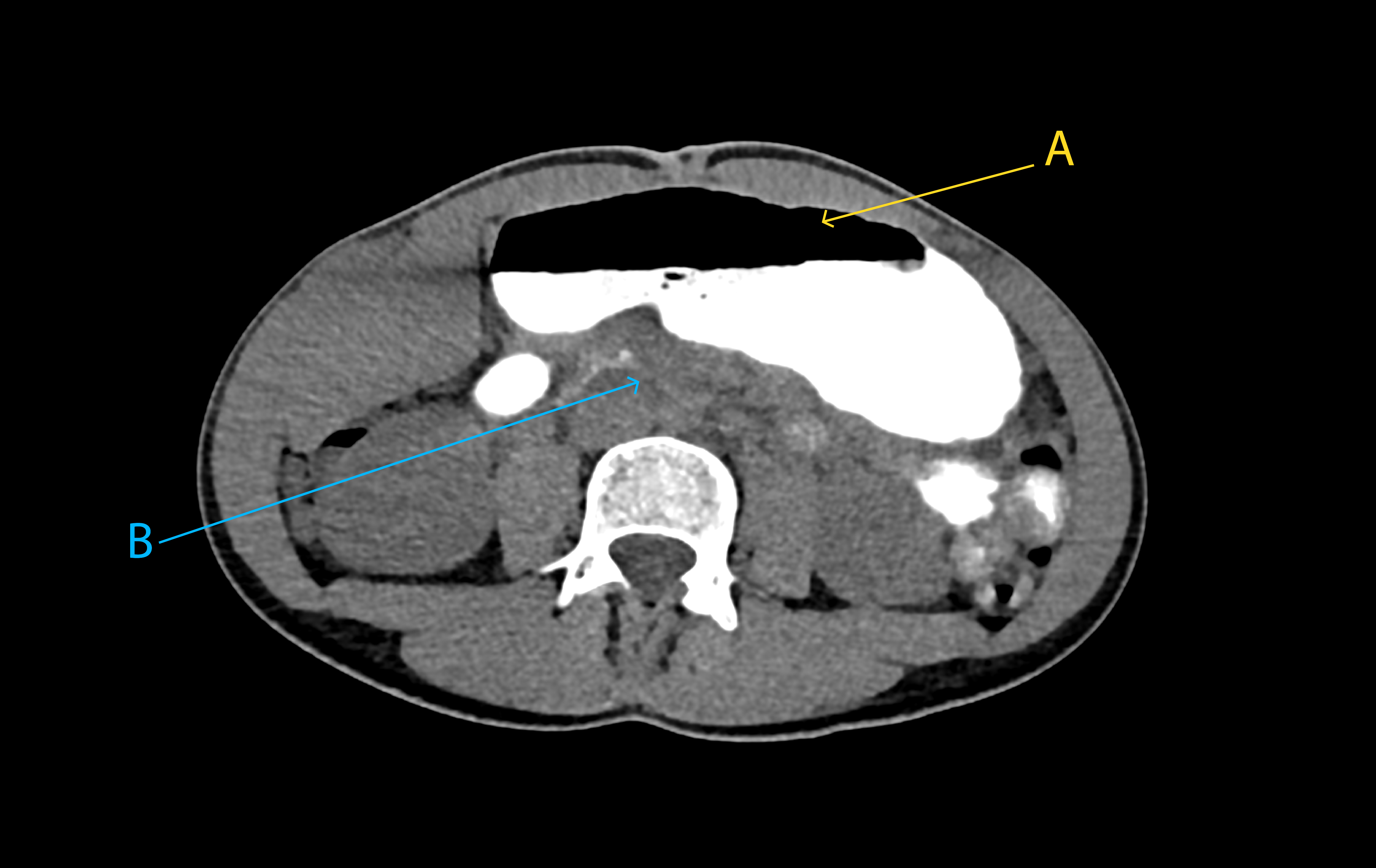
Given the persistence of symptoms, the patient was admitted for further diagnostic evaluation, including a repeat CTA with oral contrast, which showed marked gastric dilatation and aortomesenteric compression at the level of the third portion of the duodenum, findings suggestive of SMAS (Figure 1).
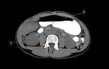
Abdominal computed tomography with oral contrast (axial view) demonstrating marked gastric dilatation (yellow arrow A) and compression of the third portion of the duodenum (blue arrow B).
Physical examination revealed a body mass index of 15.4 kg/m2 (weight: 37 kg; height: 155 cm), indicating significant underweight status. The abdomen was distended and tender on palpation in the epigastric region, without palpable masses or signs of peritoneal irritation.
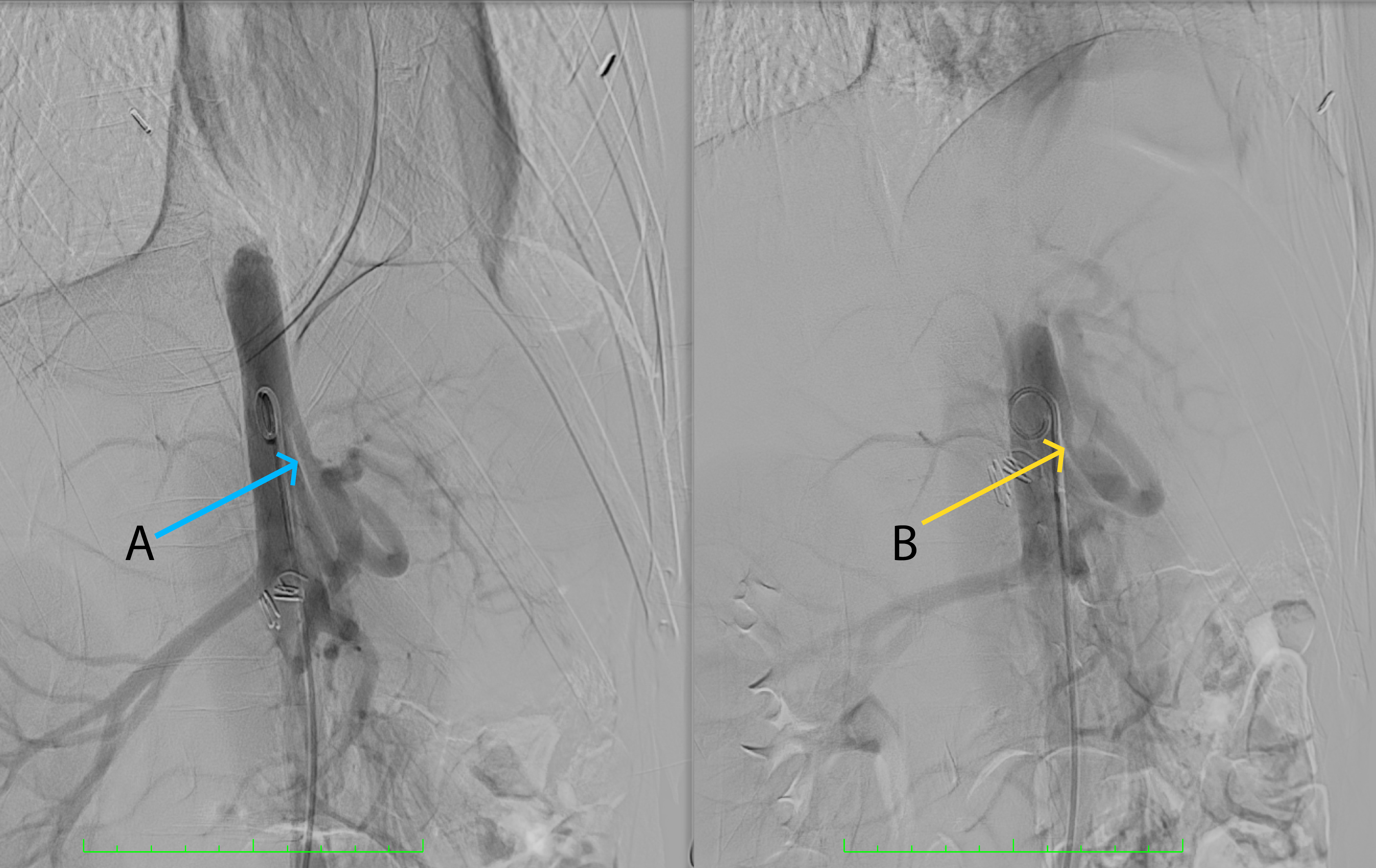
Medical management was initiated with total parenteral nutrition. Doppler ultrasonography of the mesenteric arteries was performed, suggesting the coexistence of MALS, SMAS, and NCP, with peak systolic velocity (PSV) and end-diastolic velocity (EDV) of 453 and 123 cm/s, respectively, in the proximal celiac trunk, whereas fasting PSV and EDV in the SMA were 193 and 21 cm/s, respectively. In addition, longitudinal B-mode assessment of the SMA and abdominal aorta revealed an aortomesenteric angle of 10°. Based on these findings, abdominal aortography with selective arteriography, dynamic angiography during respiratory maneuvers, and segmental pressure measurements was performed. Findings showed dynamic compression of the celiac trunk, characterized by stenosis during expiration proximal to its first major branch and an inspiratory-expiratory pressure gradient of 20 mm Hg, confirming the diagnosis of MALS (Figure 2).
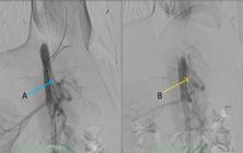
Abdominal aortography with selective celiac trunk arteriography, dynamic angiography during respiratory maneuvers, and segmental pressure measurements showing a patent celiac trunk during inspiration (blue arrow A) and celiac trunk stenosis during expiration, confirming median arcuate ligament syndrome (yellow arrow B).
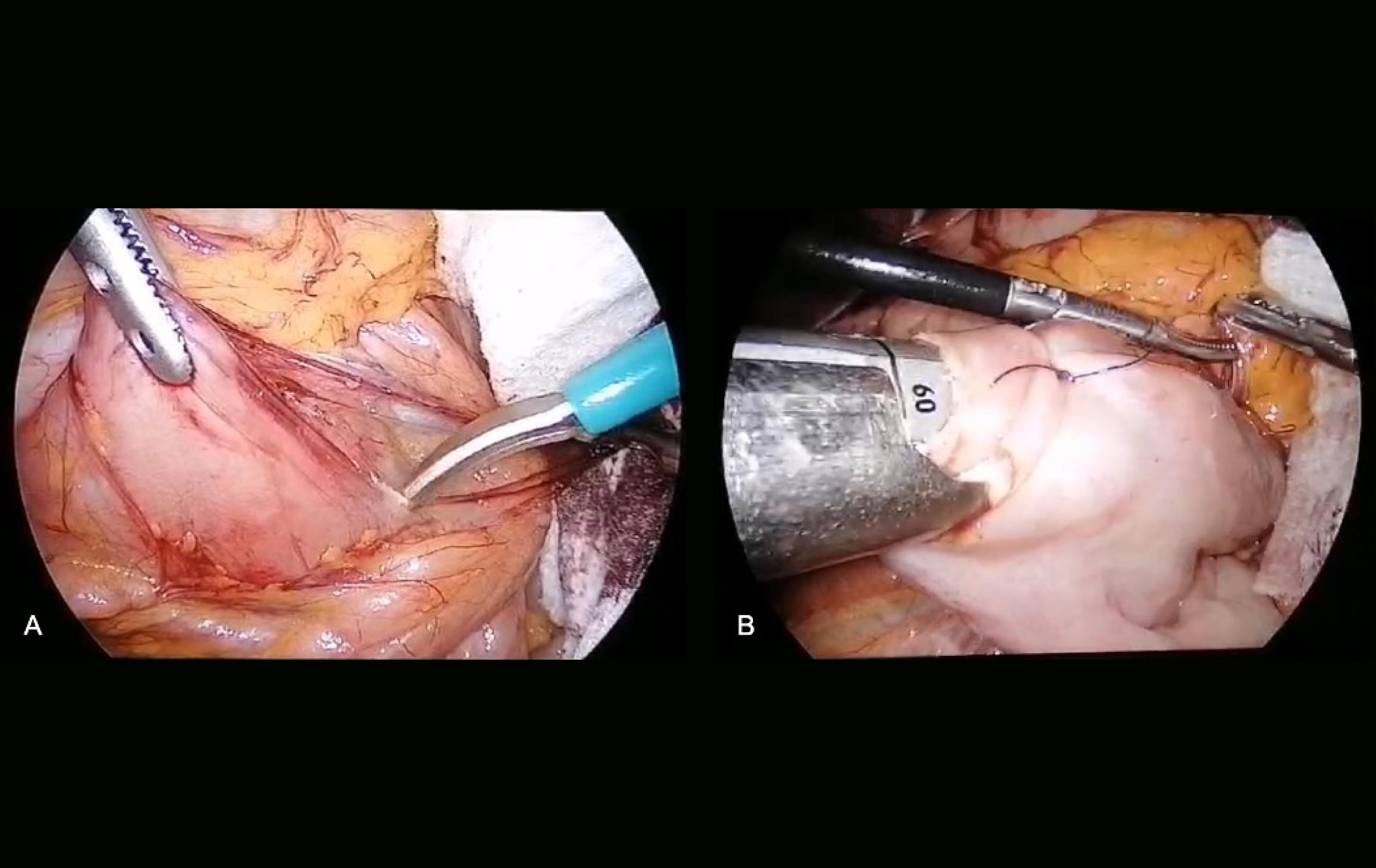
With the diagnosis of MALS, SMAS, and NCP confirmed, the patient underwent surgical treatment. The procedure was conducted by the advanced laparoscopic surgery team and included division of the ligament of Treitz (Strong procedure) along with a duodenojejunostomy (Figure 3), followed by division of the MAL for decompression of the celiac trunk. Intraoperative findings included gastric and proximal duodenal dilatation, as well as compression of the celiac trunk by the MAL.
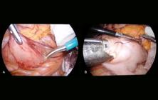
Intraoperative images of the surgical procedure. (A) Release of the ligament of Treitz (Strong procedure); (B) Duodenojejunostomy.
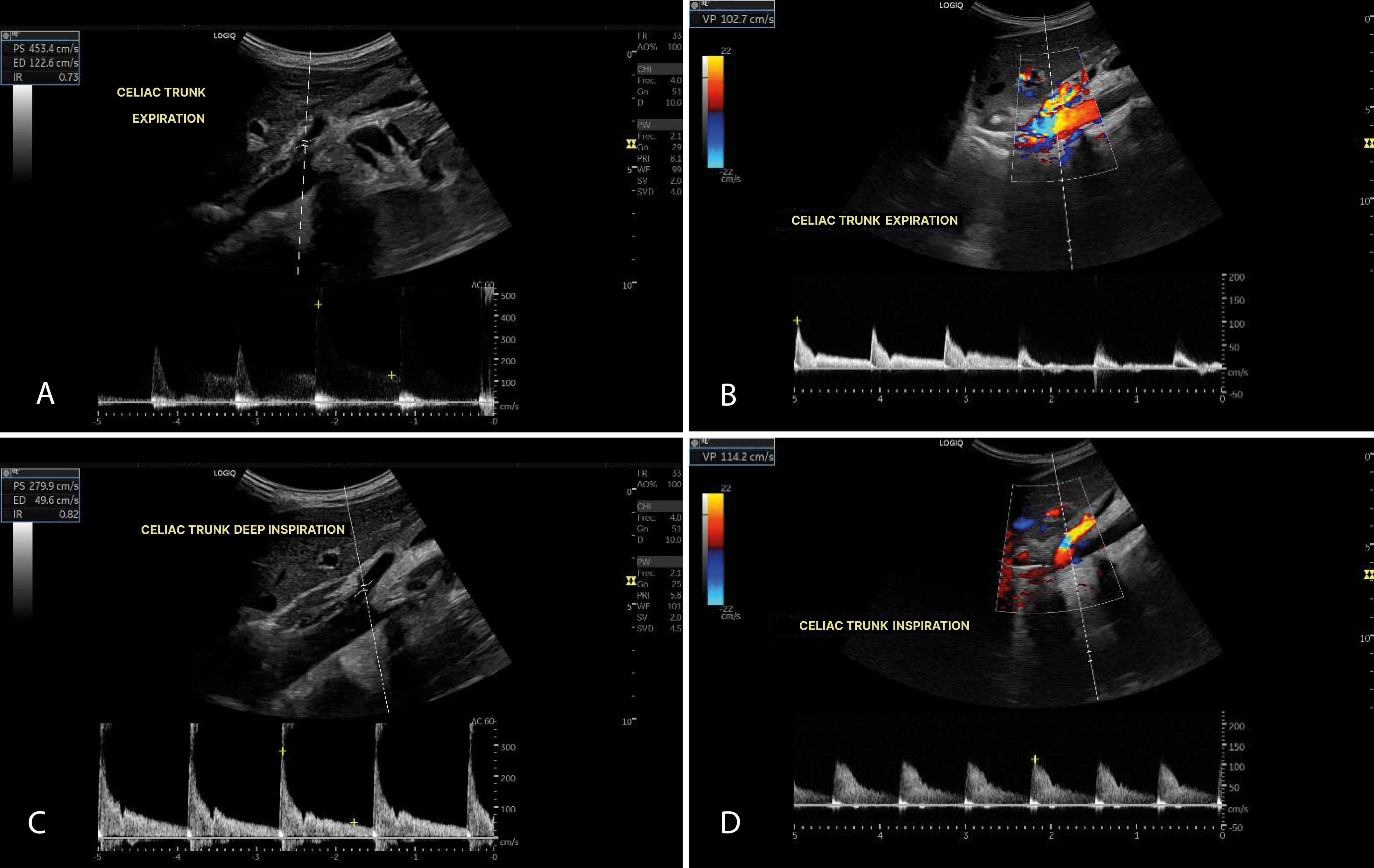
The patient had an uneventful postoperative course. Oral fluid intake was initiated on postoperative day 2 and subsequently advanced to a liquid diet. Parenteral nutrition was discontinued on postoperative day 3, and the patient was discharged with enteral nutritional supplementation. One month after surgery, follow-up Doppler ultrasonography showed a significant reduction in flow velocities, with no respiratory variation and no dynamic morphological changes in the celiac trunk (Figure 4).
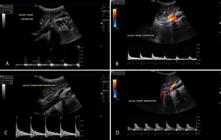
Doppler ultrasonography of the celiac trunk before and after surgical intervention during inspiration and expiration. (A) Preoperative Doppler ultrasonography of the celiac trunk during expiration; (B) Postoperative Doppler ultrasonography of the celiac trunk during expiration; (C) Preoperative Doppler ultrasonography of the celiac trunk during deep inspiration; (D) Postoperative Doppler ultrasonography of the celiac trunk during deep inspiration.
At 6 months, follow-up evaluation documented a 4-kg weight gain and complete resolution of vomiting, nausea, and postprandial pain; however, chronic abdominal pain unrelated to food intake persisted. Repeat Doppler ultrasonography of mesenteric arteries showed normal flow velocities in both SMA and celiac trunk during inspiration and expiration. Given the persistence of unexplained pain, abdominal aortography with selective celiac arteriography, dynamic angiography during respiratory maneuvers, and segmental pressure measurements was repeated. The study demonstrated a patent celiac trunk, with no signs of compression or stenosis during respiratory movements. Due to the absence of structural vascular or obstructive abnormalities, the condition was interpreted as functional chronic abdominal pain associated with severe delayed gastric emptying and gastric dilatation and concomitant anxiety and depressive symptoms, with a component of central sensitization. Multidisciplinary management and outpatient follow-up were recommended.
DISCUSSION AND LITERATURE REVIEW
Overview and epidemiology
SMAS, also known as Wilkie syndrome, occurs predominantly in young, thin women and has an estimated prevalence of 0.013-0.3% among patients with chronic gastrointestinal symptoms.5-7 It is characterized by duodenal compression secondary to narrowing of the aortomesenteric space resulting from loss of the mesenteric fat pad. Although malnutrition and low body weight are recognized risk factors, they do not explain all presentations. Congenital factors, such as a high insertion of the ligament of Treitz or a low origin of the SMA, have also been described and were identified in our patient. Other associated conditions include malignancy, HIV/AIDS, malabsorption syndromes, bariatric surgery, spinal trauma, and paraplegia; however, up to 40% of cases have no identifiable cause.8,9
Asymptomatic anatomical compression of the celiac trunk by the MAL is detected in approximately 15-25% of the population.6 When associated with clinical manifestations, this condition is referred to as MALS and occurs predominantly in young patients. Concurrent SMAS and MALS should be considered in patients with persistent obstructive symptoms.9
Likewise, Nutcracker syndrome may present with hematuria, varicocele, or left renal vein thrombosis. In the absence of clinical manifestations, as observed in our patient, the condition is referred to as Nutcracker phenomenon, an anatomical variant characterized by compression of the left renal vein without significant clinical consequences.4
Pathophysiology
The third portion of the duodenum is located between the aorta and the SMA. The SMA arises from the aorta at the level of L1 and descends toward the mesentery, normally forming an acute aortomesenteric angle of 38-65°. In SMAS, this angle may decrease to as little as 6º because of loss of the mesenteric fat pad, most commonly after rapid weight loss, resulting in duodenal compression.5,10,11
In MALS, compression of the celiac trunk may be caused by a low insertion of the MAL or a high origin of the celiac trunk.9,12 In approximately 20% of the cases, the celiac trunk originates posterior to the ligament, leading to restricted blood flow. An alternative hypothesis suggests that symptoms may arise from neurogenic stimulation of the celiac ganglion secondary to arterial compression.9
The coexistence of celiac and mesenteric anatomical abnormalities, such as low insertion of the MAL, has also been proposed. These abnormalities may also share a common embryological basis, potentially explaining the association between SMAS and MALS, particularly in young women.13,14
Clinical presentation and diagnosis
The diagnosis of SMAS is primarily clinical and is supported by imaging studies. It typically presents as proximal intestinal obstruction characterized by postprandial pain, early satiety, nausea, bilious vomiting, and weight loss.15,16 Radiologically compression of the third portion of the duodenum between the SMA and the aorta is identified, most commonly by contrast-enhanced CTA. This modality allows measurement of the aortomesenteric angle and distance (≤ 25° and ≤ 8 mm, respectively) and assessment of the retroperitoneal fat pad. The three classic diagnostic criteria include obstruction of the third portion of the duodenum with preserved peristalsis, a reduced aortomesenteric angle, and abnormalities in the origin of the SMA or fixation of the duodenum.10
In MALS, mesenteric artery Doppler ultrasonography with respiratory maneuvers is a useful, cost-effective, and radiation-free initial method. Flow velocities exceeding > 200 cm/s during deep expiration suggest celiac trunk compression, whereas a PSV greater than 350 cm/s combined with a deflection angle above 50° achieves a sensitivity of 83% and a specificity of 100% for the diagnosis of this condition.2,17,18 Moreover, Doppler ultrasonography is valuable for post-treatment follow-up, as improvements in flow parameters correlate with clinical improvement. CTA and magnetic resonance angiography are also useful for assessing extrinsic compression of the celiac trunk, with or without post-stenotic dilatation.
CTA with dynamic assessment during inspiration and expiration is considered the gold standard for confirming compression of the celiac trunk by the MAL.19 Both SMAS and MALS should be diagnosed by exclusion after other gastrointestinal disorders have been ruled out.13
Treatment
The treatment of SMAS follows a stepwise approach, beginning with conservative management and reserving surgery for refractory cases. Medical management is recommended for 4-6 weeks and includes nasogastric decompression, correction of fluid and electrolyte abnormalities, nutritional support (most commonly with total parenteral nutrition), and prokinetic agents.7,10 Surgical intervention is indicated in patients with persistent or severe symptoms or marked duodenal dilatation.9 Surgical options include Strong procedure, duodenojejunostomy, gastrojejunostomy, and Roux-en-Y reconstruction.6,12,20 Among these, duodenojejunostomy is the most commonly performed procedure, with success rates of up to 90%.9,21
In MALS, release of the MAL has been shown to be effective in most patients, resulting in significant symptomatic improvement.2
Overall, surgical treatment of SMAS and MALS leads to symptom resolution in 70-80% of patients.9,22 The coexistence of both conditions appears to be associated with a reduced response to conservative treatment, supporting the use of a combined surgical approach.
Although surgical decompression remains the standard treatment for MALS and endovascular therapy alone does not correct extrinsic compression of the celiac trunk, the latter may be considered in cases of persistent arterial stenosis following release of the MAL, including interventions such as angioplasty with or without stent placement.2
Timely surgical intervention is essential, as chronic obstruction may worsen gastric dilatation and impair gastrointestinal motility, thereby contributing to the development of chronic abdominal pain.8,23 Our patient successfully underwent a Strong procedure to mobilize and release the proximal jejunum, followed by a duodenojejunostomy to ensure adequate intestinal transit for the management of SMAS. Moreover, release of the MAL was required for the management of MALS. These procedures were justified by the marked gastric dilatation and the anatomical and pathophysiological abnormalities described above, secondary to SMAS.
CONCLUSION
The coexistence of SMAS and MALS in a young patient highlights the need for a comprehensive diagnostic approach to chronic gastrointestinal symptoms of unknown etiology. Although these conditions are uncommon, their timely recognition and appropriate management can have a substantial impact on patients’ quality of life. Current evidence supports the use of advanced imaging techniques and a stepwise therapeutic approach, with surgery reserved for selected cases. Given the possibility of a shared embryological origin, the coexistence of these conditions should be considered when there is a high index of clinical suspicion, with combined surgical intervention representing a potential treatment option. In the present case, postprandial pain related to MALS may have contributed to rapid weight loss and the subsequent development of SMAS.
Ethics statement
This manuscript was approved by the Research Ethics Committee of Clínica Imbanaco S.A.S. (the institution where the case was managed) under approval number CEI-316. The study was conducted in accordance with applicable Colombian regulations, the International Council for Harmonisation Good Clinical Practice Guidelines (ICH-GCP E6), and the ethical principles of the World Medical Association outlined in the Declaration of Helsinki. Written informed consent was obtained from the patient for publication of the clinical information and images included in this report, ensuring the protection of her privacy and confidentiality. Copies of the informed consent form and all documents required by the relevant Research Ethics Committee will be retained by the authors.
How to cite:
Financial support:
Ethics committee approval:
DATA AVAILABILITY
Data sharing does not apply to this article, as no data were generated or analyzed.
References
-
1 Loukas M, Pinyard J, Vaid S, Kinsella C, Tariq A, Tubbs RS. Clinical anatomy of celiac artery compression syndrome: a review. Clin Anat. 2007;20(6):612-7. https://doi.org/10.1002/ca.20473 PMid:17309066.
» https://doi.org/10.1002/ca.20473 -
2 Goodall R, Langridge B, Onida S, Ellis M, Lane T, Davies AH. Median arcuate ligament syndrome. J Vasc Surg. 2020;71(6):2170-6. https://doi.org/10.1016/j.jvs.2019.11.012 PMid:31882314.
» https://doi.org/10.1016/j.jvs.2019.11.012 -
3 Lee TH, Lee JS, Jo Y, et al. Superior mesenteric artery syndrome: where do we stand today? J Gastrointest Surg. 2012;16(12):2203-11. https://doi.org/10.1007/s11605-012-2049-5 PMid:23076975.
» https://doi.org/10.1007/s11605-012-2049-5 -
4 Farina R, Gozzo C, Foti PV, et al. A man with the rare simultaneous combination of three abdominal vascular compression syndromes: median arcuate ligament syndrome, superior mesenteric artery syndrome, and nutcracker syndrome. Radiol Case Rep. 2021;16(6):1264-70. https://doi.org/10.1016/j.radcr.2021.02.065 PMid:33854661.
» https://doi.org/10.1016/j.radcr.2021.02.065 -
5 Danushka PGN, Jayasinghe R, Wijemanne A. Superior mesenteric artery syndrome treatment strategies: a case report. SAGE Open Med Case Rep. 2023;11:X231184587. https://doi.org/10.1177/2050313X231184587 PMid:37425139.
» https://doi.org/10.1177/2050313X231184587 -
6 Navandhar PS, Shinde RK, Gharde P, Nagtode T, Badwaik N. Understanding superior mesenteric artery syndrome: etiology, symptoms, diagnosis, and management. Cureus. 2024;16(6):e61532. https://doi.org/10.7759/cureus.61532 PMid:38957238.
» https://doi.org/10.7759/cureus.61532 -
7 Castro BN, Ferreira AR, Graça S, Oliveira M. Combined superior mesenteric artery syndrome and nutcraker syndrome presenting as acute pancreatitis: a case report. J Vasc Bras. 2023;22:e20220161. https://doi.org/10.1590/1677-5449.202201612 PMid:37416092.
» https://doi.org/10.1590/1677-5449.202201612 -
8 Oka A, Awoniyi M, Hasegawa N, et al. Superior mesenteric artery syndrome: diagnosis and management. World J Clin Cases. 2023;11(15):3369-84. https://doi.org/10.12998/wjcc.v11.i15.3369 PMid:37383896.
» https://doi.org/10.12998/wjcc.v11.i15.3369 -
9 Tseng CK, Su WB, Lai HC, et al. Superior mesenteric artery syndrome caused by celiac axis compression syndrome: a case report and review of the literature. Eur J Gastroenterol Hepatol. 2008;20(6):578-82. https://doi.org/10.1097/MEG.0b013e3282f172fa PMid:18467920.
» https://doi.org/10.1097/MEG.0b013e3282f172fa -
10 Vásquez-Arango JN, Durán-Meléndez MA, Vásquez-Maya C. Tratamiento mínimamente invasivo del síndrome de Wilkie. Rev Colomb Cir. 2018;33(3):299-306. https://doi.org/10.30944/20117582.75
» https://doi.org/10.30944/20117582.75 -
11 England J, Li N. Superior mesenteric artery syndrome: a review of the literature. J Am Coll Emerg Physicians Open. 2021;2(3):e12454. https://doi.org/10.1002/emp2.12454 PMid:34179879.
» https://doi.org/10.1002/emp2.12454 -
12 Lakkanna A, Reddy D, Babu B, Pentakota N, Subbiah Nagaraj S. Abdominal pain associated with celiac artery compression and Superior Mesenteric Artery (SMA) syndrome: a case report. Cureus. 2024;16(9):e70520. https://doi.org/10.7759/cureus.70520 PMid:39479099.
» https://doi.org/10.7759/cureus.70520 -
13 Sianesi M, Soliani P, Arcuri MF, Bezer L, Iapichino G, Del Rio P. Dunbar’s syndrome and superior mesenteric artery’s syndrome: a rare association. Dig Dis Sci. 2007;52(1):302-5. https://doi.org/10.1007/s10620-006-9438-0 PMid:17160476.
» https://doi.org/10.1007/s10620-006-9438-0 -
14 Mathenge N, Osiro S, Rodriguez II, Salib C, Tubbs RS, Loukas M. Superior mesenteric artery syndrome and its associated gastrointestinal implications. Clin Anat. 2014;27(8):1244-52. https://doi.org/10.1002/ca.22249 PMid:23959808.
» https://doi.org/10.1002/ca.22249 -
15 Ganss A, Rampado S, Savarino E, Bardini R. Superior mesenteric artery syndrome: a prospective study in a single institution. J Gastrointest Surg. 2019;23(5):997-1005. https://doi.org/10.1007/s11605-018-3984-6 PMid:30291587.
» https://doi.org/10.1007/s11605-018-3984-6 -
16 Doyle AJ, Chandra A. Chronic mesenteric ischemia in a 26-year-old man: multivessel median arcuate ligament compression syndrome. Ann Vasc Surg. 2012;26(1):108.e5-9. https://doi.org/10.1016/j.avsg.2011.10.001 PMid:22176881.
» https://doi.org/10.1016/j.avsg.2011.10.001 - 17 Gruber H, Loizides A, Peer S, Gruber I. Ultrasound of the median arcuate ligament syndrome: a new approach to diagnosis. Med Ultrason. 2012;14(1):5-9. PMid:22396932.
- 18 Ozel A, Toksoy G, Ozdogan O, Mahmutoglu AS, Karpat Z. Ultrasonographic diagnosis of median arcuate ligament syndrome: a report of two cases. Med Ultrason. 2012;14(2):154-7. PMid:22675717.
-
19 Narwani P, Khanna N, Rajendran I, Kaawan H, Al-Sam R. Median arcuate ligament syndrome diagnosis on Computed Tomography: what a radiologist needs to know. Radiol Case Rep. 2021;16(11):3614-7. https://doi.org/10.1016/j.radcr.2021.06.093 PMid:34646405.
» https://doi.org/10.1016/j.radcr.2021.06.093 -
20 Wasef K, Hudnall A, Schmidt CR, Marsh JW, Boone BA. Robotic modified Strong procedure for superior mesenteric artery syndrome. Clin Case Rep. 2023;11(7):e7651. https://doi.org/10.1002/ccr3.7651 PMid:37465243.
» https://doi.org/10.1002/ccr3.7651 -
21 Kirby GC, Faulconer E, Robinson S, Perry A, Downing R. Superior mesenteric artery syndrome: a single centre experience of laparoscopic duodenojejunostomy as the operation of choice. Ann R Coll Surg Engl. 2017;99(6):472-5. https://doi.org/10.1308/rcsann.2017.0063 PMid:28660836.
» https://doi.org/10.1308/rcsann.2017.0063 -
22 Ben Abdallah I, Cerceau P, Pellenc Q, Huguet A, Corcos O, Castier Y. Laparoscopic surgery in chronic mesenteric ischemia: release of the superior mesenteric artery from the median arcuate ligament using the transperitoneal left retrorenal approach. Ann Vasc Surg. 2019;59:313.e5-10. https://doi.org/10.1016/j.avsg.2019.01.009 PMid:31009713.
» https://doi.org/10.1016/j.avsg.2019.01.009 -
23 Lu SB, Guo Y, Chen R, Zhang Y. Laparoscopic surgery for superior mesenteric artery syndrome. J Invest Surg. 2024;37(1):2387524. https://doi.org/10.1080/08941939.2024.2387524 PMid:39191411.
» https://doi.org/10.1080/08941939.2024.2387524
Conflicts of interest:
Correspondence
Correspondence Bárbara Dieck-Gutiérrez Clínica Medellín de Occidente – CMDO Cra. 65B #30-95, Barrio Belén 050010 - Medellín (Antioquia), Colombia Tel.: +57 3113628472 E-mail: dieckbarbara@gmail.comEditor-in-Chief responsible