Variations in hepatic circulation: a study of 500 abdominal computed tomography scans
Abstract
Background Knowledge of the vascular anatomy of the liver and other abdominal organs helps surgeons improve preoperative planning, achieve greater surgical success, prevent complications, and reduce morbidity and mortality.
Objectives To report the prevalence of anatomical variation in the proper hepatic artery and portal vein observed through computed tomography.
Methods This retrospective study was based on 500 3-phase abdominal computed tomography scans. Variations in arterial anatomy were classified according to the Michels system (1966), while those in regarding portal vein anatomy were classified according to the Cheng system (1996).
Results A total of 31.2% of the cases showed variations in arterial vascularization, the most prevalent being type V (8.2%). No participants were identified with type X, and 0.4% could not be classified. A total of 21.8% showed variation in venous vascularization, with type IV being the most prevalent (8%).
Conclusions Medical knowledge of these variations and their prevalence is fundamental for the correct surgical management of upper abdomen pathologies and lower rates of postoperative complications. Variations not classified by previous trials should be categorized according to their clinical importance, and new studies should clarify national population patterns to reduce mortality rates from surgical procedures that involve these vessels.
Keywords:
anatomic variation; liver circulation; portal vein; hepatic artery; tomography
Resumo
Contexto O conhecimento sobre a anatomia vascular do fígado e outros órgãos abdominais auxilia o cirurgião a realizar um melhor planejamento pré-operatório, obter maior sucesso cirúrgico, prevenir complicações e diminuir a morbimortalidade.
Objetivos Relatar a prevalência de variações anatômicas da artéria hepática própria e da veia porta, observadas em exames de tomografia computadorizada.
Métodos Trata-se de um estudo retrospectivo no qual foram avaliados 500 exames de tomografia computadorizada de abdome trifásico. As variações foram classificadas de acordo com Michels (1966) para a anatomia arterial e Cheng (1996) para a portal.
Resultados Entre os casos estudados, 31,2% apresentaram variação quanto à vascularização arterial, sendo a mais prevalente a do Tipo V, com 8,2%. Nenhum participante foi identificado com o Tipo X, e 0,4% não puderam ser classificados. Com respeito à venosa, 21,8% eram variados, com maior prevalência do Tipo IV, observado em 8% das imagens analisadas.
Conclusões Pode-se agregar conhecimento médico sobre as variações descritas e demonstrar sua prevalência na população estudada, o que é fundamental para o correto manejo das patologias cirúrgicas do abdome superior e para a redução das taxas de complicações pós-operatórias. Variações não classificadas por ensaios prévios devem ser categorizadas conforme sua importância clínica. Além disso, novos estudos devem ser encorajados a fim de compreender melhor os padrões populacionais do país e reduzir as taxas de mortalidade em procedimentos cirúrgicos relacionados aos vasos citados.
Palavras-chave:
variação anatômica; circulação hepática; veia porta; artéria hepática; tomografia
INTRODUCTION
Knowledge of the vascular anatomy of the upper abdomen, both normal and abnormal, is essential for quality health care, especially invasive surgical or radiological procedures in the hepatic hilum.1-4 Abnormalities in the arteries and veins are frequently found in imaging tests or cadaver dissections. They are generally considered to be of embryological origin and they can manifest as absent, duplicate, accessory, replaced, etc.5,6 Variations of the proper hepatic artery (HA) and portal vein (PV), which are essential vessels for the adequate blood supply to the liver, have been studied for decades by anatomists, since they affect between 0.1% and 47.8% of the population.4,7-10
In classical anatomy, the celiac trunk branches off from the abdominal part of the aorta and then divides into the left gastric, splenic, and common HAs. The latter divides into the gastroduodenal, right gastric, and proper HAs. The proper HA, located anterior to the PV and contained in the hepatoduodenal ligament, irrigates approximately 25% of the liver through the left and right hepatic branches.1-3,11,12
The anatomical variations of the hepatic arterial system can be explained embryologically by the persistence of segments that should be discontinued, by the regression of vessels that should persist, or by both simultaneously.13 Among all possible variations of the hepatic pedicle, arterial variations are the most frequent, representing between 13% and 45% of the cases reported in the literature. 2,4,7-10
In 1966, after 200 dissections in cadavers, Michels14 established an internationally recognized classification system involving 10 HA variations: type I - normal anatomy; type II - left HA (LHA) originating from the left gastric artery (LGA); type III - right HA (RHA) originating from the superior mesenteric artery (SMA); type IV - the coexistence of types II and III; type V - accessory LHA originating from the LGA; type VI - accessory LHA originating from the SMA; type VII - coexistence of types V and VI; type VIII - replaced LHA originating from the LGA plus accessory LHA originating from the SMA or accessory LHA originating from the LGA plus replaced LHA originating from the SMA; type IX - common LHA originating from the SMA; and type X - common HA originating from the LGA (Figure 1).7,9,13,15

Schematic illustration of variations in hepatic vascularization observed in this study. PHA = proper hepatic artery; CHA = common HA; LHA = left HA; aLHA = accessory LHA; RHA = right HA; aRHA = accessory RHA; LGA = left gastric artery; SMA = superior mesenteric artery; CT = celiac trunk; SA = splenic artery; GDA = gastroduodenal artery.
The hepatic portal system receives blood from a large part of the gastrointestinal tract, as well as from the accessory glands (pancreas and gallbladder) and the spleen, directing it to the hepatic sinusoids.12 The PV, responsible for approximately 75% of hepatic vascularization, originates at the junction of the splenic and superior mesenteric veins, forming a main trunk that bifurcates into right and left branches.13,16,17
In terms of prevalence, anatomical variations in the venous component of the hepatic hilum are the most frequent, ranging from 0.1% to 47.8% of previously described cases. 2,9,12,17-19
The Cheng classification system (1996) separates PV types into: type I - bifurcation into left and right branches, with the latter dividing into anterior and posterior branches; type II - trifurcation of the PV; type III - early origin of the right posterior branch, with subsequent bifurcation of the main trunk into right and left anterior branches; type IV - right anterior branch originating from the left branch of the PV (Figure 2).7,13,18,20

Schematic illustration of variations in the intrahepatic portal system observed in this study. PV = main trunk of the portal vein; LBPV = left branch of the PV; RBPV= right branch of the PV; RABPV = right anterior branch originating directly from the PV; RPBPV = right posterior branch originating directly from the PV.
In the present study, we used the 1966 Michels classification system, since it specifically establishes the difference between replaced and accessory arteries, important concepts from a surgical point of view.1,21 In 1996, Cheng divided variations of the intrahepatic branches of the PV into 4 classes.1
Regarding anatomical variations, replaced arteries are so named because the normal artery is absent and they can supply an entire hepatic lobe on their own. However, accessory or aberrant arteries are additional, non-essential arteries that are functionally responsible for only part of a lobe.5
Regarding their clinical implications, complications, such as segmental or lobar hepatic necrosis, which result from the inadvertent ligation of one of these vessels (especially replaced arteries), can be avoided by recognizing such anatomical variations. Such discernment is even more relevant in videolaparoscopy, cadaveric or live liver transplants, and in the recent increase in gastroplasties.3,5,6,22
Misunderstanding changes in the intrahepatic course of the PV (eg, PV embolization, hepatectomies, organ donation for transplantation, etc.) can impair the surgical technique or even make surgeries and procedures unfeasible.7,12,18,23
To assess liver anatomy, technological advances in morphological or imaging studies have allowed more research participants, since these methods are faster than cadaver dissection techniques.6 Furthermore, such examinations enable better planning for liver surgeries and, thus, lower morbidity and mortality rates.4,22
The gold standard for assessing vascular structures is angiography. However, computed tomography (CT) has proven to be a faster and less expensive alternative that can obtain relevant clinical data. Furthermore, different techniques, such as maximum intensity projection, multiplanar reconstruction and 3-dimensional reconstruction, can be applied to facilitate the characterization of vessels and, consequently, anatomical variations.9,12,19
Due to the complexity of hepatic irrigation, the great arterial and venous variability, the importance of access to these vessels, and the scarcity of studies correlating the structures evaluated in 3-phase CT, our objective was to evaluate the presence and the association of arterial and portal hepatic variations in the local population.
METHODOLOGY
This basic retrospective study with a descriptive quantitative approach examined the medical records of 500 Brazilian adults who underwent a 3-phase abdominal CT scan after adequate medical evaluation (for reasons unrelated to the study) at a hospital in the midwestern region of the state of Santa Catarina, Brazil between January 2021 and June 2022.
This study was approved by the institutional research ethics committee (decision 5,616,116; certificate 61836122.7.0000.5636), which waived the requirement for informed consent. The inclusion criteria were individuals of either sex aged ≥ 18 years who underwent abdominal CT for various clinical indications. The exclusion criteria were individuals whose extreme abdominal abnormalities made it difficult to assess the hepatic vascular network.
All images were obtained from the authors’ personal archives using a 16-channel multidetector CT scanner (Somaton Emotion, Siemens AG, Munich, Germany). The examination protocol consisted of 1-mm axial slices with a pitch of 1.0. The contrast agent was Omnipaque 350 (active ingredient iohexol; GE Healthcare, Chicago, IL, USA), used at a concentration of 755 mg/ml and administered intravenously using an injection pump (MEDRAD Vistron, Bayer AG, Leverkusen, Germany) at a flow rate of 3.5 ml/s with bolus tracking delay time. The standard field of view was 250 mm. The reconstruction thickness of the images was 1 mm. The images were exported in standard DICOM format and processed in OsiriX MD (Pixmeo SARL, Bernex Switzerland) and RadiAnt (Medixant, Poznan, Poland).
The anatomy of the celiac trunk, LGA, splenic artery, common HA, proper HA, RHA, LHA, SMA, accessory hepatic arteries, PV, and the left and right branch of the PV were analyzed. The images were classified by a radiologist with 10 years of experience. To determine the arterial pattern, analyses were performed in the axial plane, multiplanar reconstruction techniques were performed in the coronal and sagittal planes, as well as 3-dimensional reconstructions in maximum intensity projection.
Anatomical normality for the arterial system was considered a proper HA originating from the common HA and bifurcating into the right and left hepatic arteries, without accessory arteries for arterial vascularization. Anatomical normality for the venous system was considered bifurcation of the main trunk of the PV into left and right branches, with the latter dividing into the anterior right and posterior right branches for portal vascularization. Arterial and portal vein variations were classified according to the Michels14 and Cheng et al.24 systems, respectively. All observed data were manually organized into tables in Google Sheets, which generated automatic graphs that were visually analyzed by the researchers.
RESULTS
Regarding sample characterization, 307 (61.4%) were women and 193 (38.6%) were men. The mean age of the participants was 50.4 (SD, 16.7) years (minimum of 18, maximum 92). HA anatomy and variation were classified according to the Michels system (1966) (Table 1), while PV anatomy and variation were classified according to the Cheng et al.24 system (Table 2).
Normal anatomical distribution of the HA, or Michels type I (Figure 1a), is a common HA which emits a proper HA that bifurcates into LHA and RHA. This was the predominant type in this study, corresponding to 344 (68.8%) cases. Type V (Figure 1e) was the second most predominant type, observed in 41 (8.2%) cases. Types III (Figure 1c), IX (Figure 1i), and II (Figure 1b) followed, with 27 (5.4%), 25 (5%), and 24 (4.8%) cases, respectively.
None of the samples were identified as type X (Figure 1j), and 2 (0.4%) participants could not be classified. One corresponded to a case of RHA originating directly from the aorta, and the other to a case simultaneously involving a common HA originating from the SMA and accessory LHA originating from the LGA.
Regarding venous variations, 391 (78.2%) of the tomographies indicated a normal intrahepatic PV (ie, Cheng type I) (Figure 2a). The prevalence of types IV (Figure 2d) and III (Figure 2c) was similar, with 40 (8%) and 39 (7.8%) cases, respectively. Finally, type II (Figure 2b) was the rarest, with 30 (6%) occurrences.
In the overall sample, the proportion of PV type I (78.3%) (Table 2) was significantly higher than HA type I (69.1%) (Table 1) according to the χ2 test (p = 0.0009). When analyzing the sample according to sex, we found a non-significant proportion of men (PV type I, 79.7%, and HA type I, 71.4%; p = 0.058) and a significant proportion of women (PV Type I, 77.5%, and HA type I, 67.6%; p = 0.006). Finally, 258 (51.6%) individuals had normal HA and PV anatomy, while 33 (6.6%) had simultaneous variation in both.
DISCUSSION
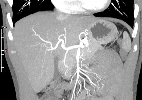
Other studies on the prevalence of HA variations have been published (Table 3). Dazzi et al.25 used angiotomography and intraoperative association to assess a sample of 66 liver donors, revealing results with 100% sensitivity and 100% specificity. Our Michels type I results (Figure 3) are comparable to theirs (68.8% and 68.2%, respectively).

Abdominal computed tomography image of the abdomen reconstructed in maximum intensity projection in the arterial phase, coronal plane, showing a proper hepatic artery originating from a common hepatic artery and bifurcating into right and left hepatic arteries.
In 479 liver transplants, Fonseca-Neto et al.8 found a high prevalence of normal anatomy (86.8%) and a low prevalence of types II, III, and IV (2.7%, 5.6% and 0.8%, respectively), which was similar to our results (4.8%, 5.4% and 1.4%, respectively). Brasil et al.4 evaluated 100 angiotomography scans, finding prevalence rates of 1% for type VI and 4% for type IX. These results are also similar to ours (2.2% and 5%, respectively).
Finally, Imam et al.26 also found similar results to ours regarding HA types VII and VIII (0.4% and 2.9%, respectively vs 0.2% and 3.6%, respectively). However, their incidence of Michels type V was higher than ours (8.2% vs 4.5%) (Figure 4).

Abdominal computed tomography image with maximum intensity projection reconstructed in the arterial phase, coronal plane, showing an accessory left hepatic artery originating from the left gastric artery.
Of the 500 CT scans we analyzed, only 2 (0.4%) could not be classified according to the Michels system. In other studies, classification failure has been as high as 12.9% (Table 3).26 Since anatomical variations derive from embryological alterations, it is difficult to develop a standardized model that addresses all possibilities.8
Both of the unclassifiable conditions in the present study have been previously documented in the literature, ie, a RHA originating directly from the aorta and a common HA originating from the SMA with an accessory LHA simultaneously originating from the LGA. The first case is relevant due to the difficult access, requiring angiography and transarterial chemotherapy to treat hepatocarcinoma, while the second has been associated with increased complication rates after pancreaticoduodenectomy.26
We performed a comparative analysis of studies on intrahepatic PV variation (Table 4), finding that our sample had a higher incidence of structural variation (21.8%) than that of Lee et al.27 (14.5%), Anwar et al.9 (4.8%), or Vidya et al.28 (12.5%). Our sample also had a higher prevalence of Cheng type IV (8%) than these studies (1%, 0.8%, and 0%, respectively).
The clinical importance of the high prevalence of Cheng type IV (Figure 5) in our sample is due to the contraindication for certain surgical procedures, such as donation of the right hepatic lobe for organ transplantation or tumor resection, since it makes adequate reconstruction impossible.9 Type II was the most prevalent variation in Lee et al.27 and Vidya et al.28 (7.1% and 10%, respectively), while type III was the most prevalent (2.4%) in Anwar et al.9

Abdominal computed tomography image in the portal phase, coronal plane, showing the right anterior branch of the portal vein originating from the left branch.
FINAL CONSIDERATIONS
This study found a high prevalence of variation in hepatic vascularization, both in the proper HA and the intrahepatic PV. Similar data have been found in other studies worldwide. Tomographic analysis and knowledge of the vascular anatomy of the liver and abdominal organs, both normal and abnormal, facilitate preoperative planning, lead to greater surgical success, prevent complications, and reduce morbidity and mortality. Variations not classified by previous trials should be categorized according to their clinical importance, and further Brazilian studies should be encouraged to clarify the patterns found in the national population.
-
How to cite:
Biz MEZ, Salame JP, Correia GG, Moreira RS. Variations in hepatic circulation: a study of 500 abdominal computed tomography scans. J Vasc Bras. 2025;24:e20240110. https://doi.org/10.1590/1677-5449.202401102
-
Financial support:
None.
-
The study was carried out at Centro Universitário de Brusque (UNIFEBE), Brusque, SC, Brazil.
-
Ethics committee approval:
This study was approved by the research ethics committee of the Centro Universitário de Brusque - UNIFEBE under the number of Opinion 5.616.116, as well as the waiver of the informed consent form.
References
-
1 López-Andújar R, Moya A, Montalvá E, et al. Lessons learned from anatomic variants of the hepatic artery in 1,081 transplanted livers. Liver Transpl. 2007;13(10):1401-4. http://doi.org/10.1002/lt.21254 PMid:17902125.
» http://doi.org/10.1002/lt.21254 -
2 Bianchi Homero F, Algieri Rubén F, Sanjurjo D, Ottone NE, Fuentes R. Multiple anatomical variations of the hepatic pedicle: case report and clinical application. Int J Morphol. 2014;32(3):782-5. http://doi.org/10.4067/S0717-95022014000300006
» http://doi.org/10.4067/S0717-95022014000300006 -
3 Araujo SA No, Mello CF Jr, Franca HA, Duarte CMA, Borges RF, Magalhães AGX. Angiotomografia multidetectores do tronco celíaco e sistema arterial hepático: anatomia normal e suas principais variantes. Radiol Bras. 2016;49(1):49-52. http://doi.org/10.1590/0100-3984.2014.0041
» http://doi.org/10.1590/0100-3984.2014.0041 -
4 Brasil IRC, Araujo IF, Lima AALA, Melo ELA, Esmeraldo RM. Computed tomography angiography study of variations of the celiac trunk and hepatic artery in 100 patients. Radiol Bras. 2018;51(1):32-6. http://doi.org/10.1590/0100-3984.2016.0179 PMid:29540943.
» http://doi.org/10.1590/0100-3984.2016.0179 -
5 Pulakunta T, Potu BK, Gorantla VR, Vollala VR, Thomas J. Surgical importance of variant hepatic blood vessels: a case report. J Vasc Bras. 2008;7(1):84-5. http://doi.org/10.1590/S1677-54492008000100016
» http://doi.org/10.1590/S1677-54492008000100016 -
6 Ruiz CR, Nascimento SRR, Vidsiunas AK, Andrades L, Souza CC. Dupla variação anatômica vascular em um único indivíduo: estudo morfométrico. ABCS Health Sci. 2017;42(3):170-3. http://doi.org/10.7322/abcshs.v42i3.919
» http://doi.org/10.7322/abcshs.v42i3.919 -
7 Freitas AC, Godoy JL, Matias JE, Stadnik LG, Coelho JC. Comparação entre exames de imagem e achados operatórios em doadores para transplante hepático intervivos. Arq Gastroenterol. 2007;44(4):325-31. http://doi.org/10.1590/S0004-28032007000400009 PMid:18317652.
» http://doi.org/10.1590/S0004-28032007000400009 -
8 Fonseca-Neto OCLD, Lima HCS, Rabelo P, Melo PSV, Amorim AG, Lacerda CM. Anatomic variations of hepatic artery: a study in 479 liver transplantations. Arq Bras Cir Dig. 2017;30(1):35-7. http://doi.org/10.1590/0102-6720201700010010 PMid:28489166.
» http://doi.org/10.1590/0102-6720201700010010 -
9 Anwar AS, Srikala J, Papalkar AS, Parveez MQ, Sharma A. Study of anatomical variations of hepatic vasculature using multidetector computed tomography angiography. Surg Radiol Anat. 2020;42(12):1449-57. http://doi.org/10.1007/s00276-020-02532-5 PMid:32691112.
» http://doi.org/10.1007/s00276-020-02532-5 -
10 Choi TW, Chung JW, Kim HC, et al. Anatomic variations of the hepatic artery in 5625 patients. Radiol Cardiothorac Imaging. 2021;3(4):e210007. http://doi.org/10.1148/ryct.2021210007 PMid:34498005.
» http://doi.org/10.1148/ryct.2021210007 - 11 Moore KL, Dalley AF, Agur AMR. Anatomia orientada para clínica. Rio de Janeiro: Grupo GEN; 2018.
-
12 Carneiro C, Brito J, Bilreiro C, et al. All about portal vein: a pictorial display to anatomy, variants and physiopathology. Insights Imaging. 2019;10(1):38. http://doi.org/10.1186/s13244-019-0716-8 PMid:30900187.
» http://doi.org/10.1186/s13244-019-0716-8 -
13 Seco M, Donato P, Costa J, Bernardes A, Caseiro-Alves F. Vascular liver anatomy and main variants: what the radiologist must know. JBR-BTR. 2010;93(4):215-23. http://doi.org/10.5334/jbr-btr.319 PMid:20957897.
» http://doi.org/10.5334/jbr-btr.319 -
14 Michels NA. Newer anatomy of the liver and its variant blood supply and collateral circulation. Am J Surg. 1966;112(3):337-47. http://doi.org/10.1016/0002-9610(66)90201-7 PMid:5917302.
» http://doi.org/10.1016/0002-9610(66)90201-7 -
15 Chen H, Yano R, Emura S, Shoumura S. Anatomic variation of the celiac trunk with special reference to hepatic artery patterns. Ann Anat. 2009;191(4):399-407. http://doi.org/10.1016/j.aanat.2009.05.002 PMid:19540742.
» http://doi.org/10.1016/j.aanat.2009.05.002 -
16 Freitas ACT, Matias JEF, Coelho JCU. Anomalia anatômica da veia porta: uma causa rara de impossibilidade de doação do lobo direito em transplante hepático intervivos. Rev Col Bras Cir. 2006;33(4):260-1. http://doi.org/10.1590/S0100-69912006000400014
» http://doi.org/10.1590/S0100-69912006000400014 -
17 Katsourakis A, Chytas D, Filo E, et al. Incidence of extrahepatic portal vein anatomic variations and their clinical implications in daily practice. J Clin Med Res. 2021;13(9):460-5. http://doi.org/10.14740/jocmr4581 PMid:34691319.
» http://doi.org/10.14740/jocmr4581 -
18 Iqbal S, Iqbal R, Iqbal F. Surgical implications of portal vein variations and liver segmentations: a recent update. J Clin Diagn Res. 2017;11(2):AE01-05. http://doi.org/10.7860/JCDR/2017/25028.9453 PMid:28384848.
» http://doi.org/10.7860/JCDR/2017/25028.9453 - 19 Bayrak AH, Dogan SN, Öztürkmen Akay H. Clinical importance of main portal vein and right portal vein variations: a prevalence study with 128-slice multidetector computed tomography. Exp Clin Transplant. 2021. No prelo. http://doi.org/10.6002/ect.2020.0539. PMid:33736589.
-
20 Guler N, Dayangac M, Yaprak O, et al. Anatomical variations of donor portal vein in right lobe living donor liver transplantation: the safe use of variant portal veins. Transpl Int. 2013;26(12):1191-7. http://doi.org/10.1111/tri.12190 PMid:24252057.
» http://doi.org/10.1111/tri.12190 -
21 Noussios G, Dimitriou I, Chatzis I, Katsourakis A. The main anatomic variations of the hepatic artery and their importance in surgical practice: review of the literature. J Clin Med Res. 2017;9(4):248-52. http://doi.org/10.14740/jocmr2902w PMid:28270883.
» http://doi.org/10.14740/jocmr2902w -
22 Sebben GA, Rocha SL, Sebben MA, Parlissolo PR Fo, Gonçalves BHH. Variações da artéria hepática: estudo anatômico em cadáveres. Rev Col Bras Cir. 2013;40(3):221-6. http://doi.org/10.1590/S0100-69912013000300010 PMid:23912370.
» http://doi.org/10.1590/S0100-69912013000300010 -
23 Sureka B, Patidar Y, Bansal K, Rajesh S, Agrawal N, Arora A. Portal vein variations in 1000 patients: surgical and radiological importance. Br J Radiol. 2015;88(1055):20150326. http://doi.org/10.1259/bjr.20150326 PMid:26283261.
» http://doi.org/10.1259/bjr.20150326 - 24 Cheng YF, Huang TL, Lee TY, Chen TY, Chen CL. Variation of the intrahepatic portal vein: angiographic demonstration and application in living-related hepatic transplantation. Transplant Proc. 1996;28(3):1667-8. PMid:8658830.
-
25 Dazzi FL, Ribeiro MA Jr, Mancero JM, et al. Are the imaging findings used to assess the portal triad reliable to perform living-donor liver transplant? Arq Bras Cir Dig. 2013;26(4):296-301. http://doi.org/10.1590/S0102-67202013000400009 PMid:24510038.
» http://doi.org/10.1590/S0102-67202013000400009 -
26 Imam A, Karatas C, Mecit N, et al. Anatomical variations of the hepatic artery: a closer view of rare unclassified variants. Folia Morphol. 2022;81(2):359-64. http://doi.org/10.5603/FM.a2021.0024 PMid:33749803.
» http://doi.org/10.5603/FM.a2021.0024 -
27 Lee KK, Lee SK, Moon IS, Kim DG, Lee MD. Surgical techniques according to anatomic variations in living donor liver transplantation using the right lobe. Transplant Proc. 2008;40(8):2517-20. http://doi.org/10.1016/j.transproceed.2008.07.079 PMid:18929785.
» http://doi.org/10.1016/j.transproceed.2008.07.079 -
28 Vidya CS, Manjunatha B, Das SK. Radiological study of portal vein variations and its morphometry with special reference to its surgical implications of liver. Braz J Morphol Sci. 2021;38:201-5. http://doi.org/10.51929/jms.38.36.2021
» http://doi.org/10.51929/jms.38.36.2021