 Primary Mesenteric Neuroendocrine Tumor: A Rare and Unexpected Finding
Abstract
Neuroendocrine tumors (NETs) are rare malignancies originating from enterochromaffin cells, with up to 90% arising in the gastrointestinal tract. Those located in the mesentery are usually metastatic, and primary mesenteric NETs are exceedingly rare. We herein report the case of an 82-year-old man referred to general surgery after an incidental mesenteric mass detected on a fluorodeoxyglucose-positron emission tomography (18F-FDG PET-CT) scan performed for the evaluation of a pulmonary lesion. An abdominal computed tomography (CT) scan revealed a 30-mm mass in the mesentery, adjacent to the ileocolic vessels. The patient underwent exploratory laparoscopy and excision of the mass. A histopathological analysis confirmed a well-differentiated NET. The patient was started on octreotide therapy, which continues to date. At 24 months of follow-up, the patient remains asymptomatic.
Given the rarity of primary mesenteric NETs and the predominance of metastatic lesions at this site, comprehensive exclusion of other potential primary-tumor sites is of utmost importance.
Keywords
neuroendocrine tumor; mesenteric mass; primary mesenteric
Introduction
Neuroendocrine tumors (NETs) are rare malignancies that arise from enterochromaffin cells, which are predominantly located in the epithelium of the gastrointestinal tract.1 However, due to the widespread distribution of neuroendocrine cells throughout the body,2 these tumors can also occur in the respiratory tract and urogenital system.3
Up to 90% of NETs originate in the gastrointestinal tract, with the small bowel, particularly the terminal ileum, being the most frequently affected site.4 Other common primary locations within the gastrointestinal system include the appendix, rectum and pancreas. When found in the mesentery, NETs are usually metastatic, with primary NETs in this location being exceedingly rare.5
Clinical Case
An 82-year-old patient with a 21-mm pulmonary nodule in the right upper lobe, was referred to general surgery following the incidental finding of a mesenteric mass on a fluorodeoxyglucose-positron emission tomography (18F-FDG PET-CT) scan performed for evaluation of the pulmonary mass.
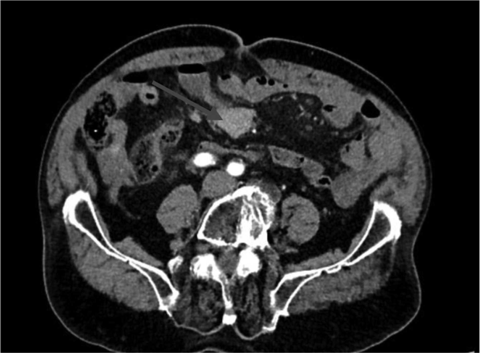
A computed tomography (CT) scan of the abdomen demonstrated a 30-mm solid mass with poorly-defined borders in the mesentery, adjacent to the ileocolic vessels. No other pathological findings were identified. The patient denied any gastrointestinal or other symptoms. The physical examination was unremarkable. The patient had recently been submitted to an upper endoscopy and colonoscopy that did not show any abnormalities.
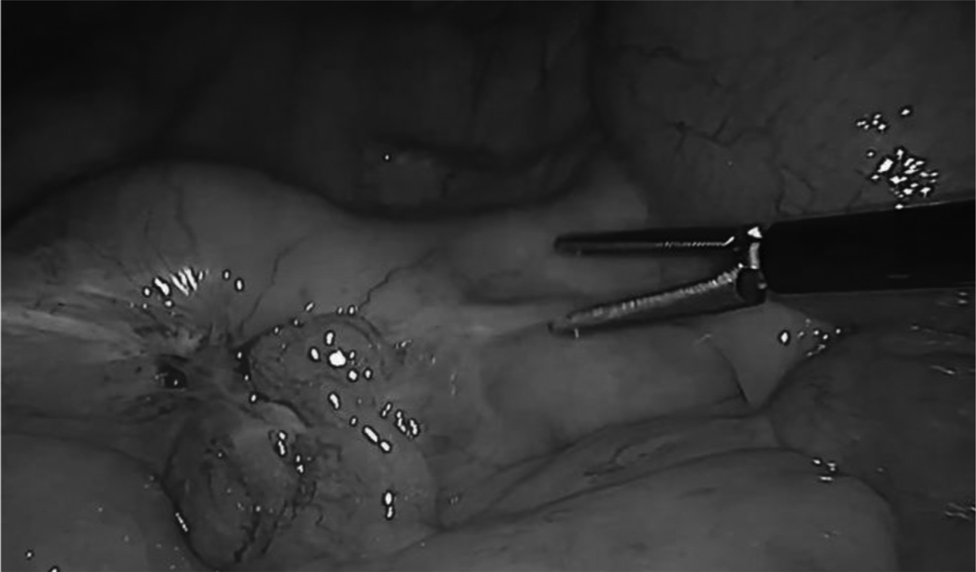
The patient underwent exploratory laparoscopy and excision of the mesenteric mass. The intraoperative exploration of the abdominopelvic cavity, with particular attention to the small bowel loops, revealed no additional lesions. The mass arising from the mesentery near the ileum was identified, and a bottom-up mobilization of the right colon and mesentery was performed, enabling the identification of the duodenum, ileocolic vessels and superior mesenteric vein, none of which showed signs of invasion.
The lesion was excised using a harmonic scalpel. Adequate vascularization of the small bowel was confirmed with indocyanine green fluorescence. The resulting mesenteric defect was closed with STRATAFIX 2-0 sutures (Ethicon, Inc.). The postoperative course was uneventful, and the patient was discharged on postoperative day 4.
The surgical specimen was submitted to a histopathological examination. Immunohistochemistry testing demonstrated positivity for synaptophysin, CDX2 (Caudal homeobox 2) and chromogranin A, and negativity for TIF-1 (Transcription Intermediary Factor 1). The Ki-67 (Ki-67 index) index was of 5%. These findings were consistent with a diagnosis of a well-differentiated NET.
The Multidisciplinary Tumor Board recommended additional workup, including repeat upper gastrointestinal endoscopy and colonoscopy, as well as the performance of capsule endoscopy, Ga-DOTANOC PET, and measurement of chromogranin levels.
A 5-mm sessile polyp in the descending colon was resected during the colonoscopy and tested negative for malignancy. No other lesions were identified. The PET-DOTANOC demonstrated somatostatin receptor hyperexpression in mesenteric lymphadenopathy, suggestive of nodal metastases from the NET. No radiotracer uptake was observed in the pulmonary lesion nor anywhere else. The chromogranin A level was of 21.3 ng/mL (normal: < 100 ng/mL). The patient was started on octreotide therapy, which is still being maintained.
A biopsy of the pulmonary nodule was performed, showing no evidence of malignancy. The Multidisciplinary Tumor Board decided that no further follow-up of the pulmonary lesion was necessary. At 24 months follow-up, the patient is asymptomatic and recurrence free. Figs. 1,2,3.

Axial preoperative computed tomography (CT) scan showing a 30-mm solid mesenteric mass (arrow).

Discussion
Primary mesenteric NETs are rare, with only about 20 cases described in the literature.5 The mesentery is most commonly involved secondarily, either through direct extension from a small-bowel primary tumor or as a site of metastasis.6 Therefore, a thorough diagnostic workup is crucial to exclude a primary lesion elsewhere.5
Clinically, these tumors are often asymptomatic and discovered incidentally during unrelated procedures or imaging studies, as occurred in the patient herein reported. When symptomatic, they may present as an abdominal mass, with pain or features of carcinoid syndrome.7
Surgical resection remains the cornerstone of therapy for NETs.8 Larger lesions, particularly those larger than 2 cm, are frequently associated with locally-advanced disease or distant metastases.9,10 However, in the case herein reported, the tumor measured 3 cm, was well-circumscribed, and showed no evidence of infiltration into the adjacent small bowel or other intra-abdominal structures.
Most of the previously-reported cases1,8–10 describe the tumor as being adherent to or involving a loop of small bowel, thus requiring resection of the affected bowel segment along with the mesenteric mass. In contrast, in the case herein presented, the absence of such adhesion or involvement enabled the excision of the mesenteric lesion alone.
Given the rarity of primary mesenteric NETs,11 this diagnosis should only be considered after exclusion of the more common primary sources, particularly within the small intestine.12 During surgery, the entire abdominal cavity, with particular attention to the small intestine, was meticulously examined, and no other lesions were identified. In addition, postoperative investigations, including upper endoscopy, colonoscopy and video capsule endoscopy, revealed no lesions, thereby supporting the diagnosis of a primary mesenteric NET. The combination of imaging, endoscopic studies and histopathological results is crucial for accurate diagnosis, treatment, and follow-up.
Conclusion
Primary mesenteric NETs are extremely rare, with only a few cases reported in the literature. Due to their rarity and the fact that mesenteric lesions are mostly metastatic, it is important to thoroughly exclude other potential primary sites, as was done in the case herein reported.
Data Availability
Data will be available upon request to the corresponding author.
References
-
1 González-Muñoz A, Aguirre-Salamanca EJ, Rivera-Rincón NA, Rodríguez-Narvaez JG, González-Sierra P, Ramírez-Giraldo C. Primary mesenteric neuroendocrine tumor: Case report. Int J Surg Case Rep 2023;109:108517. Doi: 10.1016/j.ijscr.2023.108517
» https://doi.org/10.1016/j.ijscr.2023.108517 -
2 Mashoori N, Rabani AH, Kazemeini AR. Ectopic Cushing's syndrome due to a mesenteric neuroendocrine tumour. Ann R Coll Surg Engl 2012;94(08):e251–e253. Doi: 10.1308/ 003588412X13373405387492
» https://doi.org/10.1308/003588412X13373405387492 -
3 Shogbesan O, Abdulkareem A, Pappachen B, Altomare J. Primary Mesenteric Carcinoid Tumor Presenting with Carcinoid Syndrome. Case Rep Gastroenterol 2018;12(02):396–401. Doi: 10.1159/000490522
» https://doi.org/10.1159/000490522 -
4 Moghazy KM, Zakaria HM. Bowel perforation secondary to mesenteric carcinoid tumor: a case report and review of the literature. Biomed Res (Tokyo) 2009;20(02):148–150 Available from: https://www.alliedacademies.org/articles/bowel-perforation-secondary-to-mesenteric-carcinoid-tumor-a-case-report-and-review-of-the-literature.pdf
» https://www.alliedacademies.org/articles/bowel-perforation-secondary-to-mesenteric-carcinoid-tumor-a-case-report-and-review-of-the-literature.pdf -
5 Symeonidou E, Fouza A, Gkoutziotis I, Nikolaidou C, Petras P, Mpallas K. Two synchronous primary mesenteric neuroendocrine tumors in a patient: a case report. Front Surg 2024;11:1323468. Doi: 10.3389/fsurg.2024.1323468
» https://doi.org/10.3389/fsurg.2024.1323468 -
6 Rani E, Nibhoria S, Shilpa S, Kaur N. Primary neuroendocrine tumor of mesentry – a rare case report. Scholars J Med Case Rep. 2021;9(09):909–911. Doi: 10.36347/sjmcr.2021.v09i09.022
» https://doi.org/10.36347/sjmcr.2021.v09i09.022 -
7 Agarwal A, Kaman L, Gupta A, Ramavath K, Vaiphei K. Primary mesenteric neuroendocrine tumour with liver metastasis: a common presentation of an uncommon tumour. Trop Doct 2020;50(01):65–68. Doi: 10.1177/0049475519887657
» https://doi.org/10.1177/0049475519887657 -
8 Aggarwal S, Agarwal S, Ramrakhiani D. Primary mesenteric carcinoid tumor: a rare entity. J Interdiscip Histopathol 2016;4 (01):23–25. Doi: 10.5455/jihp.20160116101011
» https://doi.org/10.5455/jihp.20160116101011 -
9 Park IS, Kye BH, Kim HS, et al. Primary mesenteric carcinoid tumor. J Korean Surg Soc 2013;84(02):114–117. Doi: 10.4174/ jkss.2013.84.2.114
» https://doi.org/10.4174/jkss.2013.84.2.114 -
10 Kim SW. Primary neuroendocrine tumor of the colon mesentery. Korean J Clin Oncol 2014;10(01):49–52. Doi: 10.14216/ kjco.14010
» https://doi.org/10.14216/kjco.14010 -
11 Morishita S, Yoshida S, Kamatani Y, Suzuhigashi S, Kitou M, Nasu T. Primary grade 2 neuroendocrine tumor of the ileal mesentery: a case report. Surg Case Rep 2022;8(01):146. Doi: 10.1186/ s40792-022-01482-x
» https://doi.org/10.1186/s40792-022-01482-x -
12 Bhandari S, Kandu H, Kandel L, Pradhan S, Pujara S, Gurung R. Mesenteric neuroendocrine tumor: a case report. IJSIT 2018;7 (04):836–841 Available from: http://www.ijsit.com/admin/ijsit_files/MESENTERIC%20NEUROENDOCRINE%20TUMOR-CASE%20REPORT_IJSIT_7.4.6.pdf
» http://www.ijsit.com/admin/ijsit_files/MESENTERIC%20NEUROENDOCRINE%20TUMOR-CASE%20REPORT_IJSIT_7.4.6.pdf
Edited by
-
Editor-in-Chief:
Henrique Sarubbi Fillmann.