 The role of probiotics in modulating the gut microbiota as a potential inhibitor of diabetic kidney disease progression
Abstract
Introduction: Gut dysbiosis is commonly observed in patients with diabetic kidney disease (DKD) and may contribute to its pathogenesis. Among microbial metabolites, butyrate plays a key role in regulating antioxidant proteins in type 2 diabetes mellitus (T2DM). Based on this, we hypothesized that the administering probiotics to diabetic rats modulates redox status and thereby attenuates renal disease progression.
Methods: An in vivo study was performed using 15 male Wistar rats (8 weeks old, 250–300 g) randomized into three groups (n = 5/group): Control (vehicles: 0.9% saline and 0.1 M citrate, pH 4.2, i.p., on day 1), T2DM (nicotinamide 100 mg/kg, i.p., followed by streptozotocin 60 mg/kg, i.p., in 0.1 M citrate buffer, pH 4.2), and T2DM + Prob (T2DM protocol plus a multistrain probiotic—Bifidobacterium longum, Bifidobacterium bifidum, and Lactobacillus rhamnosus—1010 CFU/mL by gavage for 6 weeks). The parameters evaluated were: serum creatinine, inulin clearance, microalbuminuria, urinary and lipid peroxides, glutathione, and nuclear factor erythroid 2–related factor 2 (Nrf2).
Results: Probiotic treatment significantly increased Nrf2 expression and glutathione levels, reduced urinary and lipid peroxidation, and—beyond attenuating oxidative stress—improved renal function, with lower serum creatinine and microalbuminuria and higher inulin clearance.
Conclusion: These findings indicate that probiotics prevented DKD progression, likely by modulating oxidative stress via the gut microbiota. These results suggest that probiotics may serve as renoprotective agents, potentially reducing DKD morbidity in T2DM.
Keywords:
Diabetic kidney disease; Type 2 diabetes mellitus; Probiotics; Oxidative stress
RESUMO
Introdução: A disbiose intestinal é comumente observada em pacientes com doença renal diabética e pode contribuir para sua patogênese. Entre os metabólitos microbianos, o butirato desempenha papel fundamental na regulação de proteínas antioxidantes no diabetes mellitus tipo 2. Com base nisso, levantamos a hipótese de que a administração de probióticos em ratos diabéticos poderia modular o perfil redox, atenuando, assim, a progressão da doença renal.
Métodos: Estudo in vivo utilizando ratos Wistar machos (8 semanas, 250–300 g; n = 15), randomizados em três grupos (n = 5/grupo): Controle (veículos: solução salina 0,9% e citrato 0,1 M; pH 4,2, i.p., no dia 1); DM2 (nicotinamida 100 mg/kg, i.p., seguida de estreptozotocina 60 mg/kg, i.p., em tampão citrato 0,1 M, pH 4,2); e DRD + Prob (protocolo D + probiótico multicepas Bifidobacterium longum, Bifidobacterium bifidum e Lactobacillus rhamnosus, 1010 UFC/mL via gavagem por 6 semanas). Parâmetros avaliados: creatinina sérica, clearance de inulina, microalbuminúria, peróxidos urinários e lipídicos, glutationa e Fator 2 Relacionado ao Eritroide Nuclear 2.
Resultados: O tratamento probiótico aumentou significativamente a expressão de Nrf2 e os níveis de glutationa. Por outro lado, reduziu a peroxidação urinária e lipídica. Além de atenuar o estresse oxidativo, os probióticos melhoraram a função renal, com redução da creatinina sérica e da microalbuminúria e aumento do clearance de inulina.
Conclusão: Os achados indicam que os probióticos preveniram a progressão da DRD, provavelmente modulando o estresse oxidativo por meio da microbiota intestinal. Esses resultados sugerem que os probióticos podem atuar como agentes renoprotetores, reduzindo potencialmente a morbidade da DRD no DM2.
Descritores:
Doença renal diabética; Diabetes mellitus tipo 2; Probióticos; Estresse oxidativo
Introduction
Type 2 diabetes mellitus (T2DM) is defined as a constellation of metabolic abnormalities marked by persistently elevated blood glucose levels resulting from reduced pancreatic insulin secretion and/ or decreased insulin sensitivity. It accounts for approximately 91% of the global diabetes burden and is the leading cause of chronic kidney disease (CKD) and end-stage kidney disease (ESKD). T2DM is associated with major cardiovascular risk factors, including hypertension, dyslipidemia, obesity, insulin resistance, and impaired glucose tolerance1,2.
Although complications of T2DM have a multifactorial origin, sustained hyperglycemia, by increasing inflammatory cytokines and oxidative stress and thereby promoting long-term injury, appears to be a principal driver of tissue damage in the diabetic kidney. This leads to alarming chronic complications such as retinopathy, neuropathy, cardiovascular disease, and diabetic kidney disease (DKD)3,4.
Clinically, DKD is defined by persistent albuminuria ≥30 mg/g (creatinine) and/or a glomerular filtration rate (eGFR) < 60 mL/min/1.73 m2 for at least 3 months, irrespective of etiology. It is a chronic complication, resulting in progressive damage and impairment of renal function5,6.
DKD is a significant concern worldwide, affecting millions of people with T2DM. Approximately 50% of patients with DKD present microalbuminuria that progresses to macroalbuminuria, one of the most important microvascular complications of DKD, with half of all ESKD cases related to the complication of DM5.
Thus, DKD is one of the main causes for initiating renal replacement therapy and is associated with increased morbidity and mortality. Therefore, it is urgent to explore effective, low-cost treatments to reduce morbidity and mortality in patients with DKD3,4.
Beyond traditional treatment, such as glycemic control and pharmacotherapy, new therapeutic approaches are being explored. Among these, the promising role of probiotics in the management of DKD stands out. Recent studies have shown that probiotics can play an important role in the prevention and treatment of DKD through different mechanisms, such as modulation of the gut microbiota7,8.
Probiotics are live microorganisms that confer health benefits to the host. These beneficial microorganisms are found in different foods and fermented products and are also available as supplements7.
Current research has demonstrated alterations in the gut microbiota in DKD, revealing reductions in beneficial strains such as Bifidobacterium and Lactobacillus and an increase in pathogenic bacterial species; these alterations have been associated with elevated levels of inflammatory cytokines, increased oxidative stress, and greater endotoxin translocation factors that contribute to the progression of renal dysfunction8.
On the other hand, studies indicate that the reduction of short-chain fatty acids (SCFAs), such as butyrate, acetate, and propionate, worsens the inflammatory state and impairs metabolic homeostasis, intensifying renal damage. In view of this, probiotic intervention emerges as a promising therapeutic strategy for the management of DKD, not only by modulating the gut microbiota and reducing systemic inflammation, but also through its positive effects in suppressing the progression of DKD7,8,9. The worsening of DKD leads to the irreversible need for renal replacement therapies.
Considering this scenario, experimental studies in animal models are essential to provide pathophysiological evidence that can underpin clinical research and contribute to the development of effective therapeutic protocols. Thus, this study aimed to evaluate the effects of probiotics on renal function and the redox profile of rats with DKD.
Methods
Male Wistar rats, eight weeks old, weighing 250–300 g, were allocated to the following groups: Control group (CT, n = 5), which received the vehicles for nicotinamide and streptozotocin, consisting of 0.9% saline and 0.1 M citrate buffer at pH 4.2 on day 1 of the experimental protocol, single dose; type 2 Diabetes Mellitus group (T2DM, n = 5), which received a single dose of nicotinamide (NA; 100 mg/kg) intraperitoneally (i.p.) diluted in 0.9% saline, and after 15 minutes received a single dose of streptozotocin (STZ; 60 mg/kg), i.p., diluted in 0.1 M citrate buffer at pH 4.2 on day 1 of the experimental protocol; and Type 2 Diabetes Mellitus + Probiotic group (T2DM+Prob, n = 5), which were T2DM animals that received probiotic strains (Bifidobacterium longum, Bifidobacterium bifidum, and Lactobacillus rhamnosus; 1010 CFU/ mL) by gavage for 6 weeks.
In the sixth week of the experimental protocol, animals from the different groups were placed in metabolic cages for 24-hour urine collection for studies of renal function and oxidative stress. Morphine was administered at a dose of 3 mg/kg, and 30 minutes after, the animals were removed from the metabolic cages and anesthetized with isoflurane (5% for induction and 3% for maintenance). Subsequently, animals underwent laparotomy and terminal blood collection via puncture of the abdominal aorta. The left kidney was removed, conditioned, and stored in a freezer at -80°C for redox studies. All procedures involving animals were carried out in accordance with the ethical standards established by the Animal Use Ethics Committee (CEUA) of the Faculty of Medical Sciences of Santa Casa de São Paulo (approval number 2023/07)10,11,12.
Renal Function Biomarkers
Serum creatinine (CrS) levels were determined by the Jaffé colorimetric method. This method is based on the reaction of creatinine with picric acid in an alkaline medium, forming a yellowish complex whose intensity is proportional to the creatinine concentration in the sample. The reading was performed on a spectrophotometer at 520 nm13. The quantification of microalbuminuria was carried out using an enzyme-linked immunosorbent assay (ELISA) for the detection of rat albuminuria (Bethyl Laboratories Inc, Montgomery, USA). This assay was performed on urine samples prepared according to the experimental protocols described by the manufacturer, and the results were expressed in mg/24 h. The glomerular filtration rate (GFR) was estimated by inulin clearance (CIn). After anesthesia, catheterization of the jugular vein was performed with a polyethylene tube (PE 60) for continuous infusion of inulin. In parallel, the carotid artery was catheterized with a polyethylene tube (PE 60) for blood sampling and monitoring of hemodynamic parameters. The concentration of inulin was analyzed according to methodology previously described in the literature by the anthrone method14.
Redox Markers and Antioxidant Expression
Peroxides are found in all body fluids, especially urine. Changes in their levels are considered markers of H2O2 generation or predictors of the extent of oxidative injury in vivo. Direct measurement of peroxides was performed using the FOX-2 assay. The reading was carried out by spectrophotometry at an absorbance of 560 nm and values expressed as nmol of peroxides per gram of creatinine15.
The urinary TBARS assay consisted of adding 0.4 mL of the urine sample to 0.6 mL of distilled water. To this dilution, 1.0 mL of 17.5% TCA and 1.0 mL of thiobarbituric acid (0.6%, pH 2) were added, and all test tubes were kept on ice during this first stage of the process. The solution was homogenized and then placed in a boiling water bath for 20 minutes for reaction with thiobarbituric acid. In the next step, the solution was removed from the water bath, cooled on ice, and 1.0 mL of 70% TCA was added. The solution was homogenized and incubated for 20 minutes in a capped test tube. The solution was then centrifuged for 15 minutes at 3000 revolutions per minute, and the reading was performed by spectrophotometry at an absorbance of 534 nm. The MDA level (nmol) was calculated using the molar extinction coefficient 1.56 × 10⁵ M-1 cm-1. Values were expressed per gram of creatinine16.
Renal tissue glutathione (GSH) was analyzed by the Ellman (DTNB) colorimetric method. Renal tissue was homogenized in 0.1 M phosphate buffer (pH 7.4) containing 1 mM EDTA, followed by centrifugation at 10,000–15,000 × g for 20 minutes at 4°C to obtain the supernatant. To remove proteins, 10% trichloroacetic acid (TCA) was used and the sample was centrifuged again. The treated supernatant was then mixed with phosphate buffer and 1 mM DTNB, reacting with GSH thiols to form a yellow TNB complex, whose absorbance was measured at 412 nm. Quantification was performed based on a 100µM reduced GSH standard curve, and the results were expressed as nmol/mg of protein17.
Western Blotting for Nrf2
The Nuclear Factor Erythroid 2–Related Factor 2 (Nrf2) was assessed in renal tissue samples by western blot. Protein was quantified by the Bradford assay, and 30 µg of protein per lane were separated by electrophoresis on a 10% polyacrylamide gel. Next, proteins were transferred to a nitrocellulose membrane. The membrane was blocked with TBS (50 mM Tris-HCl, pH 7.4, and 150 mM NaCl) containing 0.05% Tween 20 and 5% skim milk (or bovine serum albumin) for one and a half hours at room temperature. The membrane was then incubated overnight at 4°C in a solution containing the primary antibody (Nrf2, Thermo Fisher) and then incubated for 1 hour with an anti-rabbit secondary antibody. Fluorescent immunoblotting was performed and scans were acquired with an infrared imaging system18. Statistical analysis: results were expressed as mean ± standard deviation. Variance among groups was analyzed using the one-way ANOVAtest, followed by Tukey’s multiple comparisons post-test in GraphPad Prism version 10 for Windows®. Values of p < 0.05 were considered significant. The data used in the analyses are available upon request.
Results
Probiotics Improve Renal Function inT2DM Rats
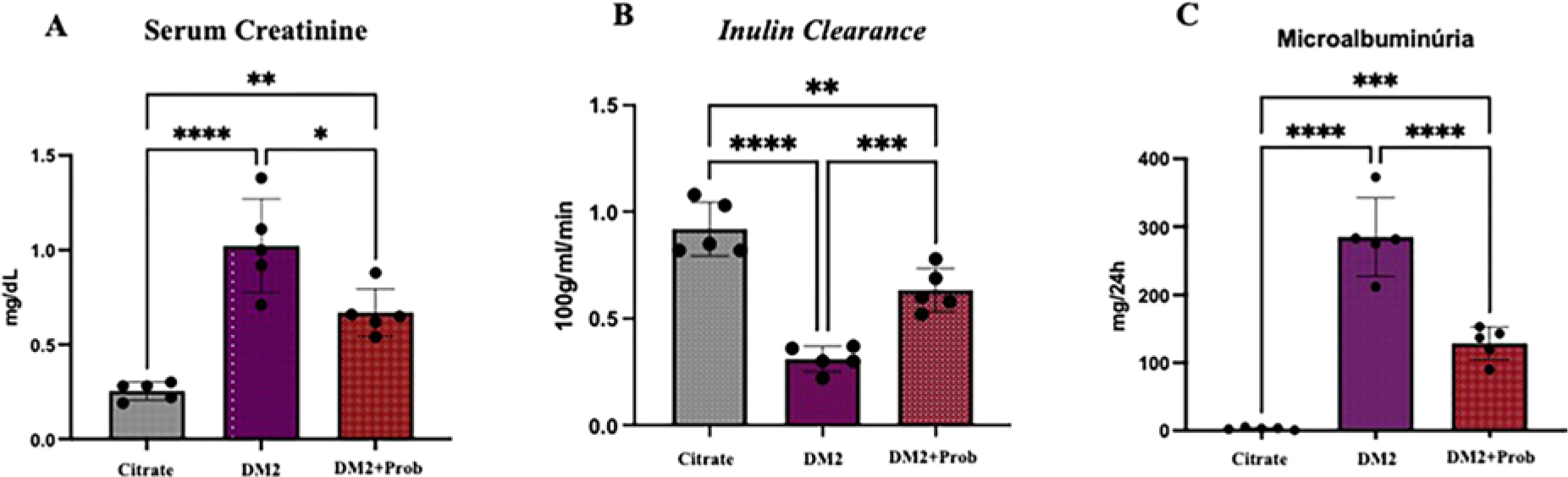
Renal function was evaluated by serum creatinine levels, microalbuminuria, and inulin clearance. As expected, the T2DM group showed reduced renal function, with elevated serum creatinine and microalbuminuria accompanied by a reduction in GFR (Figures 1A, 1B, and 1C) when compared with the Citrate group. These findings corroborate the establishment of progressive renal damage in diabetic rats, reflecting the loss of glomerular filtration capacity, characteristics of DKD. On the other hand, animals treated with probiotics demonstrated a significant improvement in renal function parameters. Inulin clearance (CIn) was partially restored, suggesting an attenuation of GFR decline (Figure 1C). Likewise, a statistically significant reduction in CrS levels and microalbuminuria was observed, indicating a renoprotective effect of probiotics (Figures 1A and 1B). These findings suggest that modulation of the gut microbiota can positively influence renal function in rats with DKD.
Probiotics improved renal function in T2DM rats. Wistar rats were chemically induced to T2DM and received Probiotics (Prob) by oral gavage for 6 weeks. Citrate: received vehicle; T2DM: Type 2 Diabetes Mellitus; T2DM + Prob: Type 2 Diabetes Mellitus + Probiotics. Group differences were tested by one-way ANOVA, followed by Tukey’s post-test. *p < 0.05 vs. Citrate; **p < 0.05 vs. T2DM; ***p < 0.05 vs. T2DM + Prob.
Probiotics Regulate Redox Signaling in DKD
Lipid and urinary peroxidation were significantly elevated in the T2DM group (Figures 2D and 2E), indicating oxidative stress in diabetic animals. In addition, there was a reduction in GSH concentration and Nrf2 expression in this group (Figures 2F and 2G), evidencing impairment of the antioxidant system. Administration of probiotics in the T2DM + Prob group resulted in a significant reduction in lipid and urinary peroxidation, with lower free radical production and less oxidative damage (Figures 2D and 2E). Moreover, GSH levels and Nrf2 expression showed a significant recovery compared with the T2DM group, indicating an increase in antioxidant defenses via modulation of the gut microbiota and suggesting greater production of short-chain fatty acids, especially butyrate (Figures 2F and 2G).

Probiotics regulate redox signaling in DKD. Wistar rats were chemically induced to T2DM and received Probiotics (Prob) by oral gavage for 6 weeks. Citrate: received vehicle; T2DM: Type 2 Diabetes Mellitus; T2DM + Prob: Type 2 Diabetes Mellitus + Probiotic. Group differences were tested by one-way ANOVA, followed by Tukey’s post-test. *p < 0.05 vs. Citrate; **p < 0.05 vs. T2DM; ***p < 0.05 vs. T2DM+Prob.
Discussion
DKD is a serious disease and an important complication of diabetes. The pathogenesis of DKD is complex and generally influenced by multiple factors. The gut microbiota has been found to play an important role in DKD. In this study, we analyzed renal function and the redox profile of rats with T2DM and DKD. Based on our findings, probiotic administration can beneficially modulate the gut microbiota, positively impacting both renal function and the redox profile of rats with T2DM and DKD19.
Our findings demonstrated that probiotics reduced serum creatinine (CrS) and microalbuminuria and increased inulin clearance in T2DM animals. A clinical study in patients with DKD using probiotics showed a significant improvement in renal function biomarkers compared with the placebo group, which corroborates the findings of our study19. The improvement observed in renal function parameters in our study may be partially explained by the reduction of uremic toxins derived from bacterial fermentation of amino acids in the gut, a process exacerbated by the sustained hyperglycemia characteristic of DKD. Probiotics have shown a positive effect by reducing the production of these toxins and, consequently, attenuating their deleterious influence on renal function20.
The correlation between the gut microbiota and DKD has been widely studied, although the exact mechanisms of this relationship are not yet fully understood. Our findings suggest that short-chain fatty acids (SCFAs), the main fermentation products of the intestinal microbiome – primarily acetate, propionate, and butyrate – may contribute to the improvement of renal function. One study observed that butyrate-producing bacteria are significantly reduced in the gut microbiota of patients with DKD21.
Studies have highlighted the interaction between intestinal dysbiosis and the progression of DKD. One study demonstrated that fecal microbiota transplantation can reverse intestinal dysbiosis and, consequently, improve renal function in rats with DKD, suggesting that microbiota modulation is a potential therapeutic resource for renal protection22,23. Another clinical study that analyzed the intestinal flora of patients with T2DM and DKD demonstrated that the gut microbiota plays an important role in the pathogenesis of DKD21.
Therefore, our findings demonstrate a marked impact on the SCFA elevation pathway, especially the butyrate pathway, which several studies have shown in to be associated with increased antioxidants21,22,23.
The findings of this work demonstrated that redox metabolites were modulated when T2DM animals were treated with probiotics, with significant expression of NRF2, an important transcription factor in the antioxidant cascade that prevents redox disturbances and, consequently, elevation of the inflammatory profile, advanced glycation end products (AGEs), and progression of DKD24,25. One study demonstrated that butyrate restored renal function and attenuated inflammation, apoptosis, and renal fibrosis in a preclinical model24.
We hypothesize that probiotic supplementation modulated Nrf2 redox signaling through specific SCFA receptors, inducing nuclear translocation of Nrf225. This was demonstrated in our study through the nuclear accumulation of Nrf2 in T2DM animals, as revealed by the increased levels this parameter. Probiotic supplementation, however, reduced this parameter, indicating a decrease in redox disturbances in these animals. These findings open new perspectives for the use of probiotics as therapeutic adjuvants in the prevention of T2DM complications and the progression of DKD.
Conclusion
Probiotics demonstrated potential as a therapeutic nutritional agent in renal protection. Moreover, their impact on the redox profile suggests that their supplementation may represent a complementary strategy to attenuate the progression of DKD, one of the most severe complications of T2DM. Thus, the inclusion of probiotics in the diet, together with lifestyle changes, may help reduce the need for renal replacement therapy, promoting an effective preventive and therapeutic approach.
Data Availability
The datasets generated and/or analyzed during the current study are not publicly available but are available from the corresponding author on reasonable request.
Funding
This study was funded by the Fundação de Amparo à Pesquisa da Faculdade de Ciências Médicas da Santa Casa de São Paulo.
References
-
1. Sá J, Canani L, Rangel E, Bauer A, Escott G, Zelmanovitz T, et al. Doença renal do diabetes. São Paulo: Diretriz Oficial da Sociedade Brasileira de Diabetes; 2022. doi: http://doi.org/10.29327/557753.2022-18.
» https://doi.org/10.29327/557753.2022-18 -
2. Luo W, Tang S, Xiao X, Luo S, Yang Z, Huang W, et al. Translation Animal Models of Diabetic Kidney Disease: Biochemical and Histological Phenotypes, Advantages and Limitations. Diabetes Metab Syndr Obes. 2023;16:1297–321. doi: http://doi.org/10.2147/DMSO.S408170. PubMed PMID: 37179788.
» https://doi.org/10.2147/DMSO.S408170 - 3. International Diabetes Federation. IDF diabetes atlas. 10th ed. Brussels: IDF; 2021.
-
4. Koye DN, Magliano DJ, Nelson RG, Pavkov ME. The global epidemiology of diabetes and kidney disease. Adv Chronic Kidney Dis. 2018;25(2):121–32. doi: http://doi.org/10.1053/j. ackd.2017.10.011. PubMed PMID: 29580576.
» https://doi.org/10.1053/j.ackd.2017.10.011 -
5. Lin YC, Chang YH, Yang SY, Wu KD, Chu TS. Update of pathophysiology and management of diabetic kidney disease. J Formos Med Assoc. 2018;117(8):662–75. doi: http://doi.org/10.1016/j.jfma.2018.02.007. PubMed PMID: 29486908.
» https://doi.org/10.1016/j.jfma.2018.02.007 -
6. Kidney Disease: Improving Global Outcomes (KDIGO) Diabetes Work Group. KDIGO 2020 clinical practice guideline for diabetes management in hronic kidney disease. Kidney Int. 2020;98(4S):S1–115. doi: http://doi.org/10.1016/j.kint. 2020.06.019. PubMed PMID: 32998798.
» https://doi.org/10.1016/j.kint.2020.06.019 -
7. Dai Y, Quan J, Xiong L, Luo Y, Yi B. Probiotics improve renal function, glucose, lipids, inflammation and oxidative stress in diabetic kidney disease: a systematic review and meta-analysis. Ren Fail. 2022;44(1):862–80. doi: http://doi.org/10.1080/0886 022X.2022.2079522. PubMed PMID: 35611435.
» https://doi.org/10.1080/0886022X.2022.2079522 -
8. Lau WL, Tran T, Rhee CM, Kalantar-Zadeh K, Vaziri ND. Diabetes and the gut microbiome. Semin Nephrol. 2021; 41(2):104–13. doi: http://doi.org/10.1016/j.semnephrol.2021. 03.005. PubMed PMID: 34140089.
» https://doi.org/10.1016/j.semnephrol.2021.03.005 -
9. Wu X, Zhao L, Zhang Y, Li K, Yang J. The role and mechanism of the gut microbiota in the development and treatment of diabetic kidney disease. Front Physiol. 2023;14:1166685. doi: http://doi.org/10.3389/fphys.2023.1166685. PubMed PMID: 37153213.
» https://doi.org/10.3389/fphys.2023.1166685 - 10. Brasil. Resolução normativa no 13 de 20 setembro de 2013. Institui a diretriz da prática de eutanásia do Conselho Nacional de Controle de Experimentação Animal – CONCEA. Diário Oficial da União; Brasília; 26 set. 2013. Seção 1.
- 11. Sheela N, Jose MA, Sathyamurthy D, Kumar BN. Effect of silymarin on streptozotocin-nicotinamide-induced type 2 diabetic nephropathy in rats. Iran J Kidney Dis. 2013;7(2): 117–23. PubMed PMID: 23485535.
- 12. Whiter P, Samson FE. Determination of inulin in plasm and urine by use of antrone. J Lab Clin Med. 1954;43:45–8.
-
13. Owen JA, Iggo B, Scandrett FJ, Stewart CP. The determination of creatinine in plasma or serum, and in urine; a critical examination. Biochem J. 1954;58(3):426–37. doi: http://doi.org/10.1042/bj0580426. PubMed PMID: 13208633.
» https://doi.org/10.1042/bj0580426 -
14. Vattimo MFF, Watanabe M, Fonseca CD, Neiva LBM, Pessoa EA, Borges FT, et al. Nephrotoxicity: from organ to cell damage. PLoS One. 2016;11(8):e0161057. doi: http://doi.org/10.1371/journal.pone.0161057. PubMed PMID: 27532263.
» https://doi.org/10.1371/journal.pone.0161057 -
15. Gay C, Collins J, Gebicki JM. Hydrogen peroxide assay with the ferric – xylenol orange complex. Anal Biochem. 1999; 273(2):149–55. doi: http://doi.org/10.1006/abio.1999.4208. PubMed PMID: 10469484.
» https://doi.org/10.1006/abio.1999.4208 - 16. Lima ES, Abdalla DSP. Peroxidação lipídica: mecanismos e avaliação em amostras biológicas. Braz J Pharm Sci. 2001;37(3): 293–303.
-
17. Filomeni G, Rotilio G, Ciriolo MR. Cell signally and the glutathione redox system. Biochem Pharmacol. 2002;64(5–6): 1057–64. doi: http://doi.org/10.1016/S0006-2952(02)01176-0. PubMed PMID: 12213605.
» https://doi.org/10.1016/S0006-2952(02)01176-0 -
18. Chen XL, Kunsch C. Induction of cytoprotective genes through Nrf2/antioxidant response element pathway: a new therapeutic approach for the treatment of inflammatory diseases. Curr Pharm Des. 2004;10(8):879–91. doi: http://doi.org/10.2174/1381612043452901. PubMed PMID: 15032691.
» https://doi.org/10.2174/1381612043452901 -
19. Miraghajani M, Zaghian N, Dehkohneh A, Mirlohi M, Ghiasvand R. Probiotic soymilk consumption and renal function among type 2 diabetic patients with nephropathy: a randomized controlled clinical trial. Probiotics Antimicrob Proteins. 2019;11(1):124–32. doi: http://doi.org/10.1007/s12602-017-9325-3. PubMed PMID: 28884306.
» https://doi.org/10.1007/s12602-017-9325-3 -
20. Kahraman M, Ertekin YH, Satman İ. The effects of kefir on renal tissues and functions in diabetic rats. Probiotics Antimicrob Proteins. 2021;13(2):375–82. doi: http://doi.org/10.1007/s12602-020-09698-9. PubMed PMID: 32820468.
» https://doi.org/10.1007/s12602-020-09698-9 -
21. Zhang L, Wang Z, Zhang X, Zhao L, Chu J, Li H, et al. Alterations of the gut microbiota in patients with diabetic nephropathy. Microbiol Spectr. 2022;10(4):e0032422. doi:http://doi.org/10.1128/spectrum.00324-22. PubMed PMID: 35863004.
» https://doi.org/10.1128/spectrum.00324-22 -
22. Hu ZB, Lu J, Chen PP, Lu CC, Zhang JX, Li XQ, et al. Dysbiosis of intestinal microbiota mediates tubulointerstitial injury in diabetic nephropathy via the disruption of cholesterol homeostasis. Theranostics. 2020;10(6):2803–16. doi: http://doi.org/10.7150/thno.40571. PubMed PMID: 32194836.
» https://doi.org/10.7150/thno.40571 -
23. Zhong C, Dai Z, Chai L, Wu L, Li J, Guo W, et al. The change of gut microbiota-derived short-chain fatty acids in diabetic kidney disease. J Clin Lab Anal. 2021;35(12):e24062. doi: http://doi.org/10.1002/jcla.24062. PubMed PMID: 34689373.
» https://doi.org/10.1002/jcla.24062 -
24. Bashir AM, Olaniyi KS. Butyrate alleviates renal inflammation and fibrosis in a rat model of polycystic ovarian syndrome by suppression of SDF-1. BMC Pharmacol Toxicol. 2023;24(1):48. doi: http://doi.org/10.1186/s40360-023-00692-9. PubMed PMID: 37789355.
» https://doi.org/10.1186/s40360-023-00692-9 -
25. Liu C, Hua H, Zhu H, Cheng Y, Guo Y, Yao W, et al. Aloe polysaccharides ameliorate acute colitis in mice via Nrf2/HO-1 signaling pathway and short-chain fatty acids metabolism. Int J Biol Macromol. 2021;185:804–12. doi: http://doi.org/10.1016/j.ijbiomac.2021.07.007. PubMed PMID: 34229016.
» https://doi.org/10.1016/j.ijbiomac.2021.07.007
Edited by
-
Editorial Responsibility
Editor-in-chief: Miguel Riella https://orcid.org/0000-0003-4181-613X.Associate Editor: Andrea Emília Stinghen https://orcid.org/0000-0001-8595-5321.