Preparation of PVDMA/PEI Polymeric Films Functionalized with Amino Acids and Their Study on Adhesion and Proliferation of Escherichia coli
Abstract
Bacterial infections represent a significant public health problem. Escherichia coli (E. coli) is a bacterium capable of causing multiple types of infections; therefore, preventing bacterial adhesion and proliferation is crucial. In this report, it is presented the synthesis of polymeric films based on poly(2-vinyl-4,4’-dimethylazlactone) (PVDMA) and poly(ethyleneimine) (PEI), whose surface has been modified with amino acids. A bilayer consists of a first layer of PEI and a second layer of PVDMA (15 bilayers), and in the case of 15.5 bilayers, it refers to the fact that the last layer is with the PEI, and functionalized with amino acids (NH2). The present strategy is based on the preparation of covalently crosslinked thin films by the layer-by-layer method using PVDMA/PEI. The films were placed on a glass surface, and 15.5-layer films were prepared with the PEI polymer as the final layer, which contains amino groups on the surface. The final layer was functionalized with Fmoc-protected amino acids (glycine (Gly), alanine (Ala), leucine (Leu), and phenylalanine (Phe)). Finally, the Fmoc group was removed, yielding surfaces functionalized with amino acids and the free amino group (NH2). The films were characterized by Fourier transform infrared spectroscopy (FTIR), optical microscopy, and field emission scanning electron microscopy (FESEM). They were incubated with a green fluorescent protein (GFP) -modified E. coli strain for 2 h. The order of interaction of the bacteria with the modified surface is as follows: (F-Leu > F NH2 > F-Ala > F-Gly > F-Phe). The leucine-modified films effectively prevented E. coli adhesion.
Keywords:
amino acids; Escherichia coli; breast cancer; polymeric film; azlactone
Introduction
Bacterial infections affect millions of people worldwide, triggering a wide range of diseases and systemic complications, and in some cases, leading to death. Therefore, the medical and scientific communities have joined efforts to develop more effective antibiotics. Escherichia coli (E. coli) is a Gram-negative bacterium belonging to the Enterobacteriaceae family. It naturally inhabits the gastrointestinal tract of warm-blooded animals and humans, and is often released into the environment through fecal matter.1 Despite its symbiotic function, certain strains are responsible for a variety of clinical infections, including enteritis, urinary and digestive tract infections, septicemia, and neonatal meningitis, among others.1 E. coli has a high survival rate in extraintestinal environments, mainly due to the influence of both biotic factors (such as competition with other microorganisms and the ability of the bacterium to acquire nutrients) and abiotic factors (including temperature, humidity, nutrient availability, pH, and solar radiation), all of which contribute to its reproductive success.2 Furthermore, the misuse of antibiotics and self-medication have led to the emergence of multidrug-resistant strains, which spread rapidly through genetic elements such as plasmids, integrons, or transposons that can be exchanged between bacterial organisms. This phenomenon represents a serious global public health concern and poses a potential risk of triggering a future health crisis.3 Due to E. coli’s remarkable adaptability and ability to thrive in nutrient-poor environments, it is capable of forming biofilms on a wide range of surfaces. These biofilms facilitate the adhesion and persistence of viruses, fungi, and other specific eukaryotic organisms.4 Contact with such microbial films can lead to cross-contamination of surfaces, accelerating the emergence and spread of infections and newly arising diseases.5-9 To prevent the spread of these microorganisms and to better understand their behavior, thin antibacterial polymer-based films10 have been developed and studied.10-13 Among the most widely used systems for such investigations are multilayer networks composed of poly(2-vinyl-4,4’ dimethylazlactone) (PVDMA) and poly(ethyleneimine) (PEI), which form covalently crosslinked assemblies with dynamic physicochemical properties. The multifunctionality of these films, derived from the intrinsic reactivity of the azlactone ring present in the polymer used during their synthesis, allows for the precise tailoring of surface properties through the incorporation of specific functional groups, thereby enabling targeted and application-oriented modification.14-16 Previous studies17,18 have shown that the functionalization of PVDMA-PEI surfaces with hydroxyl-containing compounds, such as D-glucamine, can inhibit bacterial growth. In contrast, the incorporation of aliphatic chains may promote scaffold-like architectures that support cell proliferation.
The observed cellular inhibition should be ascribed to physicochemical and biochemical interactions between the functionalized surface and cell membranes, including modifications in adhesion, signaling, or molecular recognition, rather than to any intrinsic cytotoxicity of the material, thereby ensuring biocompatibility.
More recently, antimicrobial peptides (AMPs) have gained attention for their ability to disrupt bacterial membranes through electrostatic interactions, physically damaging the cell structure.19-21 The reactive azlactone ring on the surface of PVDMA makes it particularly suitable for surface modification22,23 with peptides or free amino acids, thereby enhancing bacterial-surface interactions.24 This not only improves the biocompatibility of the coatings but also opens the door to the design of antibacterial surfaces for applications in medicine, biotechnology, and even everyday environments.13,25,26
In this work, films modified with amino acids were prepared. These films feature the amino acid attached to the outermost layer, a bond formed by the reaction of residual amino groups from PEI with carboxylic acid of the amino acid. The amino group of the amino acid is released after the Fmoc group is deprotected. A suspension of E. coli was inoculated onto this film, and bacterial adhesion and proliferation were evaluated. The results suggest possible applications in the coating of biological utensils, the modification of plastics used in food packaging, and the development of materials for wound protection; in all cases, the aim is to suppress bacterial proliferation.
Azlactone rings, derived from the PVDMA (5-(4H) oxazolone) polymer, contain a highly strained and electrophilic cyclic imide structure that makes them exceptionally susceptible to nucleophilic ring opening.27 This reactivity of azlactone rings favors rapid and efficient functionalization of the polymers containing them upon contact with a substrate. They are highly chemoselective to free amines, forming amide bonds, and can also react with alcohols and thiols, etc.25,28,29
Experimental
Materials and reagents
Acryloyl chloride (97% containing 400 ppm phenothiazine stabilizer), 2-methylalanine (98%), ethyl chloroformate (97%), triethylamine (99%), NaOH (reagent grad(e), HCl (reagent grade, 37%), 1,4-dioxane (99.8%), 2,2’-azobis (2-methylpropionitrile) (98%), branched poly(ethylene imine) (PEI, molecular weight (MW) = 25000 g mol-1) reagent grade, dimethyl sulfoxide (DMSO), hexane, acetone, glass microscope slides, Gly(Fmoc)-OH, Ala(Fmoc)-OH, Leu(Fmoc) OH, and Phe(Fmoc)-OH (glycine (Gly), alanine (Ala), leucine (Leu), and phenylalanine (Phe)), were purchased from commercial source (aapptec) and use without further purification. Standard methods obtained anhydrous solvents. IR808 was prepared in our laboratory.
Instrumentation
Glass substrates (76 × 25 mm) were cleaned with acetone, ethanol, methanol, and deionized water, and then dried under a stream of compressed air before the manufacture of multilayered films. The thicknesses of films deposited on glass substrates were determined using a field emission scanning electron microscopy (FESEM), and thicknesses were determined at least five different standardized locations on each substrate. Optical and fluorescence images were acquired using a Zeiss Axio Scope A1 microscope equipped with an AxioCam ICc 5 digital camera. Thin-layer chromatography (TLC) was performed on silica gel F254 plates (Merck). All compounds were detected using UV light. Melting points were obtained on an Electrothermal 88629 apparatus and are not corrected. Infrared spectra (IR) were recorded on a PerkinElmer FT-IR 1600 spectrometer. 1H and 13C nuclear magnetic resonance (NMR) spectra were recorded at 400 and 100 MHz, respectively, on a Bruker Avance III spectrometer in CDCl3 and DMSO-d6, using tetramethylsilane (TMS) as the internal standard. Mass spectra were obtained on an Agilent Technologies 5975C MS Spectrometer at 70 eV by direct insertion and an Agilent HPLC (Model 1100) coupled to MSD version SL. UV-Vis absorption spectra were obtained on a Varian Cary 50 SCAN spectrophotometer. Fluorescence spectra were recorded on a Photon Technology International Fluorescence System (USA) with a 1 cm standard quartz cell. Gel permeation chromatography (GPC) was performed on a Variant 9002 chromatograph equipped with a series of three columns (Phenogel: OH-646-K0, OH-645-K0, and OH-643-K0) and two detectors: a refractive index detector (Varian RI-4 and a triangle light scattering detector (LS detector MINI DAWN, Wyatt). The measurements were performed in tetrahydrofuran (THF) at 35 °C. Polystyrene standards were used for calibration of the LS detector. THF was used for the mobile phase at a flow rate of 0.7 mL min-1. Sample solutions were prepared at a concentration of 20 mg mL-1 and filtered through a 0.45 µm polytetrafluoroethylene (PTFE) membrane filter before analysis.
Methods
4,4-Dimethyl-2-vinyloxazol-5(4H)-one
2-Vinyl-4,4’-dimethylazlactone (VDMA) monomer was synthesized using procedures reported by Buck et al.27
2-Acrylamido-2-methylpropanoic acid (1)
2-Methylalanine (10.0 g, 97 mmol) in NaOH (8.9 g, 223 mmol), 2,3-di-tert-butyl-4-methoxyphenol (2.0 mg, 0.009 mmol) were weighed into a 250-mL round-bottomed flask equipped with a magnetic stir bar and dissolved in ultrapure water (25.0 mL). The solution was stirred in an ice bath until the internal temperature reached ca. 4 °C. Acryloyl chloride (10.9 g, 123 mmol) was added dropwise using an addition funnel over approximately 15 min. The reaction was stirred for 3 h at an ice bath temperature. Concentrated HCl (ca. 6.5 mL) was added slowly to the reaction solution until the solution reached pH 2, resulting in the formation of a white precipitate. The solution was stirred for an additional 30 min on ice. The white precipitate was filtered in a Buchner funnel and rinsed with 125 mL of cold water. The white solid was dried at ambient temperature. 9.2 g, 0.0587 mol, 60.5% yield; mp 187-189 ºC; IR (attenuated total reflectance (ATR), neat) ν / cm-1 3340 (NH), 3073 (C-H), 2991 (C-H, CH3), 1705 (C=O), 1649 (C=O), 1599 (C=(C); 1H NMR (400 MHz, DMSO-d6) d 8.35 (s, 1H, NH), 6.26 (dd, Htrans, J 17.0 Hz, J Htrans-Hcis 10.0 Hz, 1H, vinylic), 6.02 (dd, Htrans, J 17.2 Hz, J Htrans-Hgem 2.4 Hz, 1H, vinylic), 5.55 (dd, Hcis, J 10.0 Hz, J Hcis-Hgem 2.4 Hz, 1H, vinylic), 1.34 (s, 6H, C(CH3)2); 13C NMR (100 MHz, DMSO-d6) d 176.0, 132.0, 126.0, 125.9, 55.7, 25.6; electron ionization mass spectrometry (EIMS) m/z, 157.2 (4%), 112.2 (100%), 58.2 (100%).
4,4-Simethyl-2-vinyloxazol-5(4H)-one (2)
N-Acryloyl-2-methylalanine (6.0 g, 38.2 mmol), triethylamine (8.0 mL, 57.3 mol), and acetone (130 mL) were combined in a two-neck round-bottomed flask. The reaction mixture was purged with argon while cooling in an ice bath for 10 min. Ethyl chloroformate (5.5 mL, 57.3 mol) was added dropwise over 5 min using a pressure-equalizing addition funnel. Once the addition was complete, the reaction was stirred on ice under an inert atmosphere for three hours. The solution was then filtered using a Buchner funnel, and the precipitate was washed with cold acetone. The filtrate was concentrated via rotary evaporation and purified by vacuum distillation (boil point (bp) ca. 70 °C, 60 mbar) to give a clear liquid. The purified monomer was stored at 4 °C until further use. 1.8 g, 0.013 mol, 34% yield; Rf = 0.83 ethyl acetate 100%; IR (ATR, neat) ν / cm-1 2984 (C-H), 2938 (C-H, CH3), 2868 (C-H, CH3), 1818 (C=O, ester), 1666 (C=N imin(e), 1596 (C=(C); 1H NMR (400 MHz, DMSO-d6) d 6.30 (dd, Htrans, J 17.6 Hz; J Htrans Hcis 9.6 Hz, 1H, vinylic), 6.23 (dd, Htrans, J 17.6 Hz; J Htrans Hgem 2.0 Hz, 1H, vinylic), 5.92 (dd, Hcis, J 9.6 Hz, J Hcis-Hgem 2.0 Hz, 1H, vinylic), 1.47 (s, CH3, 6H); 13C NMR (100 MHz, DMSO-d6) d 180.6, 159.1, 129.0, 124.1, 65.5, 24.4; EIMS m/z, 139.2 (5%), 111.2 (45%), 95.2 (43%), 55.2 (100%), 42.2 (22%). Spectral data were identical to those reported previously.
Synthesis of poly(2-vinyl-4,4’-dimethylazlactone) (PVDMA) (3)
The initiator 2,2’-azobisisobutyronitrile (AIBN) (3.0 mg, 0.02 mmol, 0.01 equiv) was added to a 10 mL Schlenk flask equipped with a stir bar, then, anhydrous 1,4-dioxane (1.5 mL) was added to the flask, and the mixture was stirred until the AIBN was dissolved completely. After that, VDMA (0.8 g, 5.55 mmol, 1 equiv) was added to the flask, and the flask was capped with a septum and purged with argon for 5 min. The reaction mixture was stirred constantly at 70 °C for 16 h. The viscous reaction mixture was cooled in an ice bath to stop polymerization. While stirring, acetone (1 mL) was added to the mixture. Hexane was added until a precipitate formed, and the supernatant was decanted. The precipitate was redissolved in acetone (1 mL) and reprecipitated with hexane. This purification procedure was repeated three times, yielding a white solid (0.7 g, 90% yield); GPC: MW = 67,300 g mol-1; polydispersity index (PDI) = 1.03; IR (ATR, neat) ν / cm-1 2981, 2933 (C-H), 1818 (C=O), 1666 (C=N); 1H NMR (400 MHz, CD3COCD3) d 2.80 (m, 2H, C-CH), 2.20 (m, 1H, CH2-(C), 1.30 (s, 6H, -CH3).
Layer-by-layer fabrication of films
Polymeric films were prepared from PEI and PVDMA solutions at a concentration of 20 mM (based on the molecular weight of the polymer repeat unit) in acetone (Figure 1a). The first step in film synthesis is the deposition of PEI onto the glass surface. This surface is not smooth and has surface defects, resulting in empty sites that interact strongly with primary, secondary, and tertiary amino groups.27,30 The polymers were deposited onto rectangular glass substrates using the layer-by-layer deposition method31 with an automated dipping machine. From now on, we will refer to the “one bilayer” after the PEI has been deposited and subsequently, the azlactone polymer. The reaction between the two polymers forms a layer, and the 0.5 bilayer is formed when the PEI polymer is deposited at the end. Each immersion step lasted 20 s, followed by rinsing with acetone between each step to form the first bilayer: (i) PEI, (ii) acetone, (iii) acetone, and (iv) PVDMA (Figure 2c). The selection of the 15 and 15.5 film bilayers in this work was based on the previous work of Buck et al.,13 who reported a total thickness of 150 nm on the preparation of PEI/PVDMA films. These films exhibited high mechanical stability, a high content of residual groups, azlactones (15 bilayers) and free amino groups (15.5 bilayers) and excellent reproducibility in amidation functionalization and nucleophilic condensation of the outermost layer. Films with thicknesses in the range of 100 to 200 nm have been reported to provide greater stability and better reproducibility during post-manufacturing surface modification.32-34 Based on this background, we prepared 15.5 bilayer films to perform bacterial adhesion and proliferation experiments.


Schematic drawing that exhibits layer-by-layer assembly (a), (b) PEI as a functional group in the last layer, and (c) azlactones in the last layer.
This cycle was repeated iteratively to construct PVDMA/PEI films consisting of 15.5 bilayers (Figure 2b). Finally, the films were dried using compressed air and stored at room temperature in a vacuum desiccator until further use. All films were manufactured at ambient room temperature.
Post-fabrication functionalization of thin films
The 15.5-bilayer PVDMA/PEI films (Figure 2a) were functionalized post-fabrication using 20 mM solutions of the amino acids Fmoc-Gly, Fmoc-Ala, Fmoc-Leu, and Fmoc-Phe in DMSO, along with a solution of N-(3 dimethylaminopropyl)-N’-ethylcarbodiimide (EDC) (201 mg, 1.75 mmol), and 4-dimethylaminopyridine (DMAP) (5 mg, 0.04 mmol) (Figure 3b). The films were immersed in these solutions for approximately 20 h at room temperature. They were then soaked for 1 h in DMSO, followed by rinsing with ethanol (EtOH) and drying with compressed air. Fmoc group removal35 was carried out by immersing each functionalized film in a 10% piperidine solution in dichloromethane (DCM) for 10 min (Figure 3c). The films were then soaked in DCM for 1 h, rinsed with EtOH, and dried with compressed air before analysis and subsequent use in E. coli culture.

Schematic representation of the synthetic route for obtaining amino acid-functionalized PVDMA/PEI films. (a) 15.5 bilayer films. (b) Functionalization of films with Fmoc-protected amino acids. (c) Removal of the protective group.
Characterization of the reactivity of the thin films
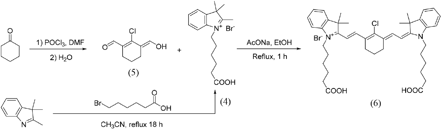
A cyanine IR808 was synthesized (Figure 4), and a calibration curve was performed (see Supplementary Information (SI) section).

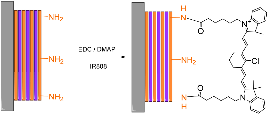
A solution of cyanine at a specific concentration (1 × 10⁻⁵ M) was used for the surface reactivity analysis. The surface with the 15.5 bilayer was immersed in dichloromethane, and an amidation reaction was performed on the surface by adding EDC and DMAP for 20 h at room temperature (Figure 5). It is worth mentioning that the surface contains amino groups, and cyanine has carboxyl groups. The amidation reaction will covalently bind cyanine and decrease the concentration of cyanine proportionally to the amide formation. After the amidation reaction was performed, it was evaluated by fluorescence analysis at 808 nm, corresponding to the cyanine emission signal. This analysis enabled us to evaluate the surface characteristics before and after the reaction, and to determine both the available amine groups and those that reacted (Figure 5).

The quantification of reactive groups was performed by determining the cyanine solution concentrations before polymer film immersion (Figure 6a), after 20 h of incubation, according to the Lambert-Beer law (Figure 6b). From these data, the corresponding molar amounts were calculated, and the difference was attributed to the moles bound to the surface. Using Avogadro’s number, the total number of surface-bound cyanine molecules was estimated, assuming that one fluorophore molecule reacts with two surface-exposed amine groups.

Monitoring of amidation reaction between PVDMA/PEI and cyanine by (a) UV-Vis and time vs. (b) absorbance relationship.
Table 1 presents the results of the interaction between cyanine and the film, indicating that the amount of free amino groups on the surface is approximately 10-17, and the two carboxyl groups of cyanine that can react with two amino groups are related. Therefore, the final concentration of the amino groups is double in relation to cyanine. The evaluation of amino groups after Fmoc release is described in Table 1 and it was determined that the release occurred at 85% based on the 15.5 bilayer film with the surface PEI.
Contact angle estimations
The contact angle estimations required a 12 MP, f/1.8, 28mm (wide), phase detection autofocus (PDAF), optical Image stabilization (OIS) camera, a 12 MP, f/2.8, 57mm (telephoto), PDAF camera, and a 2× optical zoom lens at room temperature. The static water contact angle was measured upon the deposition of a 5 µL droplet of deionized water onto selected films (2.5 × 2.5 cm2) at three different locations. Data are reported as the average of these measurements.
Characterization of E. coli adhesion on PEI/PVDMA films functionalized with amino acids
Stable transformants of E. coli ER2738 [F´proA+B+ lacIq ∆(lacZ)M15 zzf::Tn10(TetR) / fhuA2 glnV ∆(lac-proAB) thi-1 ∆(hsdS-mcrB)5]36 harboring the plasmid pQHGF301 were used in these experiments. This vector drives the expression of a super-glow green fluorescent protein (GFP) variant upon induction with isopropyl β-D-thiogalactopyranoside (IPTG).37,38 A 2-mL culture of Luria-Bertani (LB) medium supplemented with ampicillin (0.15 mg mL-1) were inoculated with 20 µL of a bacterial suspension and incubated overnight at 30 °C with shaking (300 rpm). The following day, a subculture was prepared by inoculating 50 µL of the overnight culture into 5 mL of selective LB medium and incubating at 37 °C for 2 h (300 rpm) before induction with 50 µL of 1 M IPTG. GFP overexpression was performed for 16 h at 30 °C (300 rpm). Cells were harvested by centrifugation at 12,000 rpm for 2 min, washed twice with phosphate-buffered saline (PBS), and resuspended to an optical density of 0.1 at 650 nm (OD650) per mL of PBS. This bacterial suspension was subsequently used for cell adhesion analysis.
Before the assay, the PEI/PVDMA films were sanitized by thoroughly spraying them with 70% ethanol (EtOH) in square Petri dishes, followed by two washes with PBS (each for 10 min) to remove residual EtOH. After equilibration with PBS, 25 mL of bacterial suspension was added to each Petri dish, and cell adhesion was allowed for 1 h at 37 °C with constant agitation (100 rpm). The bacterial suspension was removed, and the films were washed twice with PBS. An unmodified film was used as a control.
Optical observance of epifluorescence microscopy
After air-drying, GFP-labeled E. coli adhered to the film surface were visualized and analyzed using a Zeiss Axio Scope-A1 microscope equipped with an AxioCam ICc 5 digital camera. Five micrographs were acquired at 63× magnification, under consistent imaging settings. Cell counts per micrograph were quantified using ImageJ software.39 Following a standardized image processing workflow. Means and standard deviations were calculated from the set of captured micrographs.36
Results and Discussion
Characterization of the thin film of 15.5 bilayers
Our research has demonstrated the successful fabrication of polymeric PVDMA/PEI films using the layer-by-layer dip-coating technique. This method promotes covalent crosslinking between the amine groups of PEI and the azlactone ring present in the PVDMA polymer. The films, constructed as 15.5 bilayer polymeric surfaces supported on glass substrates, not only exhibit remarkable stability but also show a high degree of resistance to handling, instilling confidence in their durability.
Functionalization of thin films
In our laboratory, a wide variety of azlactones have been synthesized, demonstrating the robustness of our method. Azlactone isomerization and ring-opening in the presence of amino groups have been evaluated.38 Film layer preparation takes only 20 s, highlighting the efficiency of our approach. IR analysis has shown the loss of signal at 1820 cm-1, characteristic of the azlactone group, and the appearance of the signal at 1665 1650 cm-1, corresponding to the amide formed. In this novel research, the typical deprotection of the Fmoc group on the previously functionalized films (Figure 3c) enabled the exposure of free amino from the amino acids on the surface of each material. In this novel investigation, the film was functionalized with several amino acids protected with the Fmoc group (Figure 3b). Subsequently, the Fmoc group was removed using piperidine as a basic medium (Figure 3c), allowing the amino groups of the amino acids to be free on the surface of the functionalized films.
The films functionalized were subsequently characterized by Fourier-transform infrared spectroscopy (FTIR) (see Figures 7a and 7b). The FTIR analysis shows stretching vibration signals around 3300 cm-1, corresponding to the N-H bond of secondary amines now available at the surface following removal of the protecting group. Additionally, all spectra exhibit distinct bands in the 1665-1650 cm-1 region, attributed to the stretching vibrations of the C=O bond in amide groups formed through the covalent linkage between surface azlactone groups and the amino groups of the incorporated amino acids. The main difference is shown in the 3300 cm-1 signal, which corresponds to the free amino groups, due to the removal of the Fmoc group. Therefore, this signal increases significantly (Figure 7b).

Characterization by FTIR (ATR) of PVDMA/PEI (a) films functionalized with amino acids protected with Fmoc and (b) films functionalized with amino acids without the Fmoc group.
The surface roughness of the film was measured using an atomic force microscope (AFM). Figure 8 illustrates the role of the pendant functional groups on the surface in modifying the surface roughness (SR) of PVDMA/PEI films with 15.5 bilayers (4.78 nm) and those functionalized with free amino acids. This property is significantly altered by these groups, whose orientation and organization have a direct impact. This effect is consistently observed in all scaffolds modified with Fmoc-protected amino acids (see SI section). The differences in roughness values in films functionalized with Fmoc-Gly (4.10 nm) and Fmoc-Ala (4.36 nm) were minimal, as both share a similar structure. However, the use of Fmoc-Leu (5.45 nm) led to a noticeable increase in SR. This behavior is attributed to the fact that branched amino acids induce greater surface heterogeneity, while small and unbranched amino acids, such as Fmoc-Gly, promote a more uniform coating. Conversely, the use of Fmoc-Phe (2.87 nm) resulted in a smooth and uniform surface, as the conformational rigidity of the aromatic ring encourages stacking on the surface through π-π type interactions with the protective group.
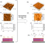
Comparative AFM analysis of the 15.5 bilayer PVDMA/PEI and PVDMA/PEI-Leu reactive films from 3D surface reconstruction (a, b), 2D surface topography image (c, d), topographic profile (e, f), and schematic representation (g, h).
The SR decreases when the Fmoc group is deprotected: Gly (1.74 nm), Ala (1.76 nm), Leu (2.28 nm), except for Phe (3.67 nm), which increases. The amino acid is covalently linked to the surface by the carboxyl group with the amine of the PEI, forming an amide bond and the deprotection of the Fmoc group, leaving the amino group of the amino acid free (Figure 8h). The PVDMA/PEI film functionalized with Phe (3.67 nm) stands out; this film presents a slightly rougher surface, due to the more voluminous aromatic ring, showing a slight increase in roughness compared to Gly (1.74 nm), Ala (1.76 nm), and Leu (2.28 nm). However, the specific combination of low SR and an apolar side chain observed in the leucine-functionalized film could, at least in part, explain its greater ability to reduce bacterial adhesion. The surface functionalized with leucine exhibited the lowest adhesion of Escherichia coli. The difference between the amino acids lies in the substituent group at the α-position. Leucine has a longer aliphatic chain than the other amino acids. This chain plays a crucial role in hindering the proper interaction between the E. coli membrane and the surface. The aliphatic chain causes repulsion between the hydrophilic head of the phospholipid and the lipophilic chain of the aliphatic amino acid, decreasing the affinity between the film and the cell membrane. These studies have demonstrated that long hydrocarbon chains significantly affect cell interactions, considering that cell membranes are composed of phospholipids. This behavior highlights the complementary role of the surface molecular architecture and its effects on interactions with biological systems. Figure 8 shows the SR of both the 15.5 bilayer PVDMA/PEI film with the PEI terminal amino groups (Figure 8a) and, in contrast, the film functionalized with the Leu-Fmoc amino acid and its subsequent deprotection. The surface with the unprotected amino acid shows the free amino group (Figure 8b). Also shown are the surface topography (Figures 8c-8d) and the profile topography (Figures 8e-8f). The latter two clearly indicate the lower surface roughness of the film with unprotected leucine (Figure 8h).
Collectively, these findings underscore the crucial role of amino acid functionalization in not only altering the surface chemistry but also the nanoscale topography of the films. The unique combination of low roughness and an apolar side chain found in the leucine-modified film, which has been shown to reduce bacterial adhesion significantly in biological assays, further underscores the importance of surface molecular architecture in biological interactions.
Multilayer thickness measurement
The multilayer thickness of the polymeric films was determined by FESEM microscopy (Figure 9). The micrographs of the non-functionalized 15.5-bilayer PVDMA/PEI film show a smooth, flat, and homogeneous surface, with an average thickness of 135.75 nm. In contrast, the functionalized films exhibited a rougher surface and a 101.10% increase in average thickness, reaching 273 nm. This increase is likely associated with the presence of surface-oriented aliphatic side chains introduced by the amino acid functionalization.

FESEM micrographs of (a) non-functionalized PVDMA/PEI film of 15.5 bilayers, and (b) PVDMA/PEI film functionalized with Leu.
Contact angle measurement
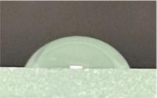
The formation of the film on the glass surface is evident from the higher contact angle values (Table 2 and Figure 10) when the film is present, which corresponds to the characteristics of the film. The most hydrophobic surface corresponds to the functionalization with Phe, which correlates with the incorporation of the aromatic system. In the Ala and Gly cases, the values showed moderately high differences, which may be influenced by surface packing and density of the exposed functional groups. Similarly, the Leu-functionalized surface exhibited a high angle compared to the glass surface and the unfunctionalized film, consistent with its non-polar aliphatic side chain. Overall, these results indicate that functionalization in the last layer is sufficient to modify the hydrophilic/lipophobic properties of the surfaces. These observations highlight the residue-specific nature of the surface energy alteration in films due to amino acid incorporation, which depends on both the size and hydrophobic character of the side chains.

These results indicate that functionalization in the final layer is sufficient to modify the hydrophilic/lipophobic properties of the surfaces. Figure 10 shows a graph of the contact angle variations relative to the amino acid. The presence of the amino acid on the surface increases the contact angle. This is due to the specific nature of each residue, which alters the surface energy of the films, which depends on both the size and the hydrophobic nature of the side chains.
Evaluation of the effect of films on E. coli proliferation
The results of the films exposed to the culture medium containing E. coli are shown in Figure 11. The images show significant implications for our understanding of bacterial adhesion. Notably, Figure 11a corresponds to the film with the surface containing the free amino groups (15.5 layers). The bright field (Figure 11b) reveals abundant and uniform bacterial adhesion. In Figure 11c, the fluorescence image clearly shows the presence and adhesion of E. coli on the surface. These results suggest that the bacteria encountered favorable sites for establishing strong interactions with the surface, such as ionic, hydrophobic, or hydrogen bonds. This interaction is attributed to the high density of accessible amine groups and the elevated surface roughness (Ra = 4.78 nm), which increases the effective contact area, potentially leading to significant advancements in our understanding of bacterial adhesion. In the film functionalized with Gly (Figure 11d), we observed moderate and evenly distributed bacterial adhesion on the bright (Figure 11e) and fluorescence (Figure 11h) field images. This finding, along with the substantial level of E. coli interaction, suggests that other factors, such as the practical exposure of available functional groups, play a role. Figure 11g shows the alanine-functionalized 15-layer film. Significant bacterial adhesion is observed, as shown in the bright-field (Figure 11h) and fluorescence (Figure 11i) images. This indicates that the incorporation of Ala alters the interfacial chemistry, reducing the availability of interactive sites. However, it is crucial to emphasize that this modification, although effective, is insufficient to inhibit bacterial adhesion, as observed in the control completely (Figure 11a). Figure 11g illustrates the alanine-functionalized 15-layer film. Significant bacterial adhesion is observed, as shown in the bright-field image (Figure 11h). This indicates that Ala incorporation alters the interfacial chemistry, reducing the availability of interactive sites; however, the modification is not sufficient to completely inhibit bacterial adhesion, as observed in the control (Figure 11a). Figure 11j corresponds to the Leu-functionalized film. In the bright-field (Figure 11k), and fluorescence (Figure 11l) images, virtually no bacterial adhesion was observed, with only 1 or 2 bacteria detected per field. This result reflects a drastic inhibition of the interaction between bacteria and the surface. Leucine has an apolar aliphatic side chain that, when positioned on the surface, can form a low-surface-energy chemical barrier, making it challenging to target bacteria effectively.

Schematic representation of the produced films with PEI at the uppermost layer (15.5 bilayers) and PVDMA (15 bilayers) before (a) and after functionalization with glycine (d), alanine (g), leucine (j), and phenylalanine (m). Optical (b, e, h, k, and n) and fluorescence images (c, f, i, l, and o) of E. coli cells cultured onto the corresponding surfaces. E. coli modified with GFP protein. Scale bars in panels (b, e, h, k, n) and (c, f, i, l, o) correspond to 200 μm.
Furthermore, its relatively low roughness and intermediate contact angle suggest a surface configuration that does not promote wetting or provide microenvironments conducive to bacterial attachment. Figure 11m shows the functionalization of the surface with the PVDMA/PEI film with the amino acid of Phe. It is observed in both brightfield (Figure 11n) and fluorescence (Figure 11o) that cell adhesion is present. The aromatic side chain of Phe likely contributes to enhanced E. coli adhesion, possibly due to π-π or hydrophobic interactions. These observations not only advance our understanding of bacterial adhesion but also inspire future studies to explore the potential of functional groups to influence bacterial interactions.
In summary, when comparing the fluorescence images of the different films (Figures 11c, 11f, 11i, 11l, and 11o), the level of bacterial adhesion follows the following ascending order: F-Leu < F-NH2 (control) < F-Ala < F-Gly < F-Phe.
Table 3 describes the quantification of bacterial proliferation, obtained by evaluating five zones. These values were averaged to calculate the mean and standard deviation for each modified film in relation to bacterial proliferation. It was confirmed that the film with leucine as the amino acid modifying the film surface exhibited the lowest bacterial proliferation value.
These findings underscore the value of molecular design based on specific amino acids as a strategy for developing surfaces with passive antibacterial properties, thus eliminating the need for biocidal or cytotoxic agents.
Possible interaction mechanism
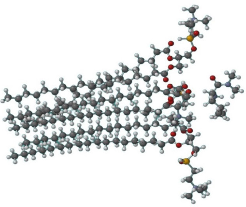
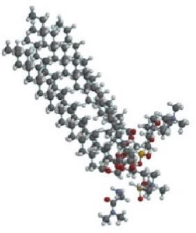
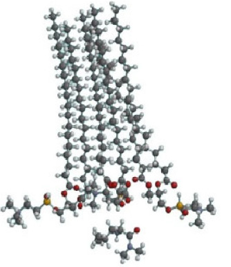
The outer membrane of Escherichia coli is composed of lipopolysaccharides, proteins, and phospholipids. The latter are characteristic of all cell membranes. Therefore, phosphatidylcholine was used as one of the main components and was made to interact with the amino acids used in this research (Gly, Ala, Leu, and Phe). To simulate their attachment to the surface, these amino acids were used in the form of N,N’-dimethyl amides, along with the free amino groups (Figure 12). Using this model, the optimized geometry of the materials phospholipids and the protected amino acid was obtained. They were positioned in the appropriate trajectory so that the phospholipid heads interacted with the amino acids. MM2 molecular dynamics were then performed between the phospholipid assembly and the amino acids in amide form, minimizing energy and consequently optimizing the interaction. Table 4 shows the results of the molecular dynamics analysis, indicating that the least effective interaction between the phospholipid and the amino acid occurred when the interaction involved leucine (Table 4). This conclusion is consistent with the experimental findings, although it should be noted that there were differences compared to those observed with the other amino acids. In the case of leucine, which has a longer aliphatic chain than the other amino acids, it distorts the geometry of the phospholipids more, increasing its energy and consequently decreasing its stability.38

Optimization by molecular dynamics between the phospholipid and the interaction with the leucine that is on the surface of the film.
Conclusions
In our work group, we have achieved significant progress in the production of bilayer (PEI/PVDMA) nanofilms. In this work, we prepared 15-and 15.5-bilayers (PEI/PVDMA)15-PEI that are pressure-resistant, resistant to contact with a physiological environment, and transparent. These nanofilms were prepared using an automated system by immersing the material in PEI polymer and PVDMA solutions. The latter was synthesized from the vinyl azlactone preparation and subsequently polymerized. The 15.5 bilayer film presents the PEI as the outermost surface layer, which has free amino groups and was functionalized with several amino acids: Gly, Ala, Leu, and Phe. All surfaces analyzed presented amino groups; the control film presented 15.5 bilayers with PEI amino groups on the surface. The films with amino acids presented free amino groups. The roughness before and after functionalization was determined using AFM, FESEM measured the thickness, and the contact angle was evaluated. Our research has shown that the type of amino acid is crucial for cell adhesion; leucine, the surface-functionalized amino acid, was the most effective at inhibiting adhesion. This result is not only decisive in this study but also has the potential to significantly reduce bacterial contamination, particularly the adhesion of bacteria such as E. coli, thereby reducing the risk of infection.
Supplementary Information
Supplementary information (UV-Vis, FTIR, 1H and 13C NMR, AFM, and ESI-MS spectra) are available free of charge at http://jbcs.sbq.org.br as a file.
Acknowledgments
We gratefully acknowledge the support for this project by Consejo Nacional de Ciencia y Tecnología (CONACyT, grants No. CF-2023-I-327 and INFR-2016-01, No. 269551), for graduate scholarship APZ (CVU 942554). Postdoctoral fellowship M.E: A-C (CVU 559644).
References
-
1 Allocati, N.; Masulli, M.; Alexeyev, M. F.; Di Ilio, C.; Int. J. Environ. Res. Public Health 2013, 10, 6235. [Crossref] [PubMed]
» Crossref» PubMed -
2 Ishii, S.; Sadowsky, M. J.; Microbes Environ. 2008, 23, 101. [Crossref] [PubMed]
» Crossref» PubMed -
3 Tamma, P. D.; Aitken, S. L.; Bonomo, R. A.; Mathers, A. J.; van Duin, D.; Clancy, C. J.; Clin. Infect. Dis. 2023, ciad428. [Crossref] [PubMed]
» Crossref» PubMed -
4 Rochelle-Newall, E.; Nguyen, T. M. H.; Le, T. P. Q.; Sengtaheuanghoung, O.; Ribolzi, O.; Front. Microbiol. 2015, 6, 308. [Crossref]
» Crossref -
5 Garrett, T. R.; Bhakoo, M.; Zhang, Z.; Prog. Nat. Sci. 2008, 18, 1049. [Crossref]
» Crossref -
6 Li, X.; Wu, B.; Chen, H.; Nan, K.; Jin, Y.; Sun, L.; Wang, B.; J. Mater. Chem. B 2018, 6, 4274. [Crossref] [PubMed]
» Crossref» PubMed -
7 Manna, U.; Lynn, D. M.; Adv. Funct. Mater. 2015, 25, 1672. [Crossref]
» Crossref -
8 Ávila-Cossío, M.; Rivero, I. A.; García-González, V.; J. Mex. Chem. Soc. 2024, 68, 135. [Crossref]
» Crossref -
9 Ávila-Cossío, M. E.; Rivero, I. A.; García-González, V.; Alatorre-Meda, M.; Rodríguez-Velázquez, E.; Calva-Yáñez, J. C.; Espinoza, K. A.; Pulido-Capiz, Á.; ACS Omega 2020, 5, 5249. [Crossref] [PubMed]
» Crossref» PubMed -
10 Buck, M. E.; Zhang, J.; Lynn, D. M.; Adv. Mater. 2007, 19, 3951. [Crossref]
» Crossref -
11 Zayas-Gonzalez, Y. M.; Lynn, D. M.; Biomacromolecules 2016, 17, 3067. [Crossref] [PubMed]
» Crossref» PubMed -
12 Buck, M. E.; Schwartz, S. C.; Lynn, D. M.; Chem. Mater. 2010, 22, 6319. [Crossref]
» Crossref -
13 Buck, M. E.; Breitbach, A. S.; Belgrade, S. K.; Blackwell, H. E.; Lynn, D. M.; Biomacromolecules 2009, 10, 1564. [Crossref] [PubMed]
» Crossref» PubMed -
14 Yang, S. Y.; Mendelsohn, J. D.; Rubner, M. F.; Biomacromolecules 2003, 4, 987. [Crossref] [PubMed]
» Crossref» PubMed -
15 Sun, H.; Hong, Y.; Xi, Y.; Zou, Y.; Gao, J.; Du, J.; Biomacromolecules 2018, 19, 1701. [Crossref] [PubMed]
» Crossref» PubMed -
16 Costa, F.; Carvalho, I. F.; Montelaro, R. C.; Gomes, P.; Martins, M. C. L.; Acta Biomater. 2011, 7, 1431. [Crossref] [PubMed]
» Crossref» PubMed -
17 Erdem Büyükkiraz, M.; Kesmen, Z.; J. Appl. Microbiol. 2022, 132, 1573. [Crossref] [PubMed]
» Crossref» PubMed -
18 Marra, I. F. S.; de Castro, P. P.; Amarante, G. W.; Eur. J. Org. Chem. 2019, 2019, 5830. [Crossref]
» Crossref -
19 Buck, M. E.; Lynn, D. M.; ACS Appl. Mater. Interfaces 2010, 2, 1421. [Crossref] [PubMed]
» Crossref» PubMed -
20 Aguilera, J. A.; García-González, V.; Alatorre-Meda, M.; Rodríguez-Velázquez, E.; Rivero, I. A.; J. Braz. Chem. Soc. 2021, 32, 2222. [Crossref]
» Crossref -
21 Cassa, M. A.; Gentile, P.; Girón-Hernández, J.; Ciardelli, G.; Carmagnola, I.; Biomater. Sci. 2024, 12, 5433. [Crossref] [PubMed]
» Crossref» PubMed -
22 Buck, M. E.; Lynn, D. M.; Polym. Chem. 2011, 3, 66. [Crossref]
» Crossref -
23 Pereira, A. A.; de Castro, P. P.; de Mello, A. C.; Ferreira, B. R. V.; Eberlin, M. N.; Amarante, G. W.; Tetrahedron 2014, 70, 3271. [Crossref]
» Crossref -
24 Lynn, D. M.; Langer, R.; J. Am. Chem. Soc. 2000, 122, 10761. [Crossref]
» Crossref -
25 Wood, K. C.; Chuang, H. F.; Batten, R. D.; Lynn, D. M.; Hammond, P. T.; Proc. Natl. Acad. Sci. USA 2006, 103, 10207. [Crossref]
» Crossref -
26 Isidro-Llobet, A.; Álvarez, M.; Albericio, F.; Chem. Rev. 2009, 109, 2455. [Crossref] [PubMed]
» Crossref» PubMed -
27 Wancura, M. M.; Anex-Ries, Q.; Carroll, A. L.; Paola Garcia, A.; Hindocha, P.; Buck, M. E.; J. Polym. Sci. A Polym. Chem. 2017, 55, 3185. [Crossref]
» Crossref -
28 Lapitskaya, V. A.; Kuznetsova, T. A.; Chizhik, S. A.; Приборы и методы измерений 2024, 15, 60. [Crossref]
» Crossref -
29 Senthilraj, H.; Kamarajan, B. P.; Ananthasubramanian, M.; Int. J. Mech. Eng. 2021, 6, 629. [Crossref]
» Crossref -
30 Lutkenhaus, J. L.; Hammond, P. T.; Soft Matter 2007, 3, 804. [Crossref] [PubMed]
» Crossref» PubMed -
31 Woodcock, D. M.; Crowther, P. J.; Doherty, J.; Jefferson, S.; Decruz, E.; Noyer-Weidner, M.; Smith, S. S.; Michael, M. Z.; Graham, M. W.; Nucleic Acids Res. 1989, 17, 3469. [Crossref] [PubMed]
» Crossref» PubMed -
32 Hammond, P. T.; Adv. Mater.2004, 16, 1271. [Crossref]
» Crossref -
33 Leveau, J. H. J.; Lindow, S. E.; J. Bacteriol. 2001, 183, 6752. [Crossref] [PubMed]
» Crossref» PubMed -
34 Kataoka, K.; Takasu, A.; Polym. J. 2021, 53, 385. [Crossref]
» Crossref -
35 ImageJ. [Link] accessed in January 2026
» Link -
36 Mares, R. E.; Meléndez-López, S. G.; Ramos, M. A.; Int. J. Mol. Sci. 2011, 12, 4625. [Crossref] [PubMed]
» Crossref» PubMed -
37 García, Á. L.; Bravo, M.; Orozco-Barrera, D.; Jara-Cortés, J.; Peon, J.; Rivero, I. A.; ChemPhotoChem 2024, 8, e202300190. [Crossref]
» Crossref -
38 Spartan’14; Wavefunction Inc, Irvine, CA, 2014. [Link] accessed in January 2026
» Link -
39 Schneider, C.; Rasband, W.; Eliceiri, K.; Nat. Methods 2012, 9, 671. [Crossref]
» Crossref