 Exploring the Link Between ITGB3 Gene Polymorphism and Diastolic Dysfunction in Arterial Hypertension Patients
Abstract
Background: Arterial hypertension (AH) is a significant risk factor for cardiovascular diseases (CVDs), including myocardial infarction and stroke. The ITGB3 gene polymorphism, particularly T1565C (rs5918), has been implicated in various cardiovascular conditions, suggesting a potential role in diastolic dysfunction (DD) among AH patients.
Objectives: The clinical-genetic relationship was studied between ITGB3 gene polymorphisms and DD in Azerbaijani patients with AH, with or without ischemic heart disease (IHD) and type 2 diabetes (T2D) mellitus.
Methods: The study included 100 Azerbaijani patients, divided into case (n = 76) and control (n = 24) groups. Echocardiography was used to assess left ventricular diastolic function. ITGB3 gene was identified using the MassArray system. Statistical analysis involved both parametric and non-parametric tests using the IBM SPSS Statistics 26.
Results: The study reported a significant association between ITGB3 gene polymorphisms and DD in AH patients. The T/C, C/C genotypes were particularly correlated with DD. The C/C variant, while more frequent in the AH group, did not reach statistical significance. A direct correlation was observed between DD and the C allele of the ITGB3 gene, with a higher E/e’ ratio and lower e’ velocity among carriers.
Conclusion: This study highlights a significant association between ITGB3 gene polymorphisms and DD in Azerbaijani patients with AH. These findings suggest the potential for genetic screening to identify individuals at higher risk of DD among hypertensive patients, underscoring the importance of personalized approaches in managing hypertension and its complications. Further research with larger and more diverse cohorts is needed to validate these results and explore the underlying mechanisms.
Keywords:
Genetic Polymorphism; Ventricular Dysfunction; Hypertension
Introduction
Arterial hypertension (AH) is the primary risk factor for cardiovascular disease (CVD). Observational studies and clinical trials have consistently shown that prolonged uncontrolled hypertension significantly increases the risk of conditions like myocardial infarction and stroke. Other modifiable risk factors for CVD include smoking, diabetes, and lipid metabolism disorders. However, AH stands out with strong cause-and-effect evidence and high prevalence of associated effects. Despite this, biologically normal AH levels in humans are typically lower than those traditionally used in clinical practice and research, leading to the underestimation of AH as a risk factor for heart failure, atrial fibrillation, chronic kidney disease, heart valve diseases, aortic syndromes, and dementia, in addition to coronary heart disease and stroke. AH can stem from various secondary causes and rare genetic variants, but most cases are categorized as essential or primary hypertension without a specific etiology.1
AH is closely linked to diastolic dysfunction (DD), which correlates with various CVDs, though its genetic underpinnings remain largely obscure.2 Diastole, the heart's phase of ventricular filling, hinges on myocardial relaxation and chamber stiffness. It's not merely a passive cycle but rather a complex interplay of physiological processes involving myocardial relaxation, stiffness, and recoil, influenced by loading conditions, heart rate, and contractile function. Diastolic function critically determines left ventricular filling and stroke volume, and dysfunction serves as a predictor of major cardiovascular events and overall mortality.3
ITGB3 encodes glycoprotein IIIa (GPIIIa), also known as the beta subunit of the platelet membrane adhesive protein receptor complex GP IIb/IIIa, which functions as a surface protein involved in cell-surface signaling and adhesion.4 This gene, encompassing exons and introns, contains polymorphic regions, including one linked to CVDs. A notable polymorphism, T1565C (rs5918), at codon 33 of exon 2 (PlA1/A2), affects amino acid residues (leucine/proline) at position 33 of the polypeptide chain. This SNP has been implicated as a risk factor in multiple diseases, such as myocardial infarction, coronary heart disease, type 2 diabetes (T2D), asthma, and various cancers including colon, non-Hodgkin lymphoma, breast, ovarian, and kidney cancers.5-13 Platelets carrying the β3 subunit with a proline at position 33 exhibit increased aggregation risk and immunogenic properties.14,15 The presence of the PlA2 allele correlates with increased platelet aggregation and binding affinity to fibrinogen, as well as altered responsiveness to aspirin and various agonists like epinephrine, adenosine diphosphate, and collagen.12 The association of the PlA1/A2 polymorphic marker of ITGB3 with arterial and venous thrombosis across different ethnic groups remains debated.10,12,16,17 Given the implications of ITGB3 gene in various diseases risk, further investigations into its polymorphic variants’ role in cardiovascular disorders is warranted.

This research aimed to explore clinical and genetic aspects in patients from Azerbaijan with AH, with or without ischemic heart disease (IHD) and T2D, focusing on carriers of genetic polymorphisms in the integrin ITGB3 gene.
Materials and Methods
This study was conducted in Baku, Azerbaijan, at the Department of Cardiology of the XMSK Hospital, the laboratory of Shafa Treatment and Diagnostics Center, and the Azerbaijan State Advanced Training İnstitute for Doctors named after Aziz Aliyev. All patients were informed about the study's purpose and objectives and provided written consent to participate. The study was carried out following the ethical principles outlined in the World Medical Association's Declaration of Helsinki.18
During patient examinations, we followed the 2018 practical guidelines of the European Society of Cardiology and the European Society of Hypertension.19
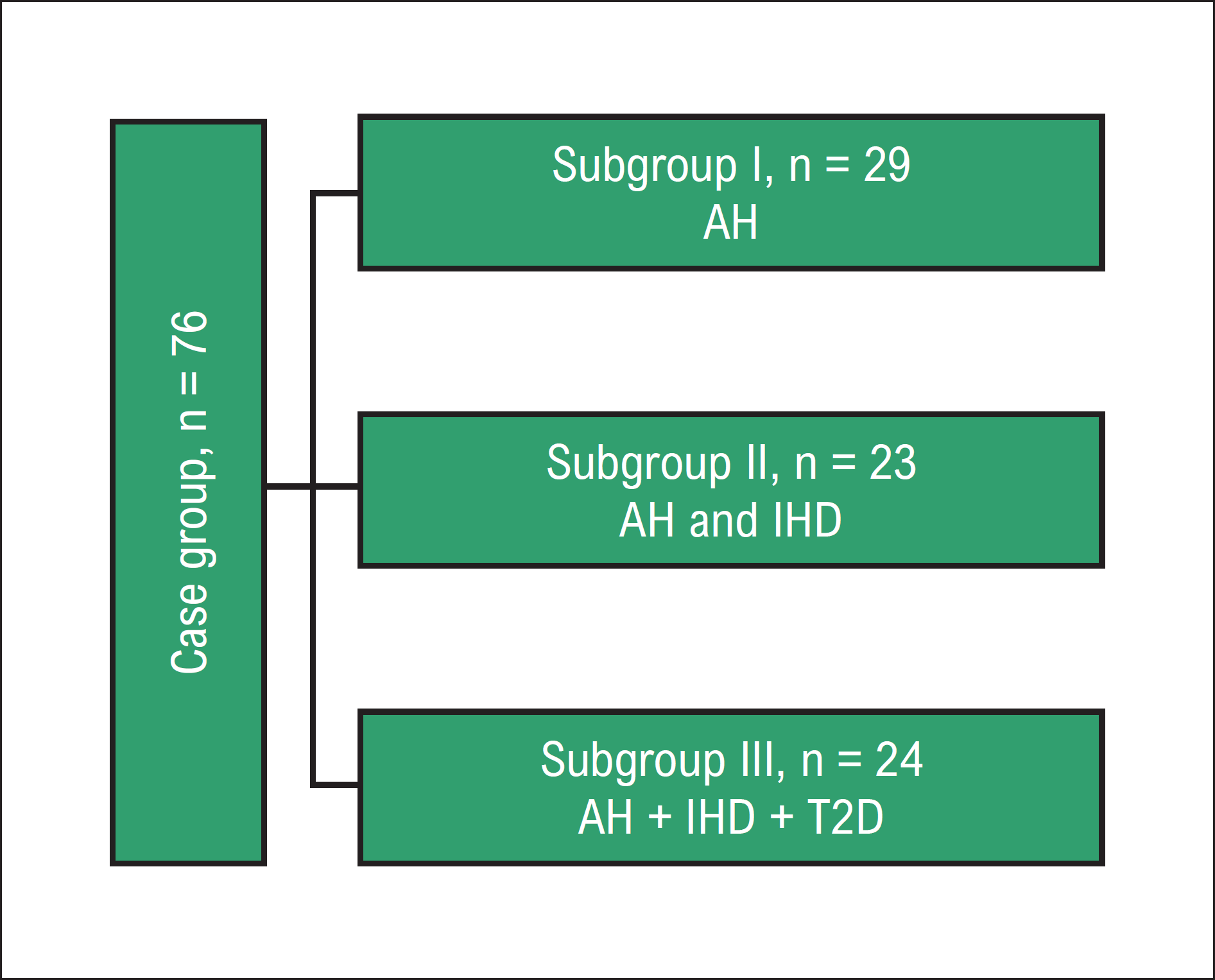
The study was conducted between 2019 and 2021, examining a total of 100 patients. Demographic characteristics and clinical data of the study population were presented, dividing them into two groups: case (n = 76) and control (n = 24). The case group with AH was further divided into three clinical subgroups based on the presence of comorbidities (IHD and T2D): subgroup I, 29 patients with AH; subgroup II, 23 patients with AH and IHD; subgroup III, 24 patients with AH, IHD, and T2D (Figure 1).

Diagram of study population. AH: Arterial hypertension; IHD: ischemic heart disease; T2D: type 2 diabetes.
Inclusion criteria
Ethnically Azerbaijani patients, aged between 20 and 77 years, of both male and female gender.
Exclusion criteria
Patients younger than 20 years or older than 77 years, pregnant patients, patients with congenital heart defects, patients with congenital or acquired blood diseases, patients undergoing oncology treatments or chemotherapy, patients with mental disorders, patients with chronic kidney failure.
Echocardiographic methods
All participants underwent echocardiography performed by the same cardiologist following a strict protocol. All subjects were positioned in a standard left-lateral posture to minimize the influence of body position. Echocardiographic recordings were taken at the end of the respiratory phase during normal breathing, using apical four- and two-chamber views.
Transmitral flow velocity was assessed using pulsed-wave Doppler, with the sample volume positioned between the mitral leaflet tips during diastole. Indicators measured to determine the diastolic function of the left ventricle included: transmitral blood flow (E peak of early diastolic blood flow and A peak of late diastolic blood flow); E/A ratio between early and late diastolic blood flow; tissue Doppler examination (septal e’ peak diastolic velocity of the mitral annulus < 7 cm/sec); ratio of the early mitral flow velocity to the velocity of the mitral annulus (septal E/e’ > 15); LA maximum volume index (> 34 mL/m2); tricuspid flow velocity (TRvel > 2/8m/s). The measurement of these indicators to assess left ventricular diastolic function was based on the 2016 recommendations of the American Society of Echocardiography.20
DNA preparation
DNA was prepared from a small aliquot of whole blood collected in ethylenediaminetetraacetic acid by using a DNA extraction matrix. ITGB3 gene polymorphism (T1565C) that corresponds to the polymorphism of amino acid residues (leucine/proline) at position 33 (PLA1/A2) of the polypeptide chain was identified using the MassARRAY system (Agena Bioscience GmbH, Germany).
Statistical analysis
The study was designed as an analytical research project using clinical methods and a scientific sample selection. It was prospective in nature, cross-sectional in duration, and conducted in a clinical setting. The sample size was determined based on the availability of eligible participants during the study period, making it a convenience sample. Qualitative data obtained during the research were analyzed using discriminataion and correlation methods. The IBM SPSS Statistics 26 software was used in statistical analysis, which included Spearman rank correlation to assess associations between variables, Pearson's Chi-square test to compare qualitative indicators and test odds ratio (OR). A significance level of p < 0.05 was adopted in all statistical procedures.
Results
Mean age (±standard deviation – SD) of the patients was 50.6±8.6, 58.3±7.6, 59.2±4.6 and 45.9±8.4 in the subgroup I, subgroup II, subgroup III and control group respectively. The predominance of male patients was observed in both case and control groups accounting for 58.3% and 67.1% respectively. High body mass index (BMI) was significantly more common in the case group (Table 1).
Allele distribution for the studied SNPs differed between cases and controls. As shown in Table 2, there was a slight difference in the prevalence of the normal variant of the polymorphism in the T/T homozygous form between AH patients and the control group.
For ITGB3 gene, a higher frequency of the heterozygous genotype T/C was observed in cases (21.1%) than in controls (20.8%). There was no significant difference in the prevalence of the heterozygous genotype between the groups. C/C homozygous variant of polymorphism was found only in patients with AH; this variant of polymorphism was not observed in the individuals of the control group. However, these differences in frequency were statistically insignificant by the bivariate analysis for C allele. Statistical analysis of the OR on the prevalence of the normal homozygous T/T genotype of the ITGB3 integrin gene showed that compared to the control group, the OR of carriers of this genotype in the group of patients with AH was 0.7 times lower (OR = 0.737, 95% CI 0.243-2.235), but the difference was not statistically significant. Compared to the control group, the OR of the mutant heterozygous C/T genotype among patients with AH was OR=1.013 (95%CI 0.328-3.134), that is, the OR for the heterozygous form in these was 1.0 times higher (Table 2).
A statistically significant difference was found in the presence of DD in AH patients (case group), especially in patients with heterozygous T/C genotype and homozygous C/C genotype of the ITGB3 gene (Central Illustration 1).
We also explored the correlation between genes and DD. A direct correlation with polymorphism in the ITGB3 gene was detected (Central Illustration 2). A direct correlation was also observed between DD and the altered genotypes of the ITGB3 gene in the AH subgroup (Central Illustration 3).
A direct correlation was observed between the C allele of the ITGB3 gene and E/e’ (Central Illustration 4). Additionally, an inverse correlation was found between e’ and the C allele of the ITGB3 gene (Central Illustration 5).
Discussion
The present study sought to investigate the relationship between ITGB3 gene polymorphisms and DD in patients with AH in an Azerbaijani cohort. The results suggested a no association between specific ITGB3 gene polymorphisms and the prevalence of DD among AH patients, highlighting the roles of the T/C heterozygous and C/C homozygous genotypes.
The ITGB3 gene, which encodes the β3 subunit of the platelet membrane adhesive protein receptor complex GP IIb/IIIa, is well-documented for its involvement in various CVDs and its influence on platelet aggregation and thrombus formation. This study adds to the growing body of evidence by demonstrating a correlation between ITGB3 polymorphisms and DD in AH patients. Specifically, the presence of the C allele was directly correlated with the E/e’ ratio and inversely correlated with e’ velocity, indicating its potential role in the mechanical and functional alterations of the left ventricle during diastole.
The higher frequency of the C/C homozygous variant in the AH group compared to controls, although not reaching statistical significance, suggests a potential trend that warrants further investigation with larger sample sizes. The observed ORs, though not statistically significant, indicate a higher likelihood of DD in carriers of the T/C and C/C genotypes. These findings are consistent with previous studies linking the ITGB3 gene to CVDs but their the implications extend to the context of DD in hypertensive patients.21,22
DD, characterized by impaired myocardial relaxation and increased ventricular stiffness, is a crucial predictor of adverse cardiovascular outcomes.23 The significant association between DD and ITGB3 polymorphisms underscores the importance of genetic factors in the pathophysiology of hypertension-induced cardiac changes. This study's findings support the hypothesis that genetic predisposition, as indicated by ITGB3 gene variants, may contribute to the development and progression of DD in hypertensive individuals.
Furthermore, the demographic characteristics of the study population, including age, BMI, and gender distribution, align with the known risk factors for both hypertension and DD. The predominance of males and the higher BMI in the case group are consistent with the epidemiology of these conditions and reinforce the study's external validity.
Limitations and future directions
While the study provides valuable insights, several limitations should be acknowledged. The sample size was relatively small, which may limit the generalizability of the findings. Additionally, the study population was restricted to Azerbaijani ethnicity, and the results may not be applicable to other ethnic groups. Future research with larger and more diverse cohorts is necessary to validate these findings and explore the broader applicability of the results.
Further investigations are also needed to elucidate the exact mechanisms through which ITGB3 polymorphisms influence diastolic function and cardiovascular risk. Longitudinal studies could provide deeper insights into the progression of DD and its clinical outcomes in patients with specific genetic profiles.
Conclusion
This study demonstrates a statistically significant association between ITGB3 gene polymorphisms and DD in patients with AH. The findings suggest that specific genetic variants, particularly the T/C and C/C genotypes, may predispose individuals to impaired diastolic function. These results highlight the potential for genetic screening in identifying at-risk hypertensive patients and underscore the need for personalized approaches in the management of hypertension and its complications. Further research with larger cohorts and diverse populations is necessary to validate these findings and explore the underlying mechanisms linking ITGB3 polymorphisms to DD.
-
Sources of Funding
There were no external funding sources for this study.
-
Study Association
This article is part of the thesis of Doctoral submitted by Vafa Nazirova, from Azerbaijan State Advanced Training Institute for Doctors named after A. Aliyev
-
Ethics Approval and Consent to Participate
This study was approved by the Ethics Committee of the Azerbaijan State Advanced Training Institute for Doctors named after A. Aliyev under the protocol number 4. All the procedures in this study were in accordance with the 1975 Helsinki Declaration, updated in 2013. Informed consent was obtained from all participants included in the study.
References
-
1 Fuchs FD, Whelton PK. High Blood Pressure and Cardiovascular Disease. Hypertension. 2020;75(2):285-92. doi: 10.1161/HYPERTENSIONAHA.119.14240.
» https://doi.org/10.1161/HYPERTENSIONAHA.119.14240 -
2 Ladeiras-Lopes R, Araújo M, Sampaio F, Leite-Moreira A, Fontes-Carvalho R. The Impact of Diastolic Dysfunction as a Predictor of Cardiovascular Events: A Systematic Review and Meta-Analysis. Rev Port Cardiol (Engl Ed). 2019;38(11):789-804. doi: 10.1016/j.repc.2019.03.007.
» https://doi.org/10.1016/j.repc.2019.03.007 -
3 Frohlich ED, Apstein C, Chobanian AV, Devereux RB, Dustan HP, Dzau V, et al. The Heart in Hypertension. N Engl J Med. 1992;327(14):998-1008. doi: 10.1056/NEJM199210013271406.
» https://doi.org/10.1056/NEJM199210013271406 -
4 Bojesen SE, Kjaer SK, Høgdall EV, Thomsen BL, Høgdall CK, Blaakaer J, et al. Increased Risk of Ovarian Cancer in Integrin Beta3 Leu33Pro Homozygotes. Endocr Relat Cancer. 2005;12(4):945-52. doi: 10.1677/erc.1.01083.
» https://doi.org/10.1677/erc.1.01083 -
5 Jin Q, Hemminki K, Grzybowska E, Klaes R, Söderberg M, Försti A. Re: Integrin Beta3 Leu33Pro Homozygosity and Risk of Cancer. J Natl Cancer Inst. 2004;96(3):234-5. doi: 10.1093/jnci/djh032.
» https://doi.org/10.1093/jnci/djh032 -
6 Wang-Gohrke S, Chang-Claude J. Re: Integrin Beta3 Leu33Pro Homozygosity and Risk of Cancer. J Natl Cancer Inst. 2005;97(10):778-9. doi: 10.1093/jnci/dji135.
» https://doi.org/10.1093/jnci/dji135 -
7 Kallio JP, Mikkelsson J, Tammela TL, Karhunen PJ, Kellokumpu-Lehtinen P. Genetic Variation in Platelet Integrin Alphabeta (GPIIb/IIIa) and the Metastatic Potential of Renal Cell Carcinoma. BJU Int. 2006;98(1):201-4. doi: 10.1111/j.1464-410X.2006.06196.x.
» https://doi.org/10.1111/j.1464-410X.2006.06196.x -
8 Feng D, Lindpaintner K, Larson MG, Rao VS, O’Donnell CJ, Lipinska I, et al. Increased Platelet Aggregability Associated with Platelet GPIIIa PlA2 Polymorphism: The Framingham Offspring Study. Arterioscler Thromb Vasc Biol. 1999;19(4):1142-7. doi: 10.1161/01.atv.19.4.1142.
» https://doi.org/10.1161/01.atv.19.4.1142 -
9 Vijayan KV, Liu Y, Dong JF, Bray PF. Enhanced Activation of Mitogen-Activated Protein Kinase and Myosin Light Chain Kinase by the Pro33 Polymorphism of Integrin Beta 3. J Biol Chem. 2003;278(6):3860-7. doi: 10.1074/jbc.M208680200.
» https://doi.org/10.1074/jbc.M208680200 -
10 Goodall AH, Curzen N, Panesar M, Hurd C, Knight CJ, Ouwehand WH, et al. Increased Binding of Fibrinogen to Glycoprotein IIIa-proline33 (HPA-1b, PlA2, Zwb) Positive Platelets in Patients with Cardiovascular Disease. Eur Heart J. 1999;20(10):742-7. doi: 10.1053/euhj.1998.1203.
» https://doi.org/10.1053/euhj.1998.1203 -
11 Papp E, Havasi V, Bene J, Komlosi K, Czopf L, Magyar E, et al. Glycoprotein IIIA Gene (PlA) Polymorphism and Aspirin Resistance: Is there Any Correlation? Ann Pharmacother. 2005;39(6):1013-8. doi: 10.1345/aph.1E227.
» https://doi.org/10.1345/aph.1E227 -
12 Cooke GE, Liu-Stratton Y, Ferketich AK, Moeschberger ML, Frid DJ, Magorien RD, et al. Effect of Platelet Antigen Polymorphism on Platelet Inhibition by Aspirin, Clopidogrel, or their Combination. J Am Coll Cardiol. 2006;47(3):541-6. doi: 10.1016/j.jacc.2005.09.034.
» https://doi.org/10.1016/j.jacc.2005.09.034 -
13 Nikolajević-Starčević J, Petrovič MG, Petrovič D. A1/A2 Polymorphism of the Glycoprotein IIIa Gene and Diabetic Retinopathy in Caucasians with Type 2 Diabetes. Clin Exp Ophthalmol. 2011;39(7):665-72. doi: 10.1111/j.1442-9071.2011.02520.x.
» https://doi.org/10.1111/j.1442-9071.2011.02520.x -
14 Franchini M, Peyvandi F, Mannucci PM. The Genetic Basis of Coronary Artery Disease: From Candidate Genes to Whole Genome Analysis. Trends Cardiovasc Med. 2008;18(5):157-62. doi: 10.1016/j.tcm.2008.04.003.
» https://doi.org/10.1016/j.tcm.2008.04.003 -
15 Ozaki K, Tanaka T. Molecular Genetics of Coronary Artery Disease. J Hum Genet 2016;61(1):71-7. doi: 10.1038/jhg.2015.70.
» https://doi.org/10.1038/jhg.2015.70 -
16 Musunuru K, Kathiresan S. HapMap and Mapping Genes for Cardiovascular Disease. Circ Cardiovasc Genet. 2008;1(1):66-71. doi: 10.1161/CIRCGENETICS.108.813675.
» https://doi.org/10.1161/CIRCGENETICS.108.813675 -
17 Bennett JS. Structure and Function of the Platelet Integrin alphaIIbbeta3. J Clin Invest. 2005;115(12):3363-9. doi: 10.1172/JCI26989.
» https://doi.org/10.1172/JCI26989 -
18 Shrestha B, Dunn L. The Declaration of Helsinki on Medical Research Involving Human Subjects: A Review of Seventh Revision. J Nepal Health Res Counc. 2020;17(4):548-52. doi: 10.33314/jnhrc.v17i4.1042.
» https://doi.org/10.33314/jnhrc.v17i4.1042 -
19 Williams B, Mancia G, Spiering W, Rosei EA, Azizi M, Burnier M, et al. 2018 ESC/ESH Guidelines for the Management of Arterial Hypertension: The Task Force for the Management of Arterial Hypertension of the European Society of Cardiology (ESC) and the European Society of Hypertension (ESH). Eur Heart J. 2018;39(33):3021-104. doi: 10.1093/eurheartj/ehy339.
» https://doi.org/10.1093/eurheartj/ehy339 -
20 Nagueh SF, Smiseth OA, Appleton CP, Byrd BF 3rd, Dokainish H, Edvardsen T, et al. Recommendations for the Evaluation of Left Ventricular Diastolic Function by Echocardiography: An Update from the American Society of Echocardiography and the European Association of Cardiovascular Imaging. J Am Soc Echocardiogr. 2016;29(4):277-314. doi: 10.1016/j.echo.2016.01.011.
» https://doi.org/10.1016/j.echo.2016.01.011 -
21 Floyd CN, Mustafa A, Ferro A. The PlA1/A2 Polymorphism of Glycoprotein IIIa as a Risk Factor for Myocardial Infarction: A Meta-Analysis. PLoS One. 2014;9(7):e101518. doi: 10.1371/journal.pone.0101518.
» https://doi.org/10.1371/journal.pone.0101518 -
22 Khatami M, Heidari MM, Soheilyfar S. Common rs5918 (PlA1/A2) Polymorphism in the ITGB3 Gene and Risk of Coronary Artery Disease. Arch Med Sci Atheroscler Dis. 2016;1(1):e9-e15. doi: 10.5114/amsad.2016.59587.
» https://doi.org/10.5114/amsad.2016.59587 -
23 Playford D, Strange G, Celermajer DS, Evans G, Scalia GM, Stewart S, et al. Diastolic Dysfunction and Mortality in 436 360 Men and Women: The National Echo Database Australia (NEDA). Eur Heart J Cardiovasc Imaging. 2021;22(5):505-15. doi: 10.1093/ehjci/jeaa253.
» https://doi.org/10.1093/ehjci/jeaa253
Edited by
-
Editor responsible for the review:
Fernando Wyss