 Qualitative and quantitative characterization of the Rhesus monkey (Macaca mulatta) penis
ABSTRACT
Background: Knowledge of the anatomy of laboratory animals is important for experimental research. Erectile dysfunction has been studied using the penises of different laboratory animals such as rats, mice, rabbits, dogs, etc. However, these animals have penises with different characteristics to the human penis. If these differences are not taken into account, the conclusions may be questionable. The Rhesus monkey (Macaca mulatta), due to its similarities to humans, could be a good model.
Objective: To characterize and quantify the components of the penis of the Rhesus monkey (Macaca mulatta), qualifying it as a model for experimental studies.
Methods: Ten adult Rhesus monkey penises were fixed in 10% buffered formalin and processed for paraffin embedding. Histological sections 5-μm thick were made and stained using histochemical techniques. We assessed the thickness of the tunica albuginea, and in the erectile tissue, the following parameters were analyzed: in the corpus cavernosum (CC): total area, area densities of collagen fibers, muscle fibers and elastic system fibers; in the corpus spongiosum (CS): area densities of collagen fibers, muscle fibers and elastic system fibers. Histomorphometric analyses were carried out on photomicrographs by using ImageJ software.
Results: The penis of the Rhesus monkey (Macaca mulatta) has a single CC. The tunica albuginea was thicker in the dorsal region (1.11 ± 0.03 mm) than in the ventral region (0.87 ± 0.01 mm). The quantitative analysis of the CC showed the following values: total area (20.33 ± 5.67 mm²), collagen fibers (24.00 ± 4.00%), muscle fibers (31.52 ± 9.93%) and elastic system fibers (8.46 ± 3.20%). The quantitative analysis of the CS showed the following values: collagen fibers (52.50 ± 11.76%), muscle fibers (10.50 ± 6.36%) and elastic system fibers (15.07 ± 4.78%).
Conclusion: The predominance of muscle tissue over connective tissue in the corpus cavernosum, similar to what is observed in humans, qualifies the Rhesus monkey penis as a good experimental model for erectile dysfunction.
Keywords:
Penis; Macaca mulatta; Models, Theoretical; anatomy and histology [Subheading]
INTRODUCTION
Different studies classify the penis into two types according to different species: fibroelastic, in which the penis increases in length and changes little in diameter; and muscle-cavernous, in which the penis increases in both length and diameter (1, 2).
The human penis is muscle-cavernous and composed of the glans, two corpora cavernosa (CC) and the corpus spongiosum (CS). The CCs are completely surrounded by the tunica albuginea, which is composed of collagen fibers and fibers from the elastic system. The elastic fibers usually form an irregular network on which the collagen fibers rest. The CCs contain smooth muscle fibers and fibrous connective tissue that form the wall of the sinusoids. The CS surrounds the urethra and, similar to the CC, is composed of smooth muscle and connective tissue fibers. Smooth muscle is an essential component of the sinusoids in the CC, CS and glans penis (3-5).
Rats and mice are widely used as experimental models for studies on the urogenital system (6-8). However, the rat penis is of the fibroelastic type, as well as has a bone component and a predominance of connective tissue in the CC (1, 5, 9).
Among non-human primates, the Rhesus monkey (Macaca mulatta) is the species most used in scientific research. This is due to the similarities between the Rhesus monkey and the human being (10). The penis of the Rhesus monkey is of the muscle-cavernous type (10).
There are several works in the literature on the structure of the Rhesus monkey's organs: lower urinary tract (11), kidneys (12-14), prostate (15), vas deferens (16), immune (17) and lymphatic systems (18), bone tissue (19) and metabolic disorders (20).
A detailed description of the Rhesus monkey penis, as well as a characterization of the different structures that compose it, has not yet been done. A study along these lines, showing very similar parameters to the human penis, could justify the use of the Rhesus monkey penis as an experimental model.
This study aims to determine, using qualitative and quantitative methods, the morphological and histological characteristics of the penis of the Rhesus monkey (Macaca mulatta), qualifying it as a model for experimental studies in humans.
MATERIAL AND METHODS
The study protocol was approved by the Animal Ethics Committee (CEUA Protocol No. 014/2015) of the Institute of Biological and Health Sciences of the Federal Rural University of Rio de Janeiro (UFRRJ).
Ten penises from adult Rhesus monkeys were collected, fixed in 4% buffered formalin and processed for paraffin embedding. Histological sections 5-μm thick were obtained from each sample and stained using the following histochemical techniques: hematoxylin and eosin to evaluate tissue integrity; Picrosirius red without polarized light to analyze the density of collagen fiber areas in the CC and CS; Picrosirius red with polarized light for qualitative analysis of collagen fibers in the tunica albuginea; Masson's trichrome for the thickness of the tunica albuginea, analysis of the area of the CC and the area density of the muscle fibers in the CC and CS; and Weigert's resorcin-fuchsin with previous oxidation for analysis of the area density of the fibers of the elastic system in the CC, CS and qualitative analysis in the tunica albuginea.
All histomorphometric analyses were carried out using ImageJ® software, version 1.50i, loaded with its own plug-in (http://www.imagej.nih.gov/ij).
Histomorphometric analyses of the thickness of the tunica albuginea and the area of the CC of the penis, including the tunica albuginea, were carried out using X12 magnification photomicrographs taken with a stereomicroscope (SteREO Discovery.V8, Zeiss, Göttingen, Germany) coupled to a digital camera (Axiocam 506 color, Carl Zeiss, Göttingen, Germany).
The "straight line" tool was used to analyze the thickness of the tunica albuginea of the CC (expressed in mm), in which three random linear measurements were taken in the dorsal and ventral regions, and the average of the measurements was taken to obtain the thickness of the tunica albuginea in each region. The "freehand" tool was used to analyze the area of the CC (expressed in mm²), in which three measurements were taken in each field, and these were averaged to obtain the area of the CC. For both analyses, five sections of each sample were analyzed, and one field of each section was observed, for a total of five fields in each sample.
Histomorphometric analyses of the area densities of collagen fibers, muscle fibers and elastic system fibers (expressed as percentages) in the CC and CS were carried out using photomicrographs at X600 magnification, obtained by using a microscope (Olympus BX51, Tokyo, Japan) equipped with a digital camera (Olympus DP71, Tokyo, Japan). The area density of these parameters was estimated using the quantification evaluation method, by superimposing a 100-point test grid (multipurpose test system) on the magnified images on the video monitor screen (21). For all these analyses, five sections of each sample were analyzed and five random fields of each section were observed, for a total of 25 fields in each sample.
RESULTS
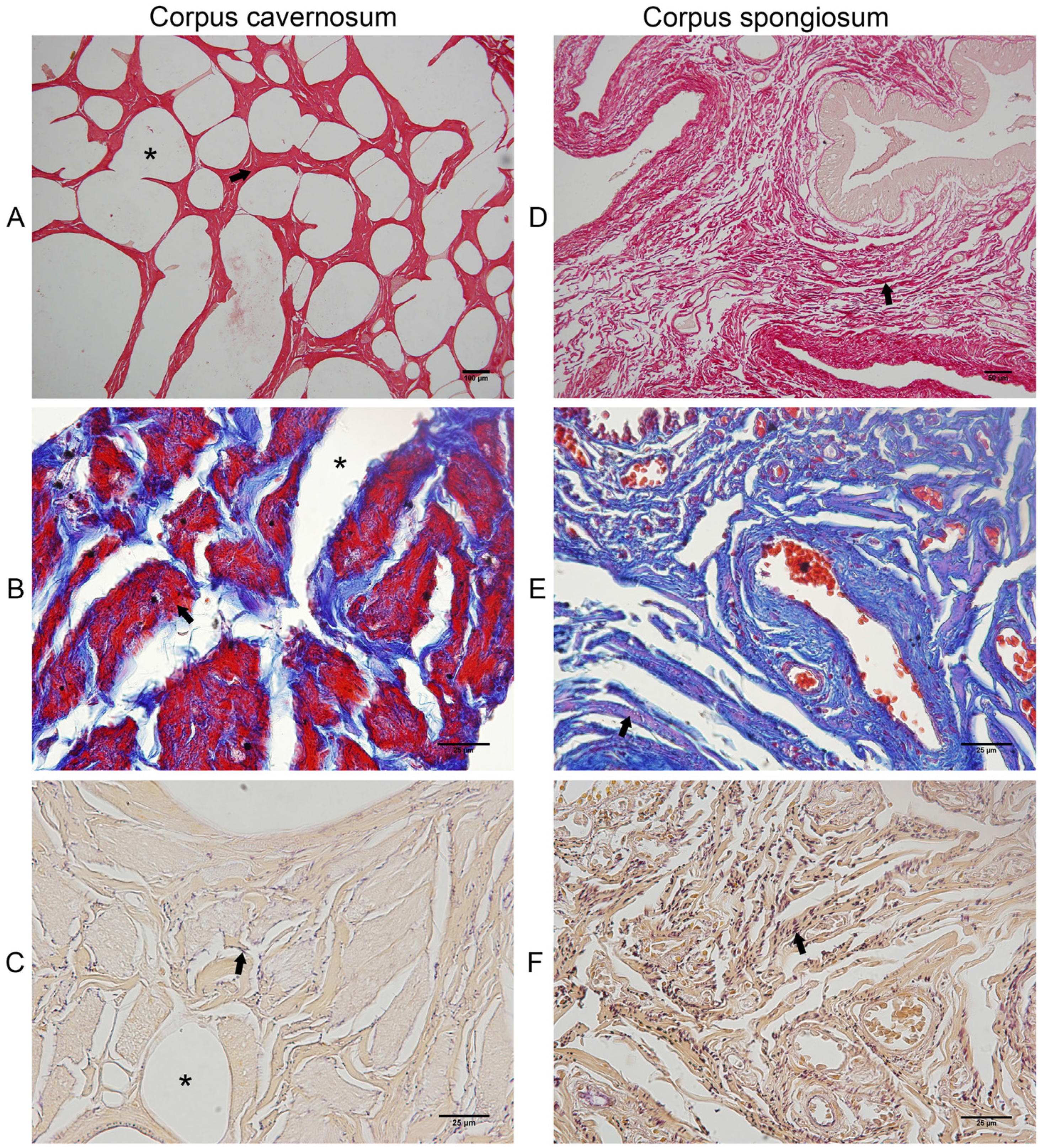
In the qualitative analysis, we observed a single CC (Figure-1), surrounded by a thick tunica albuginea, composed mainly by type-I collagen, due to the presence of thick collagen fibers with red birefringence and less numerous elastic system fibers (Figure-2). The tunica albuginea was thicker in the dorsal region (+21.62%) than in the ventral region (Table-1). The analysis of the density of smooth muscle, collagen and elastic fibers areas, the Corpus Cavernosum showed a predominance of smooth muscle fibers (49%) over collagen (38%) and elastic system fibers (13%). On the other hand, the Corpus Spongiosum showed a predominance of collagen (67%), over elastic system fibers (19%) and smooth muscle fibers (14%). The quantitative analyses of the components of the CC and CS are shown in Table-1. Figure-3 shows the different components of CC and CS.
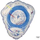
Photomicrograph of the Rhesus monkey penis. Tunica albuginea (yellow arrow), corpus cavernosum (red arrow), corpus spongiosum (black arrow), urethra (asterisk). Masson's Trichrome, X12.
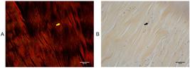
Photomicrographs of the tunica albuginea of the Rhesus monkey penis. A) Thick collagen fibers (yellow arrow), type-I collagen, picrosirius red with polarization, X600. B) Elastic system fibers are less numerous (black arrow). Weigert's resorcin-fuchsin, X600.
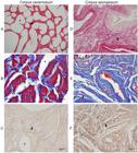
Photomicrographs of the corpus cavernosum and corpus spongiosum of the Rhesus monkey penis. A) Corpus cavernosum, sinusoids (*), collagen fibers (arrow), picrosirius red, X100. B) Smooth muscle fibers (arrow), sinusoids (*), Masson's trichrome, X600. C) Elastic system fibers (arrow), sinusoids (*), Weigert's resorcin-fuchsin, X600. D) Corpus spongiosum, collagen fibers (arrow), picrosirius red, X200. E) Smooth muscle fibers (arrow), Masson's trichrome, X600. F) Elastic system fibers (arrow), Weigert's resorcin-fuchsin, X600.
Data on the parameters analyzed in the corpus cavernosum and corpus spongiosum of the penis of the Rhesus monkey (Macaca mulatta)
DISCUSSION
Knowledge of the anatomy of laboratory animals is essential for experimental research, allowing the animal to be adapted to the study being carried out. Erectile dysfunction, for example, has been studied using the penises of different species (22), including rats and mice (23, 24), rabbits (25) and monkeys (26).
Although these species have several characteristics similar to those of the human penis, there are differences in the penile anatomy of these animals compared to the human penis (1, 5, 9).
The rat's CC is predominantly composed of connective tissue (1, 5, 9). In contrast, the male penis is predominantly muscular. This could make it difficult to make a comparison using these animals as a model. As in rats, the penises of dogs and cats also have a bony component in their cranial part, which is considered to be an ossified part of the CC and is part of the erectile components (27, 28).
The human penis has two paired corpora cavernosa (4), as is the case with other species such as rats, dogs and rabbits, in which the penis has two distinct corpora cavernosa, partially separated by the median septum (27). The penis of the Rhesus monkey was not clearly divided into two corpora cavernosa. De Siqueira et al. (29) also observed a single non-septated CC in the marmoset. Despite being unique, the components of the Rhesus monkeys CC are very similar to those of humans, such as the predominant presence of smooth muscle fibers in relation to the fibrous component. This characteristic allows for a more reliable comparison. The proportion of smooth muscle fibers in the CC of the human penis is approximately 40% (30). Similarly, the CC of the Rhesus monkey penis had 49% more muscle fibers than the other components analyzed.
The tunica albuginea of the CC in humans is a structure composed of inner circular and outer longitudinal layers which have bundles of thick collagen fibers resting on a network of irregularly arranged elastic fibers (3, 31, 32). In the CC of the penis of the Rhesus monkey, we observed a thick tunica albuginea enveloping the entire CC, which showed thick collagen fibers birefringent in red when observed under polarized light, which may characterize type-I collagen fibers. The analysis of the tunica albuginea in adult men showed that the dorsal region was thicker than the ventral region, making the ventral region a more vulnerable area, as in humans (33). Even from the human fetal period, the tunica albuginea already shows a morphological difference in relation to the dorsal and ventral regions. Gallo et al. (34) studied human fetuses between 13- and 36-weeks post-conception and reported that the thickness of the tunica albuginea was greater in the dorsal than ventral region. In our study, the tunica albuginea of the Rhesus monkey penis also showed this characteristic of being thicker dorsally than ventrally (+21.62%).
It is known that the elastic system fibers form an interconnected network in order to keep the collagen fiber bundles together (32). The density of elastic system fibers in the CS of the Rhesus monkey penis was higher than in the CC (+ 43.86%). These characteristics are very similar to those described in the human penis, where there is also a predominance of elastic fibers in the CS (31).
All the similarities between the different structures when comparing the penis of the Rhesus monkey and humans, fully justify the use of this animal as a good experimental model for the study of the human penis.
CONCLUSIONS
The predominance of smooth muscle fibers over connective tissue in the corpus cavernosum, similar to what is observed in humans, qualifies the penis of the Rhesus monkey as a good experimental model.
REFERENCES
-
1 Ribeiro ICA, Abidu-Figueiredo M, Costa FB, Pereira-Sampaio MA, Chagas MA. Stereological study of the elastic fiber and smooth muscle cell system in the bovine and buffalo penis. [Internet]. Pesq Vet Bras. 2013;33:107-12. Available at. <https://www.scielo.br/j/pvb/a/qySxxxSK5zdWLPkZzqsTNkw/?format=html>
» https://www.scielo.br/j/pvb/a/qySxxxSK5zdWLPkZzqsTNkw/?format=html -
2 Schimming BC, Moraes GN. Morphological analysis of the elastic and collagen fibers in the ram penis. [Internet]. Pesq. Vet. Bras. 2018;38:2159-65. Available at. <http://www.pvb.com.br/antigo/?link=verart&tipo=ID&campo1=2510>
» http://www.pvb.com.br/antigo/?link=verart&tipo=ID&campo1=2510 - 3 Brock G, Hsu GL, Nunes L, von Heyden B, Lue TF. The anatomy of the tunica albuginea in the normal penis and Peyronie's disease. J Urol. 1997;157:276-81.
-
4 Hsu GL, Hsieh CH, Wen HS, Hsu WL, Wu CH, Fong TH, et al. Anatomy of the human penis: the relationship of the architecture between skeletal and smooth muscles. J Androl. 2004;25:426-31. doi: 10.1002/j.1939-4640.2004.tb02810.x.
» https://doi.org/10.1002/j.1939-4640.2004.tb02810.x -
5 Cunha GR, Sinclair A, Ricke WA, Robboy SJ, Cao M, Baskin LS. Reproductive tract biology: Of mice and men. Differentiation. 2019;110:49-63. doi: 10.1016/j.diff.2019.07.004.
» https://doi.org/10.1016/j.diff.2019.07.004 -
6 Felix-Patrício B, Medeiros JL Jr, De Souza DB, Costa WS, Sampaio FJ. Penile histomorphometrical evaluation in hypertensive rats treated with sildenafil or enalapril alone or in combination: a comparison with normotensive and untreated hypertensive rats. J Sex Med. 2015;12:39-47. doi: 10.1111/jsm.12750.
» https://doi.org/10.1111/jsm.12750 -
7 Marchon RG, Gregório BM, Costa WS, Pereira-Sampaio MA, Sampaio FJ, De Souza DB. Effects of comfort food diet on the penile morphology of stressed rats. Heliyon. 2023;9:e17013. doi: 10.1016/j.heliyon.2023.e17013.
» https://doi.org/10.1016/j.heliyon.2023.e17013 -
8 Hashimoto M, Karnup S, Daugherty SL, Cho KJ, Banno E, Shimizu N, et al. Sex differences in lower urinary tract function in mice with or without spinal cord injury. Neurourol Urodyn. 2024;43:267-75. doi: 10.1002/nau.25323.
» https://doi.org/10.1002/nau.25323 -
9 Goyal HO, Braden TD, Williams CS, Dalvi P, Mansour MM, Mansour M, et al. Abnormal morphology of the penis in male rats exposed neonatally to diethylstilbestrol is associated with altered profile of estrogen receptor-alpha protein, but not of androgen receptor protein: a developmental and immunocytochemical study. Biol Reprod. 2004;70:1504-17. doi: 10.1095/biolreprod.103.026328.
» https://doi.org/10.1095/biolreprod.103.026328 -
10 Casteleyn C, Bakker J. Anatomy of the Rhesus Monkey (Macaca mulatta): The Essentials for the Biomedical Researcher. In: Rutland CS, El-Genry S, editors. Updates on Veterinary Anatomy and Physiology [Internet]. IntechOpen; 2021. 1-65. Available at. <https://www.intechopen.com/chapters/78564>
» https://www.intechopen.com/chapters/78564 -
11 Ganzer R, Köhler D, Neuhaus J, Dorschner W, Stolzenburg JU. Is the rhesus monkey (Macaca mulatta) comparable to humans? Histomorphology of the sphincteric musculature of the lower urinary tract including 3D-reconstruction. Anat Histol Embryol. 2004;33:355-61. doi: 10.1111/j.1439-0264.2004.00576.x.
» https://doi.org/10.1111/j.1439-0264.2004.00576.x - 12 Kessler MJ, Roberts JA, London WT. Adult polycystic kidney disease in a rhesus monkey (Macaca mulatta). J Med Primatol. 1984;13:147-52.
-
13 Batchelder CA, Lee CC, Martinez ML, Tarantal AF. Ontogeny of the kidney and renal developmental markers in the rhesus monkey (Macaca mulatta). Anat Rec (Hoboken). 2010;293:1971-83. doi: 10.1002/ar.21242.
» https://doi.org/10.1002/ar.21242 -
14 Batchelder CA, Keyser JL, Lee CC, Tarantal AF. Characterization of growth, glomerular number, and tubular proteins in the developing rhesus monkey kidney. Anat Rec (Hoboken). 2013;296:1747-57. doi: 10.1002/ar.22756.
» https://doi.org/10.1002/ar.22756 -
15 Wakui S, Furusato M, Sasaki S, Masaoka T, Ushigome S, Aizawa S. Immunohistochemical localization of the epidermal growth factor-receptor in rhesus-monkey prostate. Anat Histol Embryol. 1996;25:109-11. doi: 10.1111/j.1439-0264.1996.tb00066.x.
» https://doi.org/10.1111/j.1439-0264.1996.tb00066.x -
16 Wen F, Chungui T, Hualin D, Li G, Licai Z, Wen Z. Intra-abdominal vas deferens cyst in a laboratory rhesus macaque (Macaca mulatta). J Med Primatol. 2022;51:404-6. doi: 10.1111/jmp.12606.
» https://doi.org/10.1111/jmp.12606 -
17 Batchelder CA, Duru N, Lee CI, Baker CA, Swainson L, Mccune JM, et al. Myeloid-lymphoid ontogeny in the rhesus monkey (Macaca mulatta). Anat Rec (Hoboken). 2014;297:1392-406. doi: 10.1002/ar.22943.
» https://doi.org/10.1002/ar.22943 -
18 Pabst R, Miller LA, Schelegle E, Hyde DM. Organized lymphatic tissue (BALT) in lungs of rhesus monkeys after air pollutant exposure. Anat Rec (Hoboken). 2020;303:2766-73. doi: 10.1002/ar.24456.
» https://doi.org/10.1002/ar.24456 -
19 Tian H, Zhao X, Hu F, Hu H. Sex Determination According to the Lengths of Hand Bones in Rhesus Macaques (Macaca Mulatta). Anat Rec (Hoboken). 2017;300:1741-6. doi: 10.1002/ar.23637.
» https://doi.org/10.1002/ar.23637 -
20 Capitanio JP, Del Rosso LA, Yee J, Lemoy MM. An analysis of risk factors for spontaneously occurring type 2 diabetes mellitus in rhesus macaques (Macaca mulatta). J Med Primatol. 2024;53:e12695. doi: 10.1111/jmp.12695.
» https://doi.org/10.1111/jmp.12695 -
21 de Souza DB, Silva D, Cortez CM, Costa WS, Sampaio FJ. Effects of chronic stress on penile corpus cavernosum of rats. J Androl. 2012;33:735-9. doi: 10.2164/jandrol.111.014225.
» https://doi.org/10.2164/jandrol.111.014225 -
22 Gajbhiye SV, Jadhav KS, Marathe PA, Pawar DB. Animal models of erectile dysfunction. Indian J Urol. 2015;31:15-21. doi: 10.4103/0970-1591.128496.
» https://doi.org/10.4103/0970-1591.128496 -
23 Palese MA, Crone JK, Burnett AL. A castrated mouse model of erectile dysfunction. J Androl. 2003;24:699-703. doi: 10.1002/j.1939-4640.2003.tb02729.x.
» https://doi.org/10.1002/j.1939-4640.2003.tb02729.x -
24 Zhou X, Wang S, Zhou R, Zhang T, Wang Y, Zhang Q, et al. Erectile dysfunction in hypospadiac male adult rats induced by maternal exposure to di-n-butyl phthalate. Toxicology. 2022;475:153227. doi: 10.1016/j.tox.2022.153227.
» https://doi.org/10.1016/j.tox.2022.153227 -
25 Bischoff E. Rabbits as models for impotence research. Int J Impot Res. 2001;13:146-8. doi: 10.1038/sj.ijir.3900681.
» https://doi.org/10.1038/sj.ijir.3900681 -
26 Williams JK, Andersson KE, Christ G. Animal models of erectile dysfunction (ED): potential utility of non-human primates as a model of atherosclerosis-induced vascular ED. Int J Impot Res. 2012;24:91-100. doi: 10.1038/ijir.2011.56.
» https://doi.org/10.1038/ijir.2011.56 - 27 Ellenport CR. Carnivore Urogenital Apparatus. In: Getty, R, editor. Sisson and Grossman's The Anatomy of the Domestic Animals. Philadelphia: W. B. Saunders Company. 1975; pp. 1576-89.
-
28 Alaa HS. Anatomical and Histological Study of Dog Penis. [Internet]. MRVSA. 2016;5:8-14. Available at. <https://mrvsa.com/upload/article%202%20Anatomical%20and%20Histological%20Study%20of%20Dog%20Penis.pdf>
» https://mrvsa.com/upload/article%202%20Anatomical%20and%20Histological%20Study%20of%20Dog%20Penis.pdf -
29 de Siqueira GHL, Silva FE, Santana MIS. Morphological description of male genital organs of Marca's marmoset (Mico marcai). Anat Histol Embryol. 2018;47:372-84. doi: 10.1111/ahe.12365.
» https://doi.org/10.1111/ahe.12365 -
30 Claro Jde A, Aboim J, Andrade E, Alarcon G, Ortiz V, Sampaio F, et al. Histomorphometry of penile smooth muscle fiber in severe erectile dysfunction. Sao Paulo Med J. 2005;123:181-6. doi: 10.1590/s1516-31802005000400005.
» https://doi.org/10.1590/s1516-31802005000400005 -
31 Hsu GL, Brock G, von Heyden B, Nunes L, Lue TF, Tanagho EA. The distribution of elastic fibrous elements within the human penis. Br J Urol. 1994;73:566-71. doi: 10.1111/j.1464-410x.1994.tb07645.x.
» https://doi.org/10.1111/j.1464-410x.1994.tb07645.x -
32 Akkus E, Carrier S, Baba K, Hsu GL, Padma-Nathan H, Nunes L, et al. Structural alterations in the tunica albuginea of the penis: impact of Peyronie's disease, ageing and impotence. Br J Urol. 1997;79:47-53. doi: 10.1046/j.1464-410x.1997.26511.x.
» https://doi.org/10.1046/j.1464-410x.1997.26511.x -
33 Hsu GL, Brock G, Martínez-Piñeiro L, von Heyden B, Lue TF, Tanagho EA. Anatomy and strength of the tunica albuginea: its relevance to penile prosthesis extrusion. J Urol. 1994;151:1205-8. doi: 10.1016/s0022-5347(17)35214-x.
» https://doi.org/10.1016/s0022-5347(17)35214-x -
34 Gallo CB, Costa WS, Furriel A, Bastos AL, Sampaio FJ. Development of the penis during the human fetal period (13 to 36 weeks after conception). J Urol. 2013;190:1876-83. doi: 10.1016/j.juro.2013.05.050.
» https://doi.org/10.1016/j.juro.2013.05.050