 Effect of a physical therapy program analyzing the neurological state, pain and activity limitation in leprosy neural injury: a clinical trial
ABSTRACT
Leprosy affects peripheral nerves and leads to neural damage, physical disabilities, and functional impairments. Physical therapy has emerged as a crucial component in managing patients living with leprosy, offering various treatment modalities, including neural mobilization, stretching, and joint mobilizations. This study aimed to evaluate the effects of physical therapy on the neural status, pain, and functionality of patients with neural damage due to leprosy. This clinical trial selected 40 patients living with leprosy with neural damage. Simplified neurological examinations (SNE) were performed to assess neural palpation, muscle strength, and sensitivity. Pain intensity was measured using the visual analog scale (VAS) and functionality was evaluated using the screening of activity limitation and safety awareness (SALSA) scale. Participants underwent a three-week physical therapy intervention. Techniques such as stretching, Cyriax transverse massage, pumping, joint mobilizations, myofascial release, and neural mobilization were employed to improve pain, joint mobility, and neural impulse conduction. The data showed improved palpation, muscle strength, and sensitivity, particularly in the posterior tibial nerve. Pain intensity decreased after the intervention (6.4±2.5 before and 5.3±2.9 after physical therapy treatment, p<0.004). The SALSA scale showed a reduction in functional limitations, from 25% to 17.5% in severe limitations. However, the disability score failed to significantly change (1.2±0.08 before and 1.2±0.09 after, p<0.999). Physical therapy effectively enhanced neural status, reduced pain, and improved functionality in patients living with leprosy and neuritis. This study also shows the importance of the VAS, SALSA, and SNE scales in evaluating patient improvement.
Keywords
Leprosy; Neuropathy; Physical therapy; Pain; Disability
RESUMO
A hanseníase afeta os nervos periféricos e leva a danos neurais, incapacidades físicas e prejuízos funcionais. A fisioterapia emergiu como um componente crucial no manejo dos pacientes com hanseníase, oferecendo diversas modalidades de tratamento, incluindo mobilização neural, alongamentos e mobilizações articulares. Este estudo teve como objetivo avaliar os efeitos da fisioterapia no estado neural, dor e funcionalidade em pacientes com lesão neural decorrente da hanseníase. Trata-se de um ensaio clínico que selecionou 40 pacientes com hanseníase e lesões neurais. Exames neurológicos simplificados (SNE) foram realizados para avaliar palpação neural, força muscular e sensibilidade. A intensidade da dor foi medida pela escala visual analógica (EVA) e a funcionalidade foi avaliada pela escala de triagem de limitação de atividade e conscientização de segurança (SALSA). Os participantes foram submetidos a uma intervenção fisioterapêutica de três semanas. Técnicas como alongamento, massagem transversal Cyriax, bombeamento, mobilizações articulares, liberação miofascial e mobilização neural foram empregadas para melhorar a dor, a mobilidade articular e a condução do impulso neural. Os dados mostraram melhorias na palpação, na força muscular e na sensibilidade, principalmente no nervo tibial posterior. A intensidade da dor diminuiu após a intervenção (6,4±2,5 antes e 5,3±2,9 após o tratamento fisioterapêutico, p<0,004). A escala SALSA revelou uma redução nas limitações funcionais, passando de 25% para 17,5% nas limitações graves. Contudo, o escore de incapacidade não se alterou significativamente (1,2±0,08 antes e 1,2±0,09 depois, p<0,999). A fisioterapia mostrou-se eficaz na melhoria do estado neural, na redução da dor e na melhoria da funcionalidade em pacientes com hanseníase e neurite. Além disso, demonstra-se a importância das escalas VAS, SALSA e SNE na avaliação da melhora do paciente.
Descritores
Hanseníase; Neuropatia; Fisioterapia; Dor; Incapacidade
RESUMEN
La lepra afecta los nervios periféricos y provoca daños neurales, discapacidades físicas y deficiencias funcionales. La fisioterapia surgió como un componente crucial en el tratamiento de pacientes con lepra, ofreciendo varias modalidades de tratamiento, incluida la movilización neural, los estiramientos y las movilizaciones articulares. Este estudio tuvo el objetivo de evaluar los efectos de la fisioterapia en el estado neural, el dolor y la funcionalidad en pacientes con lesión neural resultante de la lepra. Se trata de un ensayo clínico que seleccionó 40 pacientes con lepra y lesiones neurales. Se realizaron evaluaciones neurológicas simplificadas (SNE) para evaluar la palpación neural, la fuerza muscular y la sensibilidad. Se midió la intensidad del dolor mediante la escala visual analógica (EVA) y se evaluó la funcionalidad mediante la escala de detección de limitación de actividad y conciencia de seguridad (SALSA). Se sometieron los participantes a una intervención fisioterápica de tres semanas. Se utilizaron técnicas como estiramiento, masaje transverso Cyriax, bombeo, movilizaciones articulares, liberación miofascial y movilización neural para mejorar el dolor, la movilidad articular y la conducción del impulso neural. Los datos mostraron mejoras en la palpación, la fuerza muscular y la sensibilidad, principalmente en el nervio tibial posterior. La intensidad del dolor disminuyó tras la intervención (6.4±2.5 antes y 5.3±2.9 después del tratamiento fisioterápico, p<0.004). La escala SALSA reveló una reducción en las limitaciones funcionales, del 25% al 17.5% en las limitaciones graves. Sin embargo, la puntuación de discapacidad no cambió significativamente (1.2±0.08 antes y 1.2±0.09 después, p<0.999). Se demostró que la fisioterapia es eficaz para mejorar el estado neural, reducir el dolor y mejorar la funcionalidad en pacientes con lepra y neuritis. Además, se demuestra la importancia de las escalas VAS, SALSA y SNE en la evaluación de la mejora del paciente.
Palabras clave
Lepra; Neuropatía; Fisioterapia; Dolor; Discapacidad
INTRODUCTION
Leprosy, a chronic infectious disease caused by Microbacterium leprae , primarily affects macrophages and the Schwann cells in peripheral nerves, leading to neural damage. This damage results in thermal, painful, and tactile sensitivity disorders, atrophies, and paresis, potentially progressing to physical disabilities 1 , 2 .
During the course of leprosy, patients often experience acute inflammatory complications that are known as leprosy reactions or reaction states and are classified as type 1 and type 2 reactions 3 . Both reactions contribute to disabilities and physical limitations 4 . Neuritis is an inflammatory condition affecting the nerves, typically occurring in reactive episodes 5 . The diagnosis of neuritis relies on evaluating patients’ medical history, palpating the affected nerve, and comparing consecutive simplified neurological assessments 4 . In addition to pharmacological interventions, comprehensive management of patients necessitates the implementation of measures focused on rehabilitating and preventing functional impairments. Consequently, the involvement of professional physiotherapists is essential due to their extensive expertise, encompassing not only rehabilitation practices but also overall care provision for patients living with leprosy, functional evaluation, and health education 6 . Physical therapy possesses a wide range of treatment available for patients with this condition. One such modality is neural mobilization, which serves as a physiotherapeutic technique to diagnose and alleviate neural pain and treat neuritis. Neural mobilization involves applying oscillatory or static movements to tension the peripheral nerve, physiologically improving neural impulse conduction. Furthermore, techniques such as stretching, joint mobilizations, and myofascial releases have been found to be advantageous for all patients suffering from leprosy 6 , 7 . Some studies have described the effect of physiotherapeutic techniques on patients living with leprosy. However, they only targeted one type of intervention 8 - 11 .
This study aims to evaluate the effects of physical therapy on the neural status, pain, and functionality of patients living with leprosy with neural lesions before and after intervention.
METHODOLOGY
Patient selection and data collection
This clinical trial was conducted to investigate the efficacy of a specific intervention in 40 patients diagnosed with leprosy. The participants eligible for the study were aged from 18 to 75 years of all sexes who had neural damage caused by leprosy at the time of enrollment. The study was conducted from August 2019 to August 2020 at the Health Specialties Center, an outpatient clinic specializing in dermatology and infectious diseases in the municipality of Várzea Grande, Mato Grosso, Brazil.
Patients with a history of irregular attendance at appointments (more than two consecutive absences from appointments), low adherence to medication as advised by medical professionals, chronic alcoholism (reported by the patients themselves), diabetes mellitus, or previous neural injuries unrelated to leprosy were excluded from the study (questioned at anamnesis and according to the medical records of the health unit).
Patients were evaluated at the beginning of the study (before the physiotherapeutic intervention) and were considered as a control group. After intervention, patients were evaluated again and the data were compared.
Ethical approval for this study was obtained and was registered with the Brazilian Registry of Clinical Trials under identifier U1111-1281-4618. This study followed the recommendations outlined in the revised version of the Consolidated Standards of Reporting Trials statement. All participants provided informed consent by signing a form.
During this study, patients received the prescribed polychemotherapy treatment, and prednisone/dexamethasone was administered for type-1 reactions, in accordance with the guidelines from the Brazilian Ministry of Health 4 . Epidemiological, socioeconomic, and clinical data were collected and analyzed.
Data were collected by patient screening, initial assessment, intervention, and final assessment. Screening was conducted by the nursing team, and the eligible candidates were assessed by the physical therapy team. Both the initial and final assessments were performed by two qualified physiotherapeutic professionals. One of the professionals was aware of the procedures and recorded the results, whereas another remained blinded to the procedural outcomes and recorded the results. At the end of the study, results were compared. In case of divergence, a third professional would be asked to evaluate.
Physical therapy intervention
The intervention consisted of 12 sessions thrice per week, totaling 30 days ( Chart 1 suppl ). Patients underwent a conventional physiotherapeutic procedure based on their individual needs. Various physiotherapeutic techniques were employed to alleviate their pain and improve their joint and neural mobility and range of motion, and reduce their limitations and disabilities 11 . The sessions began with active or passive stretches, each lasting 30 seconds, to enhance muscle tissue flexibility while respecting patients’ limits. These stretches were based on the recommendations by Seijas 12 , as stretching increases the number of sarcomeres and thus improves flexibility, preventing deformities, alleviating pain, and addressing the limitations of joint range of motion in patients living with leprosy. Subsequently, manipulation techniques such as Cyriax transverse massage, pumping, global joint mobilizations, and myofascial release—as guided by Kysner and Colby 13 —were applied.
To mobilize soft structures by realigning connective tissue, restoring its physiology and breaking down established adhesions, the Cyriax transverse massage technique was employed in the affected regions. The movements in this technique were performed in the transverse plane and the longitudinal axis, applying pressure in one direction with two fingers and gradually increasing pressure and depth while maintaining a uniform rhythm. The physiological effect of this technique reduces established muscular adhesions from neural injury, reducing pain. To promote muscle relaxation and enhance local circulation, the pumping technique described by Lederman 14 was utilized. This technique consists of three phases: first, segment tensioning (a slow, regular, and progressive movement); second, tension maintenance (for 10 to 20 seconds); and third, a slow return to the initial position. This technique aims to reduce muscle pain due to the reduction of neural impulses stemming from neural injury. Joint mobilizations were performed according to Maitland’s concepts 15 , involving the application of slow and repetitive micromovements in the joints. The technique commenced with grades I and II, using oscillatory movements with a slow rhythm at the beginning of the joint range. Then, grades III and IV were employed, which also consisted of oscillatory movements, but performed at the end of the range based on tissue resistance. Finally, grade V, classified as manipulation, involved joint mobilization, improving mobility and reducing leprosy-related disabilities. The exercises in the Manual of Disability Prevention by the Ministry of Health for patients living with leprosy were also applied 16 . As treatment to tension peripheral nerves, the neural mobilization technique based on the proposals by Butler 17 and Pereira-Júnior and Schons 18 was employed. This technique involved movements to tension the ulnar, median, radial, common fibular, and posterior tibial nerves. Then, after encountering resistance during tensioning, oscillatory movements from 10 to 40 repetitions were performed. The main physiological effect of neural mobilization involves disrupting adhesions, increasing the mobility of the tensioned nerve and enhancing blood flow, axoplasmic flow, and neural conduction, improving pain, motor function, and sensory limitation.
Neurological evaluations
All participants in this study underwent a simplified neurological examination (SNE) that followed the standardized protocol established by the Brazilian Ministry of Health 16 . This examination was conducted before and after the intervention. The assessment included the examination of the face, upper limbs, and lower limbs and consisted of three components: neural palpation, evaluation of muscle strength, and assessment of sensitivity.
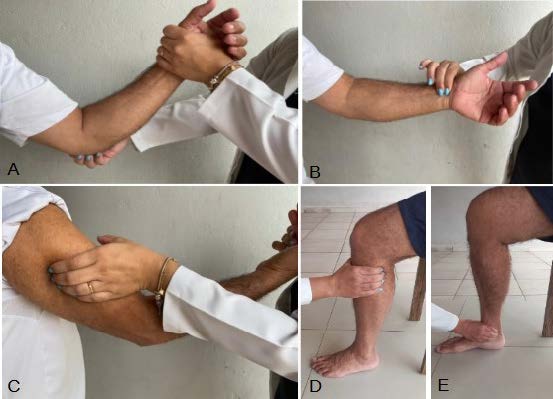
Neural palpation involved examining specific nerve points on the body surface. The ulnar nerve was palpated at the epitrochlear portion of the elbow, the radial nerve was palpated about two fingers behind the final insertion of the deltoid muscle at arm level, the median nerve was examined at the carpal tunnel in the wrist, the common fibular nerve was assessed about two finger widths behind and below the head of the fibula at leg level, and the posterior tibial nerve was examined behind and below the medial malleolus at ankle level. During palpation, pain along the course of the nerve was noted and scored as follows: pain on the right side (score 1), pain and neural thickening on the right or left side (score 2), and absence of pain or thickening (normal, score 0) ( Figure 1 A-E).
A: ulnar nerve palpation; B: median nerve palpation; C: palpation of the radial nerve; D: palpation of the common fibular nerve; E: palpation of the posterior tibial nerve
Muscle strength assessment was conducted in accordance with the guidelines by the Brazilian National Supplementary Health Agency 16 . Assessment involved analyzing specific movements related to the following nerves: fifth finger abduction (ulnar nerve), thumb abduction (median nerve), wrist extension (radial nerve), ankle dorsiflexion (common fibular nerve), and hallux extension (common fibular nerve). Each movement was assigned a score based on the observed response. A score of 5 was given if no movement or contraction was observed; 4, contraction without joint movement; 3, incomplete movement; 2, complete movement without resistance; 1, full movement with partial resistance; and 0, normal movement. Sensory evaluation was performed using Semmes-Weinstein filaments (Sorri, Brazil). Evaluation involved applying a series of monofilaments with varying weights to different points. Assessment began with the lightest monofilament, weighing 0.05 grams. In case of no response to this contact, the next monofilament (weighing 0.2 grams) was used. This sequence continued up to the 300-gram filament or until a positive response was obtained. The analyzed points included six palmar points and nine plantar points. In the sensory evaluation, hypoesthesia (score 1) was defined as the absence of sensitivity to monofilaments weighing 0.2, 2, 4, 10, and 300 grams. Anesthesia (score 2) was recorded in case of no response to the touch of any monofilament. A normal response (score 0) was assigned in case of a positive response to the 0.05-gram monofilament 16 .
The scores from the parameters assessed for each nerve were summed to analyze the functional status of each nerve, which included the scores from palpation, muscle strength, and sensitivity evaluations for the ulnar, median, radial, fibular, and tibial nerves. Based on these scores, the degree of physical disability (DPD) was determined using the guidelines from the Brazilian Ministry of Health 16 . Participants without neural impairments were classified as degree zero. Those with hypoesthesia, anesthesia, or reduced muscle strength (graded on from 1 to 4 according to the Kendal scale) were classified as grade one. Participants showing visible deficiencies such as claw fingers, bone resorption, muscle atrophy, contractures, or ulcers were classified as grade two 16 . Additionally, a score for eyes, hands, and feet (EHF) was assigned, which represents the sum of all the scores analyzed by the DPD for the bilateral assessment of EHF.
Evaluation scale
The intensity of pain experienced by participants was measured using the visual analogue pain scale (VAS) 5 , a numerical rating ranging from 0 to 10. A score of 0 indicated the absence of pain, whereas one of 10 indicated severe pain 19 . To assess participants’ perception of activity limitation, the screening of activity limitation and safety awareness (SALSA) scale 16 was employed. It consisted of a questionnaire comprising 20 items, which were divided into five areas: eyes, hands (skills and work), feet (mobility), and self-care. Participants’ responses were then categorized into five levels: no activity limitation (from 10 to 24), mild limitations (25 to 39), moderate limitations (40 to 49), severe limitations (50 to 59), and very severe limitations (60 to 80).
Statistical analysis
The methodology in this study utilized a sample calculation based on the population of new leprosy cases reported in the municipality of Várzea Grande in 2020, which amounted to 184 patients. A confidence level of 85% and a margin of error of 10% were considered in determining the minimum sample size, resulting in a requirement of 40 patients. Initially, the distribution of all discrete variables was analyzed. Since their distributions were non-normal, the Wilcoxon paired non-parametric test was employed to compare the effects of the intervention on the neural state of the same patient, as well as on the pain scales (SALSA, DPD, and EHF). To assess the effect of physical therapy on the SALSA, VAS, DPD, EHF scales, and neural state scores across different patient groups, the non-parametric Mann-Whitney test was utilized for comparative analysis. Additionally, the Pearson’s chi-square test was employed to compare the improvement and worsening of palpation, strength, and sensitivity. In all analyses, a 5% alpha value was established as the significance level. The statistical software Stata, version 12.0, was used to perform all statistical tests.
RESULTS
Clinical and socioeconomic characteristics
Table 2 shows patients’ demographic characteristics and clinical classification. All patients in this study fell into a multibacillary classification, with a predominantly borderline clinical manifestation. Most patients underwent multidrug therapy (MDT) during the intervention.
A total of 192 nerves were affected among the 40 patients. Most showed involvement of up to three affected nerves, averaging 4.8 nerves per patient. This study performed the physical therapy intervention described in the previous on all affected nerves. Considering the frequency of neural injury showed that the ulnar nerve was the most commonly affected, followed by the posterior tibial nerve.
Patients’ perception and pain assessment
The evaluation of functional capacity limitations followed SALSA in the chosen population. Most patients (32.5%) showed mild limitations. After the end of the physical therapy procedure, the proportion of patients with severe limitations notably decreased, from 25 to 17.5%. Furthermore, 10% of patients successfully concluded the physical therapy treatment without any complaints of limitation ( Table 2 ).
Regarding pain assessment, the initial measurement indicated an average intensity of 6.4±2.5 on the scale. However, after the end of the physical therapy treatment, the average intensity decreased to 5.3±2.9, a reduction in pain according to VAS (p<0.004).
Neurological evaluation
The bilateral neurological assessments of patients occurred before and after a physical therapy session. Findings showed improvements in various aspects. Firstly, palpation of the posterior tibial nerve showed significant improvement (p=0.048). Secondly, the strength test showed increased muscle strength in wrist (p=0.007) and hallux extension (p=0.043). Lastly, the evaluation of sensitivity showed improvement in the dermatomes of the ulnar (p=0.036), median (p=0.036), and posterior tibial (p=0.021) nerves ( Table 3 ).
Further analysis the overall impact of physical therapy summed and compared the scores for neural palpation, muscle strength, and sensitivity of the tested nerves before and after treatment ( Table 4 ).
DPD showed no significant changes after treatment. Patients’ disability scores, including EHF (ocular, manual, and pedal), which assessed neurological functions of the eyes, hands, and feet, failed to differ significantly before and after the procedure (1.2±0.08 and 1.2±0.09, respectively, before and after physical therapy treatment). The mean values ± standard deviation for the EHF score totaled 3.4±0.3 before the procedure and 3.7±0.4 after the procedure, with no statistically significant differences ( p <0.999).
DISCUSSION
This findings of this study focused on rehabilitating individuals living with leprosy by physical therapy interventions. Results indicate that physical therapy played a significant role in improving the condition of patients living with leprosy who had neural damage 20 . This study may have an important application in the use of physical therapy for patients living with leprosy. The therapy in this study produced analgesic, anti-inflammatory, and anti-fibrotic effects, reducing leprosy-induced inflammation. All these effects could improve the quality life of patients and may avoid disability.
Data showed that the patients referred to physical therapy primarily complained of chronic pain and averaged five affected nerves, indicating a multibacillary classification. Santos et al. 21 reported similar findings, finding a higher propensity for pain in multibacillary and reactional patients. The prevalence of pain in patients living with leprosy can be attributed to the affinity of the bacillus for peripheral nerves, particularly the ulnar, median, radial, common fibular, and posterior tibial ones 22 . The consequences of damage in these nerves have substantial economic, social, and personal impacts 7 . In this study, the ulnar nerve was the most affected bilaterally. This nerve is highly susceptible to damage, which may cause claw hands 23 .
Physiotherapeutic interventions for leprosy primarily aim to prevent deformities, alleviate existing ones, and reduce pain 8 . Thus, the demand for effective, efficient, and comprehensive therapeutic approaches in physical therapy has been growing. The goal involves enhancing patient outcomes, especially for individuals with severe peripheral neuropathy due to leprosy. In this context, physical therapy aids relieving pain, resolving inflammatory processes, and inhibiting the progression of neural damage 24 .
Rehabilitation of leprosy neural lesions configures a process that involves a therapeutic sequence related to the needs of each patient. This study utilized stretching at the initial phase of each physical therapy session to enhance tissue and joint flexibility, consistently performing static stretching, which involved the maintenance of distal range of motion for a specific duration. Diaz et al. 9 have shown the widespread use of stretching in patients with leprosy sequelae, highlighting that both static and proprioceptive stretching achieved the desired objectives.
This study investigated applying the Cyriax transverse friction therapy in patients with neural damage due to leprosy, primarily to alleviate pain. The treatment for musculoskeletal disorders often uses Cyriax transverse friction therapy . One of the observed physiological effects of this therapy reduces pain by activating diffuse noxious inhibitory controls. This pain suppression mechanism could trigger the release of endogenous opiates within the body. Additionally, the therapy is believed to disrupt strong cross-links and/or adhesions while softening scar tissue. Previous studies have reported these effects, such as Stasinopoulos and Johnson 25 , which shown vasodilation and an associated increase in blood flow.
Additionally, the pumping technique, a manual therapy maneuver involving fascial mobilization to improve local circulation and tissue nutrition, has been applied to the patients 14 . Data suggest it effectively reduced pain and yielded significant improvements.
Conventional therapies are routinely employed in the treatment of patients living with leprosy. Among these, neural mobilization stands out as a widely utilized technique in clinical practice for the diagnosis and treatment of leprosy-related injuries in muscles innervated by peripheral nerves due to the compression and entrapment of the edematous nerve. Neural mobilization is a non-pharmacological treatment option physiotherapists administer to reduce disability and pain, enhancing individuals’ quality of life. Veras et al. 10 have shown the positive efficacy of this technique in patients living with leprosy, improving electromyographic function, disability level, and pain intensity. This study found no difference in disability level after the intervention. However, patients showed a significant reduction in VAS scores, indicating an improvement in pain perception.
This study used conventional techniques such as joint mobilization, increasing synovial fluid movement and thus enhancing joint nutrition 8 . Rath et al. have shown that this technique reduces rehabilitation by 15 days in patients with leprosy 26 . Based on the physiological effects according to the used physiotherapeutic techniques, ranging from stretching to neural and joint mobilizations, these data suggest the decrease of patients’ pain. VAS measurement evinced such differences.
The SALSA scale assessed patients’ functional capacity. It can analyze foot mobility, self-care, hand functioning, and dexterity, evincing limitations and classifying them according to their severity 7 . Some individuals suffering from leprosy may fail to perceive themselves as limited, which can lead to domestic accidents. SALSA can serve as an important tool to find these limitations 27 , 28 , enhance the quality of care for patients living with leprosy, and serve as a comprehensive resource for individual attention during treatment and post-polychemotherapy discharge 28 . Tiago et al. 11 utilized SALSA to evaluate patients who underwent neural decompression surgery due to leprosy, showing its utility in assessing these patients.
Regarding the simplified neurological assessment, which included neural palpation, muscle strength assessment, and sensitivity evaluation 16 , most nerves showed no significant changes after physical therapy treatment. However, the tibial nerve showed substantial improvement, indicating the effectiveness of physical therapy in addressing pain and nerve thickening and preventing neural lesion progression. Improved nerve impulse might result in better motor function. This study observed statistically significant changes in muscle strength, particularly for wrist and hallux extension, crucial movements to prevent hand drop and walking difficulties. Additionally, sensitivity in the dermatomes of the ulnar, median, and posterior tibial nerves, which are commonly affected in leprosy 23 , showed statistically significant changes. These findings suggest the contribution of physical therapy in improving neural status, muscle strength, and sensitivity.
This study has some limitations. Firstly, a portion of the collected data rely on self-reports, such as pain and sensitivity, which might vary depending on patients’ emotional/physical state. Secondly, the effectiveness of the pharmacological treatment could be considered another constraint as some patients respond more favorably than others. Also, variations occurred in drug treatment, with most patients receiving similar doses, whereas some were prescribed higher doses. Additionally, most participants fall within the 50 to 70 year-age range, but the inclusion of younger individuals could limit this study.
While the study failed to find a significant reduction in DPD and in EHF scores, some studies 27 have reported a correlation between the EHF score and the SALSA scale, positively correlating them. This suggests that the EHF score can provide valuable information regarding patients’ functional status.
In conclusion, this study shows the effectiveness of physical therapy in improving the functional status and alleviating pain in patients living with leprosy and the importance of VAS, SALSA, and SNE in evaluating patients’ improvement.
Supplementary Chart
REFERENCES
-
1. Eichelmann K, González González SE, Salas-Alanis JC, Ocampo-Candiani J. Leprosy. An update: definition, pathogenesis, classification, diagnosis, and treatment. Actas Dermosifiliogr. 2013;104(7):554-63. doi: 10.1016/j.adengl.2012.03.028
» https://doi.org/10.1016/j.adengl.2012.03.028 -
2. Kubota RMM, Brancini VCL, Gouveia AS, Nardi SMT, Paschoal VDA, et al. Adverse effects of multidrug therapy for leprosy: use of alternative doses and post discharge evaluation. Hansen Int. 2014;39:8-21. doi: 10.47878/hi.2014.v39.35024
» https://doi.org/10.47878/hi.2014.v39.35024 -
3. Queiroz TA, Carvalho FPB, Simpson CA, Fernandes ACL, Figueirêdo DLA, et al. Perfil clínico e epidemiológico de pacientes em reação hansênica. Rev Gaúcha Enf. 2015;36:185-91. doi: 10.1590/1983-1447.2015.esp.57405
» https://doi.org/10.1590/1983-1447.2015.esp.57405 -
4. Ministério da Saúde (Brasil). Orientações para uso corticoesteroides em Hanseníase. Normas e Manuais Técnicos [Internet]. Brasília: Ministério da Saúde; 2010 [cited 2021 Aug 12]. Available from: http://bvsms.saude.gov.br/bvs/publicacoes/orientacoes_para_corticosteroides_hanseniase.pdf
» http://bvsms.saude.gov.br/bvs/publicacoes/orientacoes_para_corticosteroides_hanseniase.pdf -
5. Pimentel MIF, Nery JAC, Borges E, Rolo R, Sarno FN. Neurite silenciosa na hanseníase multibacilar avaliada através da evolução das incapacidades antes, durante e após a poliquimioterapia. An Bras Dermatol. 2004;79(2):169-79. doi: 10.1590/S0365-05962004000200005
» https://doi.org/10.1590/S0365-05962004000200005 - 6. Pereira Junior NS, Almeida RM. Manual de recursos terapêuticos manuais. João Pessoa: UFPB; 2016.
-
7. Reis BM, Fernandes LFRM. Association between the Rosén and Lundborg Score and the Screening Activity Limitation and Safety Awareness scale in hand functional evaluation of patients with leprosy diagnosis. Disabil Rehabil. 2019;41(13):1578-83. doi: 10.1080/09638288.2018.1432708
» https://doi.org/10.1080/09638288.2018.1432708 -
8. Martins RL, Albuquerque APF, Holanda AL, Pacas ASS, Galdino DMC, et al. Intervenção fisioterapêutica nos comprometimentos da hanseníase. Br J Health Rev. 2021;4(1):983-90. doi: 10.34119/bjhrv4n1-086
» https://doi.org/10.34119/bjhrv4n1-086 -
9. Diaz AF, Moro FL, Binotto JM, Fréz AR. Estudo comparativo preliminar entre os alongamentos proprioceptivo e estático passivo em pacientes com sequelas de hanseníase. Fisioter Pesqui. 2008;15(4):339-44. doi: 10.1590/S1809-29502008000400004
» https://doi.org/10.1590/S1809-29502008000400004 -
10. Véras LS, Vale RG, Mello DB, Castro JA, Lima V, et al. Electromyography function, disability degree, and pain in patients living with leprosy undergoing neural mobilization treatment. Rev Soc Bras Med Trop. 2012;45(1):83-8. doi: 10.1590/s0037-86822012000100016
» https://doi.org/10.1590/s0037-86822012000100016 -
11. Tiago LMP, Barbosa MFF, Santos DFD, Faria AD, Gonçalves MA, et al. Late follow-up of peripheral neural decompression in leprosy: functional and clinical outcomes. Arq Neuropsiquiatr. 2021;79(8):716-23. doi: 10.1590/0004-282X-ANP-2020-0032
» https://doi.org/10.1590/0004-282X-ANP-2020-0032 - 12. Seijas G. Anatomia e alongamentos essenciais. São Paulo: Ed. Manole; 2015.
- 13. Kysner C, Colby LA. Exercícios terapêuticos – fundamentos e técnicas. 6th ed. São Paulo: Manole; 2015.
- 14. Lederman E. Fundamentos da terapia manual – fisiologia, neurologia e psicologia. São Paulo: Manole; 2001.
- 15. Maitland GD. Treatment of the glenohumeral joint by passive movement. Physical therapy. 1971;57(6):261-7.
-
16. Ministério da Saúde (Brasil). Manual de prevenção de incapacidades [Internet]. Brasília: Ministério da Saúde; 2008 [cited 2021 Aug 12]. Available from: https://bvsms.saude.gov.br/bvs/publicacoes/manual_prevencao_incapacidades.pdf
» https://bvsms.saude.gov.br/bvs/publicacoes/manual_prevencao_incapacidades.pdf - 17. Butler D. Mobilização do sistema nervoso. São Paulo: Manole; 2003.
- 18. Pereira Júnior AA, Schons DG. Os efeitos da mobilização neural em pacientes com lombalgia. Fisioter Saude Func. 2015;4(2):14-20.
- 19. Galvão O, Silva MG. Validade e fidedignidade preliminares da EVA modificada para a população idosa. EssFisi Online. 2005;1(4):22-30.
-
20. Sousa JB, Carqueja CL, Baptista AF. Physical rehabilitation to treat neuropathic pain. Rev Dor. 2016;17(Suppl 1):S85-90. doi: 10.5935/1806-0013.20160056
» https://doi.org/10.5935/1806-0013.20160056 -
21. Santos VS, Santana JC, Castro FD, Oliveira LS, Santana JC, et al. Pain and quality of life in patients living with leprosy in an endemic area of Northeast Brazil: a cross-sectional study. Infect Dis Poverty. 2016;5:18. doi: 10.1186/s40249-016-0113-1
» https://doi.org/10.1186/s40249-016-0113-1 -
22. Scollard DM, Truman RW, Ebenezer GJ. Mechanisms of nerve injury in leprosy. Clin Dermatol. 2015;33(1):46-54. doi: 10.1016/j.clindermatol.2014.07.008
» https://doi.org/10.1016/j.clindermatol.2014.07.008 -
23. Manivannan G, Das P, Karthikeyan G, John AS. Reconstructive surgery in children to correct ulnar claw hand deformity due to leprosy. Lepr Rev. 2014;85(2):74-80. doi: 10.47276/lr.85.2.74
» https://doi.org/10.47276/lr.85.2.74 -
24. Souza MV, Silva MO. Laserterapia em afecções locomotoras: revisão sistemática de estudos experimentais. Rev Bras Med Esporte. 2016;22(1):76-82. doi: 10.1590/1517-869220162201144326
» https://doi.org/10.1590/1517-869220162201144326 -
25. Stasinopoulos S, Johnson MI. Cyriax physical therapy for tennis elobow/lateral epicondylitis. Br J Sports Med. 2004;38(6):675-7. doi: 10.1136/bjsm.2004.013573
» https://doi.org/10.1136/bjsm.2004.013573 -
26. Rath S, Schreuders TA, Stam HJ, Hovius SE, Selles RW. Early active motion versus immobilization after tendon transfer for foot drop deformity: a randomized clinical trial. Clin Orthop Relat Res. 2010;468(9):2477-84. doi: 10.1007/s11999-010-1342-4
» https://doi.org/10.1007/s11999-010-1342-4 -
27. Nascimento DDS, Ramos Junior AN, Araújo OD, Macêdo SF, Silva GVD, et al. Activity limitation and social participation restriction of people with leprosy: a cross-sectional analysis of magnitude and associated factors in a hyperendemic municipality in the state of Piauí, Brazil, 2001-2014. Epidemiol Serv Saude. 2020;29(3):e2019543. doi: 10.5123/s1679-49742020000300012
» https://doi.org/10.5123/s1679-49742020000300012 -
28. Barbosa JC, Ramos Jr AN, Alencar MJF, Castro CGJ. Pós-alta em hanseníase no Ceará: limitação da atividade funcional, consciência de risco e participação social. Rev Bras Enf. 2008;61:727-33. doi: 10.1590/S0034-71672008000700012
» https://doi.org/10.1590/S0034-71672008000700012
-
Financing source:
Conselho Nacional de Desenvolvimento Científico e Tecnológico (CNPq Process number 307492/2020-6)
-
Brazilian Registry of Clinical Trials (REBEC) - identifier U1111-1281-4618 – Approved by the Research Ethics Committee: 3.511.331. CAAE: 15299019.9.0000.8124.