 Updates on placental studies in bitches
ABSTRACT:
The placenta, despite being a temporary organ, is essential for the development and survival of the conceptus. During pregnancy, it performs essential functions, such as nutrition, protection, circulation, excretion, and secretion. Studies in humans already detail placental measurements, associating them with perinatal outcomes and diseases in adult life. However, in dogs, these studies are still limited. Therefore, this research aimed to review and unify the main studies already published on the evaluation of the placenta in bitches. Although, several forms of placental assessment have been described, more studies on this topic, whether macroscopic or microscopic, are needed to optimize and prioritize immediate and long-term clinical research into the condition of the placenta and its influence on canine neonates.
Key words:
neonate; dogs; placenta
RESUMO:
A placenta, apesar de ser um órgão temporário, é imprescindível para o desenvolvimento e sobrevivência do concepto. Durante a gestação, desempenha funções essenciais, como nutrição, proteção, circulação, excreção e secreção. Estudos em humanos já detalham medidas placentárias associando-as a desfechos perinatais e a doenças na vida adulta. No entanto, em cadelas, esses estudos ainda são limitados. Portanto, este trabalho tem como objetivo revisar e unificar os principais estudos já publicados sobre a avaliação da placenta de cadelas. Embora diversas formas de avaliação placentária tenham sido descritas, mais estudos sobre este tema, sejam eles macroscópicos ou microscópicos, são necessários para otimizar e priorizar a pesquisa clínica imediata e a longo prazo sobre a condição da placenta e a sua influência sobre os neonatos caninos.
Palavras-chave:
neonato; cães; placenta; volume placentário
INTRODUCTION
The placenta is a temporary organ composed of maternal endometrial membranes and fetal somatic extraembryonic mesoderm, enabling maternal-fetal connections through the umbilical cord. It is responsible for promoting exchanges such as the transport of gases and nutrients between the mother and fetus, preventing potential harm to the newborn that could extend into the postnatal period (DA SILVA et al., 2022). Other placental functions include nutrition, protection, circulation, excretion, and secretion, all of which are crucial for the survival of the conceptus (FOWDEN et al., 2006).
The placentation of bitches begins between the 14th and 18th days of gestation (HYTTEL et al., 2012). This process unfolds in three phases: 1st, apposition; 2nd, adhesion of the blastocyst to the endometrium; and 3rd, invasion (penetration) of the trophoblast into the uterus (BISCHOF & MARTELLI, 1992). In this same species, the placenta is macroscopically classified as zonary, formed by a central belt, the transfer zone, and two lateral bands called the marginal hematomas (TESI et al., 2020a). The labyrinthine zone is composed of trophoblastic lamellae, with cytotrophoblasts and syncytiotrophoblasts covered by maternal vessels (MIGLINO et al., 2006).
Information regarding placental structure during the gestational period can be significant for dating gestation and assessing fetal viability, as well as identifying potential placental alterations that could adversely affect fetal health (ALMEIDA et al., 2003). Therefore, evaluating the placenta immediately postpartum makes it possible to describe its macroscopic and microscopic characteristics to detect lesions that can act as markers of neonatal health (GREGHI et al., 2023), which can assist in the treatment and adoption of preventive measures.
In humans, the placenta has been extensively studied. Research has demonstrated that placental weight is correlated with birth weight and even with certain adult diseases. Consequently, some hospitals weigh this organ as a routine protocol (BARKER et al., 2010). Additionally, in this species, pregnant individuals with preeclampsia exhibit lower placental weight and volume, leading to newborns with lower birth weights. This is attributed to reduced uteroplacental blood flow, lower concentrations of growth factors, and alterations in angiogenesis (RUDGE et al., 2012).
Similarly, in canines, FARIAS et al. (2023) reported a positive association between neonatal birth weight and placental weight, as well as a positive correlation between placental volume and weight. However, studies addressing this topic in dogs are limited, hindering our understanding of the extent to which these parameters are correlated. Therefore, this research aimed to review and consolidate both macroscopic and microscopic assessments of the primary studies available on the evaluation of canine placenta.
Macroscopic evaluation of the placenta
Through the evaluation of fetal membranes, it is possible to obtain important information that can elucidate the causes of abortions, prematurity, and immaturity, as well as neonatal and maternal conditions. This practice helps improve treatment and the implementation of preventive measures. In humans, many studies have detailed placental dimensions such as length, width, and weight, suggesting relationships with perinatal outcomes (SALAFIA et al., 2008). Therefore, studies on the origins of the development of health and disease suggest that placental measures signal fetal and placental adaptations to environmental insults (NASCENTE et al., 2020). In veterinary medicine, these data are still scarce, but some studies have investigated the correlation between the placenta and concepts/neonates in various animal species, including dogs.
Placental morphology
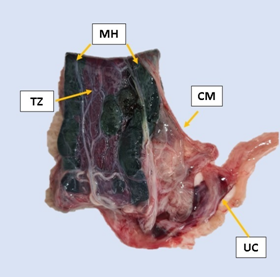
In terms of macroscopic aspect, FARIAS et al. (2023) assessed canine placentas obtained via cesarean section and reported that that all the placentas presented a dark red transfer zone and a dark green marginal hematomas, which is the normal standard for this species (Figures 1 and 2). In 77.7% of the analyzed placentas, some areas in the placental labyrinth had an oval to rounded shape and a dark green color, which corresponds to a marginal hematoma. According to AMBRÓSIO et al. (2009), marginal hematomas arise from small hemorrhages that occur physiologically during the placentation process. Therefore, due to the destruction of the hemoglobin in this extravasated blood, a green pigment called “uteroverdine” is released, which accumulates at the edges of the placenta, giving these areas a dark green color.
Macroscopic analysis of the canine placenta, demonstrating its belt shape, with marginal hematomas (MH), transfer zone (TZ), chorioallantoic membrane (CM) and umbilical cord (UC).
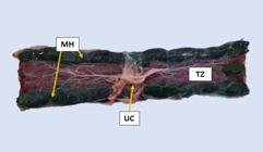
Macroscopic analysis of the canine placenta. Marginal hematoma (MH), transfer zone (TZ) and umbilical cord (UC).
Placental weight and volume
Since the placenta is the sole source of nutrients and oxygen for the fetus, it plays a crucial role in fetal growth rates. In humans, the relationship between birth weight and placental weight has been studied since the 1940s as an index of fetal growth adequacy. In this species, placental thickness and surface area influence its weight, accounting for 36.6% of neonatal birth weight variation (SALAFIA et al., 2008). This finding is relevant as it demonstrated a direct relationship between birth weight and neonatal survival. Therefore, one of the conditions that is most closely related to death in canine neonates, and that can negatively influence their future viability, is low birth weight (TESI et al., 2020a). Moreover, weight is an easily measurable parameter that is less prone to errors than other parameters that also provide information about neonatal well-being (SCHRANK et al., 2019). The instability or loss of weight is considered the best early indicator of neonatal health changes. In humans, universal weight monitoring is a routine clinical practice, as a lack of monitoring newborn weight changes can lead to significant complications such as dehydration (ZIA et al., 2022).
Recent studies have been conducted to elucidate the connection between placental weight and birth weight in dogs. TESI et al. (2020a) reported a correlation between placental weight and birth weight in small and toy dog breeds. The findings revealed that puppies with lower birth weights tended to have a corresponding lower placental weight. SARLI et al. (2021) calculated the placenta/puppy weight ratio (PPR) in neonates born by elective or emergency cesarean section who survived up to 7 days of life (Group 1) and in live-born neonates who died within 7 days postpartum (Group 2). However, no significant differences in the PPR were observed between the two groups. Moreover, they compared PPR between large and small litters, using reference values for litter size from BORGES et al. (2011). For breeds not described by these authors, litter size was classified as either large or small based on the breed’s live weight and the affinity criteria with other breeds within the same morphological group, according to the Fédération Cynologique Internationale. As a result, a significant difference was reported, the median PPR obtained was 10.6%, ranging from 7.24 to 17.16 for large litters. In small litters, the median PPR was 18.78%, ranging from 13.55 to 33.33%. This finding demonstrated that litter size can influence placental weight to appropriately supply all fetuses.
Placental weight and neonatal weight are ways of evaluating placental efficiency (PE). Placental efficiency is an index obtained by dividing the newborn’s birth weight by the weight of its placenta, indicating the number of grams of fetus produced per gram of placenta. One of the factors that alters placental efficiency is the vascularization of the placenta (SARLI et al., 2022). The correlation between placental efficiency, placental vascularization and neonatal viability was assessed via placental and neonatal data from 75 pups. Placental efficiency is correlated with growth rate and neonatal mortality, suggesting that placental efficiency could be a useful parameter in the evaluation of canine neonates (GLORIA et al., 2024).
FARIAS et al. (2023) also reported that neonatal weight was positively correlated with both placental weight and placental volume in a population of various dog breeds and neonates delivered by elective or emergency cesarean section. To date, this study stands out as the only one focusing on placental volume in dogs. In the same work, placental volume demonstrated a positive correlation with placental weight, providing an alternative assessment of the placenta, particularly in cases where weighing the placenta on a scale may not be feasible. In humans, studies have shown that low placental volume and birth weight in infants from mothers with preeclampsia are attributed to reduced uteroplacental blood flow, lower concentrations of growth factors, and potential alterations in placental angiogenesis associated with this condition (RUDGE et al., 2012). Hence, placental volume, similar to weight, has emerged as an effective means of assessing placental health and is directly linked to fetal and neonatal development. However, further studies on this topic in bitches are needed for a more comprehensive understanding of the impact of placental characteristics on immediate and long-term postnatal development (FARIAS et al., 2023).
Ultrasonographic evaluation
Biometry through ultrasound is considered an important technique for morphometric analysis and monitoring the development and growth of maternal organs as well as embryonic and fetal gestational structures, which can be useful in reducing prenatal mortality due to the early diagnosis of disorders that may negatively impact pregnancy (SIMÕES et al., 2020). Therefore, placental measurements are also useful as they allow safe and painless access to pregnancy. The evaluation of placental thickness (PT), for example, has a strong correlation with gestational age and can be assessed from the second third of the bitch’s gestation in breeds of different sizes via the formula: gestational age = (0.021 × PT) - 0.314 (ALMEIDA et al., 2003; MALDONADO et al., 2012). From days 26 to 27 of gestation, the placenta is visualized as a parallel structure under the uterus, and its zonal appearance is identified only from days 29 to 31 of gestation. Furthermore, its edges curve inward from 32 to 53 days of gestation (FROES & GIL, 2019).
Elastographic assessment of the placenta has been performed in humans to characterize structural stiffness and identify anomalies such as preeclampsia, intrauterine growth-restriction, and perinatal death (GUPTA et al., 2008). SIMÕES et al. (2020) conducted elastographic and biometric evaluations of canine placentas and reported that the fetal part of the placenta increased in thickness until 50 days of gestation and then stabilized, whereas the maternal part increased in thickness until 40 days of gestation. With respect to elastography, no intraobservational differences were observed. However, the values and patterns described in this study, such as placental thickening and shear wave velocity (SWV) via elastography, elucidate the structural and biochemical adaptations that occur throughout gestational stages. These findings highlighted the potential of elastography as a valuable technical tool in veterinary obstetrics.
Another way to assess the placenta during the gestational period is through contrast-enhanced ultrasound. Contrast-enhanced ultrasound (CEUS) is a new diagnostic method that allows the evaluation of hemodynamic changes by studying vascular perfusion characteristics. This method assesses tissue perfusion through the intravenous administration of microbubbles, which, due to their size similar to that of red blood cells, diffuse into both the macro and microcirculation. They also enhanced the Doppler signal when associated, improving the quality of the examination (FELICIANO et al., 2019). SILVA et al. (2021) evaluated, through CEUS, the placentas of brachycephalic bitches at three time points: M1 (25 days of gestation - postimplantation), M2 (45 days of gestation - fetogenesis), and M3 (53 days of gestation - final third). They reported that the contrast distribution was homogeneous in the walls of embryonic vesicles and placental tissue and was not detected in embryos/fetuses. Visually, no differences were observed in the enhancement pattern regarding the different moments evaluated, the assessed area of the placenta, or the quantity of fetuses.
Conversely, Doppler ultrasound allows an assessment of maternal-fetal hemodynamics, thus assisting in the identification of possible vascular alterations (DI SALVO et al., 2006; BLANCO et al., 2009). The main parameters evaluated through this examination are the resistance index (RI), pulsatility index (PI), peak systolic velocity (PSV), peak diastolic velocity (PDV), and end-diastolic velocity (EDV). According to NAUTRUP (1998), atypical formation in the Doppler wave in the uteroplacental arteries correlated with an underdeveloped canine fetus, which dies in the first days of life. Thus, there is a need to identify these maternal-fetal alterations as early as possible to institute appropriate treatment, either pre or postpartum. SILVA et al. (2021), in addition to the above mentioned indices (RI, PI, PSV, and PDV), also assessed the mean time of maximum (TaMax) and minimum (TaMin) velocities in pregnant bitches. They reported that the variables PSV, PDV, TaMax, and TaMin significantly increased from M2 to M3, whereas the values of PI and RI decreased from M2 to M3. However, when these two time points were compared, no significant changes were reported. These findings demonstrated that there is a change in fetal-placental hemodynamics to support fetal growth and development.
Microscopic evaluation of the placenta
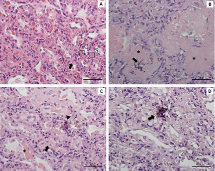
Histologically, the region of the placental labyrinth is composed of three distinct zones. The first is the glandular zone, where projections of endometrial glands are observed. The next zone is the junctional zone, where the fusion of maternal and fetal tissues occurs; this zone contains the terminal part of the lamellar zone, cellular debris, maternal vessels, and glandular secretions. Finally, the lamellar zone is characterized by the presence of cytotrophoblasts and syncytiotrophoblasts (Figure 3A). At the edges of the placental girdle, there are marginal hematomas, which are formed by degeneration of the maternal endothelium (AMBRÓSIO et al., 2009; HYTTEL et al., 2012).

Histological evaluation of the fetal face of canine placentas. A) Normal placental labyrinth, consisting of chorionic lamellae between maternal and fetal vessels, showing a synciotrophoblast (S) and cytotrophoblasts (C), and demonstrating a capillary (arrow); B) Large necrotic area with score 2 (*) and calcification with score 1 (arrow); C) Placental labyrinth with more extensive (*) and less extensive (arrow) area of hemorrhage in score 1, and area of calcification in score 1 (arrowhead); D) Placental labyrinth with focal area of score calcification (arrow). HE staining, 40x magnification.
Studies have demonstrated that some microscopic alterations can be found in the placenta, which can be correlated with perinatal outcomes. TESI et al. (2021b) conducted a comprehensive macroscopic and microscopic evaluation of the placenta and the amnion of full-term neonates, correlating the findings with the birth weights of the puppies. Thus, out of the 69 evaluated placentas, 43 presented necrosis, 36 presented calcification, and 25 presented vascular congestion. However, none of these alterations affected the birth weight of the neonates, similar to the findings of FARIAS et al. (2023), who investigated the presence of necrosis (Figure 3B), calcification (Figure 3C and D), and hemorrhage in canine placentas from cesarean sections. On the other hand, SARLI et al. (2021) reported a negative correlation between the presence of necrosis and neonatal viability in dogs. This type of alteration is more prevalent in the group of animals that died within 7 days of life.
Another study evaluated the biochemical parameters of amniotic fluid (AF) and histological aspects of canine placentas, both from elective (EL) and emergency (EM) cesarean sections. They observed a divergence in the arrangement of collagen fibers in the placenta between the two groups. However, no significant differences were observed concerning the presence or extent of histopathological lesions. A negative correlation (r = -0.609, P = 0.003) was also observed between the AF biochemical results and placental histology, specifically between the glucose concentration and the presence of necrosis in the placental labyrinth. The composition of AF changed due to the influence of the type of cesarean section, possibly caused by prolonged hypoxia in patients with dystocia. However, no significant correlations were found between placental histopathological findings and neonatal viability (GREGHI et al., 2023).
Placental efficiency, defined as the amount of fetal weight produced per gram of placenta, can be compromised due to changes in surface area, thickness of the barrier between maternal and fetal circulations, and/or density and architectural arrangements between maternal and fetal vascularization (MEIRELLES et al., 2017). Hence, adequate blood perfusion in the placenta is clearly related to proper fetal and neonatal development. FARIAS et al. (2023) quantified microvascular density (MVD) in canine placentas and correlated it with placental and neonatal parameters. However, no statistically significant results were found. SARLI et al. (2021) also assessed MVD in placentas from normal neonates (Group 1) and those who died within 7 days of life (Group 2) and reported no significant correlation between the two groups. However, the MVD was negatively correlated with placental efficiency. MVD is commonly evaluated in tumors from humans and other animals, but there are limited data on its assessment in placentas, especially in the canine species (FARIAS et al., 2023).
TESI et al. (2020a) aimed to evaluate the vascular area of the placenta through immunohistochemistry and calculated the vascularization index (VI) and total vascular area (TVA), which were obtained by multiplying the VI by the transfer zone area (TZA). They reported a positive correlation between TVA and birth weight, as well as between TVS and placental weight. However, a negative correlation was observed between VI and birth weight as well as VI and placental weight.
The placenta is responsible for hormonal production, leading to the presence of specific hormone receptors within this organ.However, in the canine species, the placenta does not have the ability to produce steroid hormones such as progesterone and estrogen. Nonetheless, studies indicate that placental tissue possesses receptors for both hormones. The gene expression of estrogen receptor (ERα) and oxytocin receptor (OTR) in the placenta, for example, is modulated according to the gestational phase and labor. ERα is expressed in greater quantities up to 40 days of gestation. OTR is consistently produced during gestation and at the beginning of labor (VEIGA et al., 2009). COSTA et al. (2015) investigated the expression of ERα and progesterone receptor (PR) in placentas from three distinct groups of neonates: placentas obtained from normal delivery (ND), placentas from cesarean section (CS), and placentas from bitches with primary uterine atony (PUA). It was observed that PR expression was nuclear and present only in maternal tissues. Intense PR expression was also observed in the ND and CS placentas. This suggested that, despite a decrease in plasma progesterone concentrations prior to delivery, the availability of receptors persists. Thus, serum progesterone concentration is more important for triggering labor than the availability of placental receptors, according to the literature (VERMEIRSCH et al., 2000). In the same research, COSTA et al. (2015) reported that progesterone receptor expression was evident throughout the placental labyrinth, likely in deciduous cells, as they are the only ones capable of expressing PR. In PUA placentas, the labeling was weak, or there was no labeling for PR.
CONCLUSION
As the placenta plays a crucial role in embryonic/fetal and neonatal development, comprehensive studies are indispensable for a thorough understanding of its function in both physiological and pathological contexts. This knowledge can be effectively applied in obstetric and neonatal clinics within the field of veterinary medicine. Furthermore, various methods of placental assessment have been described to optimize and prioritize immediate and long-term clinical research on placental conditions. However, additional studies in the canine species are essential to establish a clearer understanding of the correlation between placental parameters and perinatal outcomes, considering both macroscopic and microscopic aspects.
In clinical practice, macroscopic aspects are more easily used. Therefore, it is necessary to understand greater depth how they correlate with microscopic aspects and neonatal data. Therefore, soon, a method for macroscopic evaluation of the placenta in the postpartum period could be established as a way of evaluating neonatal health, predicting possible conditions and the identifying of most appropriate treatment methods
ACKNOWLEDGEMENTS
The authors would like to thank Coordenação de Aperfeiçoamento de Pessoal de Nível Superior (CAPES), Brazil, which funded part of this research and Conselho Nacional de Desenvolvimento Científico e Tecnológico (CNPq) for the fellowship granted.
REFERENCES
-
ALMEIDA, A. H. D. et al. Ultrasonographic measurement of placental dimensions normal pregnant Boxers and its correlation to the gestational period. Brazilian Journal of Veterinary Research and Animal Science, v.40, p.136-140, 2003. Available from: <Available from: https://doi.org/10.1590/S1413-95962003000800007 >. Accessed: Aug. 26, 2023. doi: 10.1590/S1413-95962003000800007.
» https://doi.org/10.1590/S1413-95962003000800007.» https://doi.org/10.1590/S1413-95962003000800007 -
AMBRÓSIO, C. E. et al. Development biology in dogs: Behavior of embryo/fetus related to placenta. Revista Brasileira de Reprodução Animal, v.6, p.133, 2009. Available from: <Available from: http://www.cbra.org.br/pages/publicacoes/rbra/download/p133.pdf >. Accessed: Jul. 18, 2023.
» http://www.cbra.org.br/pages/publicacoes/rbra/download/p133.pdf -
BARKER, D. J. P. et al. The surface ffs of the placenta and hypertension in the offspring in later life. International Journal of Developmental Biology, v.54, p.525-530, 2010. Available from: <Available from: https://www.ncbi.nlm.nih.gov/pmc/articles/PMC3923649/pdf/nihms240451.pdf > Accessed Jul. 18, 2023. doi: 10.1387/ijdb.082760db.
» https://doi.org/10.1387/ijdb.082760db.» https://www.ncbi.nlm.nih.gov/pmc/articles/PMC3923649/pdf/nihms240451.pdf -
BISCHOF, P.; MARTELLI, M. Current topic: proteolysis in the penetration phase of the implantation process. Placenta, v.13, n.1, p.17-24, 1992. Available from: <Available from: https://doi.org/10.1016/0143-4004(92)90003-C >. Accessed: Jul. 20, 2023. doi: 10.1016/0143-4004(92)90003-C.
» https://doi.org/10.1016/0143-4004(92)90003-C.» https://doi.org/10.1016/0143-4004(92)90003-C -
BORGES, K. S. et al. Litter size at birth in purebred dogs- A retrospective study of 224 breeds. Theriogenology, v.75, p.911-919, 2011. Available from: <Available from: https://doi.org/10.1016/j.theriogenology.2010.10.034 >. Accessed: Nov. 20, 2023.
» https://doi.org/10.1016/j.theriogenology.2010.10.034 -
BLANCO, P. G. et al. An experimental model to study resistance index and systolic/diastolic ratio of uterine arteries in adverse canine pregnancy outcome. Reproduction in Domestic Animals, v.44, p.164-166, 2009. Available from: <https://doi.org/10.1111/j.1439-0531.2009.01369.x>. Accessed: Sept. 02, 2023.
» https://doi.org/Sept. 02, 2023» https://doi.org/10.1111/j.1439-0531.2009.01369.x -
CASALS, J. B. et al. Pluripotent stem cells proliferation is associated with placentation in dogs. Animal Reproduction, v.17, 2020. Available from: <Available from: https://www.scielo.br/j/ar/a/9Bwv4kN4tTV8HD9WGWVxXPH/?format=pdf⟨=en >. Accessed: Apr. 03, 2024. doi: 10.1590/1984-3143-AR2020-0040.
» https://doi.org/10.1590/1984-3143-AR2020-0040» https://www.scielo.br/j/ar/a/9Bwv4kN4tTV8HD9WGWVxXPH/?format=pdf⟨=en -
COSTA, I. F. et al. Analysis of the expression of receptors for progesterone and estrogen α in canine placentas from eutocia and dystocia. Revista Brasileira de Reprodução Animal, v.39, n.4, p.394-398, 2015. Available from: <Available from: http://cbra.org.br/pages/publicacoes/rbra/v39/n4/p394-398%20(RB587).pdf >. Accessed: Aug. 29, 2023.
» http://cbra.org.br/pages/publicacoes/rbra/v39/n4/p394-398%20(RB587).pdf -
DA SILVA, C. S. et al. Systematic review of the assessment of placental insufficiency. e-Acadêmica, v.3, n.2, p.e4232201-e4232201, 2022. Available from: <Available from: https://www.eacademica.org/eacademica/article/view/201/160 >. Accessed: Aug. 30, 2023. doi: 10.52076/eacad-v3i2.201.
» https://doi.org/10.52076/eacad-v3i2.201» https://www.eacademica.org/eacademica/article/view/201/160 -
DI SALVO, P. et al. Doppler evaluation of maternal and fetal vessels during normal gestation in the bitch. Veterinary Science, v.81, p.382-388, 2006. Available from: <Available from: https://pubmed.ncbi.nlm.nih.gov/16697431/ >. Accessed: Sept. 02, 2023. doi: 10.1016/j.rvsc.2006.03.004.
» https://doi.org/10.1016/j.rvsc.2006.03.004.» https://pubmed.ncbi.nlm.nih.gov/16697431/ -
FARIAS, S. F. S. T. et al. Macro and microscopic characteristics of the placenta and its relationship with the weight and the Apgar score of canine neonates. Theriogenology, v.202, p.21-27, 2023. Available from: <Available from: https://www.sciencedirect.com/science/article/pii/S0093691X23000614?via%3Dihub >. Accessed: Jul. 12, 2023. doi: 10.1016/j.theriogenology.2023.02.018.
» https://doi.org/10.1016/j.theriogenology.2023.02.018.» https://www.sciencedirect.com/science/article/pii/S0093691X23000614?via%3Dihub - FELICIANO, M. A. R. et al. Ultrassonografia em cães e gatos. MedVet, 45, 2019, v.1. 726 p.
-
FOWDEN, A. L. et al. Programming placental nutrient transport capacity. The Journal of Physiology, v.572, n.1, p.5-15, 2006. Available from: <Available from: https://physoc.onlinelibrary.wiley.com/doi/epdf/10.1113/jphysiol.2005.104141 >. Accessed: Jul. 12, 2023. doi: 10.1113/jphysiol.2005.104141.
» https://doi.org/10.1113/jphysiol.2005.104141.» https://physoc.onlinelibrary.wiley.com/doi/epdf/10.1113/jphysiol.2005.104141 -
FROES, T. R.; GIL, E. M. U. Advances of gestational ultrasonography in bitches. Revista Brasileira de Reprodução Animal, v.43, n.2, p.248-260, 2019. Available from: <Available from: http://www.cbra.org.br/portal/downloads/publicacoes/rbra/v43/n2/p248-260%20(RB806).pdf >. Accessed: Jul. 20, 2023.
» http://www.cbra.org.br/portal/downloads/publicacoes/rbra/v43/n2/p248-260%20(RB806).pdf -
GLORIA, A. et al. Does placental efficiency and vascularization affect puppy health? A study in Boxer and Dobermann dogs. Animals, v.14, n.3, p.423, 2024. Available from: <Available from: https://www.mdpi.com/2076-2615/14/3/423 >. Accessed: Jul. 17, 2024. doi: 10.3390/ani14030423.
» https://doi.org/10.3390/ani14030423.» https://www.mdpi.com/2076-2615/14/3/423 -
GUPTA, L. M. et al. Detection of fetal growth restriction with preterm severe preeclampsia: experience at two tertiary centers. American Journal of Perinatology, v.25, p.247-249, 2008. Available from: <Available from: https://www.thieme-connect.de/products/ejournals/abstract/10.1055/s-2008-1075034 >. Accessed: Aug. 28, 2023. doi: 10.1055/s-2008-1075034.
» https://doi.org/10.1055/s-2008-1075034.» https://www.thieme-connect.de/products/ejournals/abstract/10.1055/s-2008-1075034 -
GREGHI, J. R. et al. Emergency cesarean section in dogs: Usefulness of amniotic fluid biochemical parameters and placental morphology as indicators of neonatal viability. Theriogenology, v.211, p.115-124, 2023. Available from: <Available from: https://doi.org/10.1016/j.theriogenology.2023.08.011 >. Accessed: Nov. 20, 2023. doi: 10.1016/j.theriogenology.2023.08.011.
» https://doi.org/10.1016/j.theriogenology.2023.08.011» https://doi.org/10.1016/j.theriogenology.2023.08.011 - HYTTEL, P. et al. Embriologia Veterinária. 2ª ed. Rio de Janeiro: Elsevier, 2012. p.455.
-
MALDONADO, A. L. L. et al. Ultrasound determination of gestational age using placental thickness in female dogs: an experimental study. Veterinary Medicine International, 2012. Article ID 850867. Available from: <Available from: https://doi.org/10.1155/2012/850867 > Accessed: Ago. 28, 2023. doi: 10.1155/2012/850867.
» https://doi.org/10.1155/2012/850867.» https://doi.org/10.1155/2012/850867 - MCGEADY, T. A. et al. Forms of implantation and placentation. In.: MCGEADY, T. A. et al. Veterinary Embryology. 2 ed. Blackwell Publishing, 2017, cap.10, p.78-104.
-
MEIRELLES, M. G. et al. What is the placental and maternal role in equine foal development?. Revista Brasileira de Reprodução Animal, v.41, n.1, p.68-73, 2017. Available from: <Available from: http://www.cbra.org.br/portal/downloads/publicacoes/rbra/v41/n1/p068-073%20(RB637).pdf >. Accessed: Sept. 13, 2023.
» http://www.cbra.org.br/portal/downloads/publicacoes/rbra/v41/n1/p068-073%20(RB637).pdf -
MIGLINO, M. A. et al. The carnivore pregnancy: the development of the embryo and fetal membranes. Theriogenology, v.66, n.6-7, p.1699-1702, 2006. Available from: <Available from: https://www.sciencedirect.com/science/article/pii/S0093691X06001051 >. Accessed: Apr. 03, 2024. doi: 10.1016/j.theriogenology.2006.02.027.
» https://doi.org/10.1016/j.theriogenology.2006.02.027.» https://www.sciencedirect.com/science/article/pii/S0093691X06001051 -
NAUTRUP, C. P. Doppler ultrasonography of canine maternal and fetal arteries during normal gestation. Journal of Reproduction Fertility, v.112, p.301-314, 1998. Available from: <Available from: https://rep.bioscientifica.com/view/journals/rep/112/2/jrf_112_2_012.xml >. Accessed: Jul. 20, 2023. doi: 10.1530/jrf.0.1120301.
» https://doi.org/10.1530/jrf.0.1120301.» https://rep.bioscientifica.com/view/journals/rep/112/2/jrf_112_2_012.xml -
RUDGE, M. V. C. et al. Influence of weight, volume and absolute placental density on the weight of newborns of pregnant women with arterial hypertension or Diabetes Mellitus. Diabetes Clínica, v.5, p.362-366, 2012. Available from: <Available from: https://www.researchgate.net/publication/301298224_Influencia_do_peso_volume_e_densidade_absoluta_da_placenta_no_peso_dos_recem-nascidos_de_gestantes_portadoras_de_hipertensao_arterial_ou_Diabetes_Mellitus >. Accessed: Aug. 30, 2023.
» https://www.researchgate.net/publication/301298224_Influencia_do_peso_volume_e_densidade_absoluta_da_placenta_no_peso_dos_recem-nascidos_de_gestantes_portadoras_de_hipertensao_arterial_ou_Diabetes_Mellitus -
SALAFIA, C. M. et al. Placental characteristics and birthweight. Paediatric and Perinatal Epidemiology, v.22, p.229-239, 2008. Available from: <Available from: https://onlinelibrary.wiley.com/doi/10.1111/j.1365-3016.2008.00935.x >. Accessed: Jul. 15, 2023. doi: 10.1111/j.1365-3016.2008.00935.x
» https://doi.org/10.1111/j.1365-3016.2008.00935.x» https://onlinelibrary.wiley.com/doi/10.1111/j.1365-3016.2008.00935.x -
SARLI, G. et al. Canine Placenta Histological Findings and Microvascular Density: The Histological Basis of a Negative Neonatal Outcome?. Animals, v.11, n.5, p.1418, 2021. Available from: <Available from: https://pubmed.ncbi.nlm.nih.gov/34063427/ >. Accessed: Ago. 28, 2023. doi: 10.3390/ ani11051418.
» https://doi.org/10.3390/ ani11051418.» https://pubmed.ncbi.nlm.nih.gov/34063427/ -
SCHRANK, M. et al. Bodyweight at birth and growth rate during the neonatal period in three canine breeds. Animals, v.10, n.1, p.8, 2019. Available from: <Available from: https://doi.org/10.3390/ani10010008 > Accessed: Sept. 02, 2023. doi: 10.3390/ani10010008.
» https://doi.org/10.3390/ani10010008.» https://doi.org/10.3390/ani10010008 -
SILVA, P. et al. Contrast-enhanced ultrasound evaluation of placental perfusion in brachicephalic bitches. Theriogenology, v.173, p.230-240, 2021. Available from: <Available from: https://www.sciencedirect.com/science/article/pii/S0093691X21002727?via%3Dihub >. Accessed: Sept. 02, 2023. doi: 10.1016/j.theriogenology.2021.08.010.
» https://doi.org/10.1016/j.theriogenology.2021.08.010.» https://www.sciencedirect.com/science/article/pii/S0093691X21002727?via%3Dihub -
SIMÕES, A. P. R. et al. Placental ARFI elastography and biometry evaluation in bitches. Animal Reproduction Science, v.214, p.106289, 2020. Available from: <Available from: https://www.sciencedirect.com/science/article/pii/S0378432019304671?via%3Dihub >. Accessed: Sept. 03, 2023. doi: 10.1016/j.anireprosci.2020.106289.
» https://doi.org/10.1016/j.anireprosci.2020.106289.» https://www.sciencedirect.com/science/article/pii/S0378432019304671?via%3Dihub -
TESI, M. et al. Relationship between placental characteristics and puppies’ birth weight in toy and small sized dog breeds. Theriogenology, v.141, p.1-8, 2020a. Available from: <Available from: https://www.sciencedirect.com/science/article/pii/S0093691X19303577?via%3Dihub >. Accessed: Jul. 12, 2023. doi: 10.1016/j.theriogenology.2019.08.017.
» https://doi.org/10.1016/j.theriogenology.2019.08.017.» https://www.sciencedirect.com/science/article/pii/S0093691X19303577?via%3Dihub -
TESI, M. et al. Gross and histological findings in the canine placenta and amnion at term: What’s normal, abnormal or pathological?. Reproduction in Domestic Animals, v.56, n.5, p.691-702, 2021b. Available from: <Available from: https://onlinelibrary.wiley.com/doi/10.1111/rda.13905 >. Accessed: Jul. 12, 2023. doi: 10.1111/rda.13905.
» https://doi.org/10.1111/rda.13905.» https://onlinelibrary.wiley.com/doi/10.1111/rda.13905 -
VEIGA G. A. L. et al. Endocrinology of pregnancy and parturition in bitches. Revista Brasileira de Reprodução Animal, v.33, p.3-10, 2009. Available from: <Available from: http://www.cbra.org.br/pages/publicacoes/rbra/download/RB154%20Veiga%20pag3-10.pdf >. Accessed: Apr. 10, 2024.
» http://www.cbra.org.br/pages/publicacoes/rbra/download/RB154%20Veiga%20pag3-10.pdf -
VERMEIRSCH, H. et al. Imunohistochemical detections of the estrogen receptor-α and progesterone receptor in the canine pregnant uterus and placental labyrinth. The Anatomical Record, v.260, p.42-50, 2000. Available from: <Available from: https://anatomypubs.onlinelibrary.wiley.com/doi/epdf/10.1002/1097-0185%2820000901%29260%3A1%3C42%3A%3AAID-AR50%3E3.0.CO%3B2-8 >. Accessed: Sept. 02, 2023. doi: 10.1002/1097-0185(20000901)260:1<42::AID-AR50>3.0.CO;2-.
» https://doi.org/10.1002/1097-0185(20000901)260:1<42::AID-AR50>3.0.CO;2-.» https://anatomypubs.onlinelibrary.wiley.com/doi/epdf/10.1002/1097-0185%2820000901%29260%3A1%3C42%3A%3AAID-AR50%3E3.0.CO%3B2-8 -
ZIA, M. T. K. et al. Weight loss monitoring reduces the occurrence of neonatal hypernatremic dehydration in breastfeeding neonates. International Journal of Pediatrics and Adolescent Medicine, v.9, n.1, p.22-26, 2022. Available from: <Available from: https://www.sciencedirect.com/science/article/pii/S2352646721000211 >. Accessed: Apr. 15, 2024. doi: 10.1016/j.ijpam.2021.02.004.
» https://doi.org/10.1016/j.ijpam.2021.02.004.» https://www.sciencedirect.com/science/article/pii/S2352646721000211
Edited by
-
Editors
Rudi Weiblen (0000-0002-1737-9817)Bernardo Garziera Gasperin (0000-0002-3715-2345)