 Ultrasound-guided intraovarian injection in female cats
ABSTRACT:
Chemical sterilization is well established for male dogs and cats and serves as a cost-effective population control method worldwide. However, minimally invasive techniques for ovarian access remain scarce, hindering advances in female sterilization research. This study described and evaluated the feasibility of ultrasound-guided percutaneous intraovarian injection in female cats. Twenty-one mixed-breed females (6 months to 5 years, 1.9-3.5 kg) were sedated and underwent abdominal ultrasonography. With the animals in dorsal recumbency, 0.1 mL of 40% patent blue dye was injected into each ovary under ultrasound guidance. Ovarian dimensions were measured by ultrasonography before and immediately after injection to assess success, and the time required for each procedure was recorded. Following both injections, the animals were anesthetized for exploratory celiotomy to evaluate dye leakage and inspect adjacent organs for damage. Ovariohysterectomy was then performed for macroscopic evaluation of the ovaries. Statistical analyses assessed associations between procedural success and age, weight, parity, and estrous cycle phase. Of 42 ovaries, 24 injections were successful, with the left ovary showing the highest accuracy (58%). The procedure was performed significantly faster on the left ovary (P < 0.05). Technique success was more frequent in animals in estrus and interestrus (P < 0.05). Additionally, ultrasonographic measurements effectively confirmed successful delivery, demonstrated by significant post-injection increases in ovarian dimensions (P < 0.001). These findings showed that female cat ovaries can be accurately assessed for compound deposition without surgical intervention. Furthermore, no complications occurred, confirming the safety and practicality of the technique.
Key words:
minimally invasive technique; non-surgical sterilization; females; reproductive control; feline
RESUMO:
A esterilização química está bem estabelecida para cães e gatos machos. Tem sido utilizada em muitos países como um método de baixo custo para controle populacional. No entanto, métodos minimamente invasivos para acesso ovariano ainda são escassos e são necessários para possibilitar o progresso de estudos que visam inibir a reprodução em gatas e cadelas. O objetivo deste estudo é descrever e avaliar o sucesso da técnica da injeção intraovariana percutânea guiada por ultrassonografia em gatas. Para o estudo, 21 gatas, sem raça definida, com idade entre 6 meses e 5 anos e pesando de 1,9 a 3,5 kg foram submetidas a sedação e ultrassonografia abdominal. Em decúbito dorsal, o ovário esquerdo foi localizado e 0,1 mL de corante azul patente a 40% foi injetado. As medidas de largura, comprimento e diâmetro dos ovários foram avaliadas por ultrassonografia antes e imediatamente após as injeções para identificar o sucesso da injeção. O tempo gasto até a injeção foi coletado. O mesmo procedimento foi repetido para o ovário direito. Subsequentemente, os animais foram submetidos à anestesia geral e a celiotomia exploratória para detectar possíveis extravasamentos do conteúdo injetado e a presença de perfurações ou lacerações de órgãos adjacentes. Foi também realizada a ovariohisterectomia e os ovários foram coletados para avaliação macroscópica. Além disso, idade, peso, número de partos e fase do ciclo estral foram investigados estatisticamente em associação com o sucesso da técnica. A técnica foi realizada em 42 ovários, dos quais 24 injeções foram bem-sucedidas. A maior taxa de sucesso foi no ovário esquerdo, com 58% de acerto. O tempo necessário para executar a técnica diferiu estatisticamente entre os ovários, sendo realizado de forma mais eficiente no ovário esquerdo (P < 0,05). Os resultados indicam que a técnica é viável, particularmente em animais nas fases de estro e interestro (P < 0,05). Além disso, as medidas ovarianas pré e pós-injeção demonstraram a capacidade da ultrassonografia de determinar o sucesso da técnica, evidenciado pelo aumento significativo nas dimensões ovarianas após a injeção (P < 0,001). Estes resultados evidenciam que é possível acessar os ovários de gatas para a deposição de compostos sem intervenção cirúrgica. Contudo, ainda não foram observadas complicações durante o estudo, comprovando assim, a segurança da técnica.
Palavras-chave:
técnica minimamente invasiva; esterilização não cirúrgica; fêmeas; controle reprodutivo; feline
INTRODUCTION
Ultrasound-guided percutaneous injection is a widely used technique for delivering drugs to target organs in both clinical and experimental settings (BERGAMINO et al., 2015; HOEBER et al., 2016; THIN et al., 2020; DAI et al., 2024). By localizing drug delivery, this approach enhances pharmacological efficacy while minimizing systemic side effects (DELANEY et al., 2022). In reproductive medicine, intraovarian injection is a well-established technique in humans (CAKIROGLU et al., 2020), cattle (MALARD et al., 2020), and horses (GRADY et al., 2019), typically performed via transvaginal ultrasonography. More recently, ultrasound-guided percutaneous intraovarian injection has been adopted for experimental animal studies (GOMES et al., 2021; DAI et al., 2024), enabling localized administration of compounds for various purposes (DAI et al., 2024; CAKIROGLU et al., 2020; GOMES et al., 2022).
Chemosterilization has attracted increasing attention as an alternative to surgical castration, especially for addressing the overpopulation of stray animals in developing countries (JANA & SAMANTA, 2007). Non-surgical methods appeal to public health programs because they circumvent many drawbacks associated with surgery, such as high costs, need for operating facilities and specialized personnel and instruments, as well as postoperative care (ADIN, 2011).
Sclerosant-based chemical castration has shown considerable promise for large-scale sterilization and has been extensively studied in males (JANA & SAMANTA, 2011; KARMAKAR & DAS, 2017; LEOCI et al., 2014). In females, CAVALIERI & HAYES (2017) demonstrated ultrasound-guided intraovarian injection of calcium chloride and zinc gluconate in cattle but identified ovarian positioning as a limiting factor. They emphasized the importance of refining ovarian volumetric parameters and preventing reflux of the injected material.
Building on previous research, our study determined that an injection volume of 0.1 mL is appropriate for feline ovaries (SANTOS et al., 2022) and standardized the ultrasound-guided percutaneous intraovarian injection technique in female rats (GOMES et al., 2021). Further studies using varying concentrations of calcium chloride investigated its potential as a sterilizing agent in experimental models (GOMES et al., 2022).
Despite these advances, no minimally invasive technique has been validated for compound delivery into feline ovaries. The present study evaluated the technical feasibility of ultrasound-guided percutaneous intraovarian injection in cats. We also examined whether age, body weight, parity, or estrous cycle phase affect procedural success, and whether ultrasonography can reliably confirm compound deposition based on changes in ovarian dimensions.
MATERIALS AND METHODS
Animals and experimental design
Twenty-one clinically healthy, mixed-breed female cats were selected from the elective ovariohysterectomy waiting list at the Veterinary University Hospital (HUVet) of the Universidade Federal do Pampa (UNIPAMPA). Before surgery, each cat underwent a complete clinical evaluation, including anamnesis, physical examination, complete blood count, serum biochemistry (kidney and liver function), electrocardiography, and owner-signed informed consent.
We included only animals with normal clinical and laboratory findings. Data on age, body weight, and reproductive history were recorded. Cats were categorized as < 2.5 kg or ≥ 2.5 kg; < 1 year or ≥ 1 year of age; and as nulliparous, primiparous, or multiparous. Vaginal cytology was used to classify animals by estrous cycle phase at the time of injection.
The study was carried out between April and October 2023. All ultrasonographic evaluations and ovarian punctures were performed by the same examiner to ensure procedural consistency. Following ultrasound-guided injection, exploratory celiotomy, ovariohysterectomy, and macroscopic ovarian evaluation were performed.
Acclimation and animal preparation
Animals were admitted to the HUVet cattery 12 hours before acclimation. A synthetic feline facial pheromone (Feliway Classic® - Ceva Saúde Animal, Paulínia, Brazil) was sprayed beforehand to reduce stress- and fear-related behaviors. An 8-hour fasting period was instituted with free access to water. To minimize handling reactivity and stress, each cat received 100 mg gabapentin orally 2 hours before handling (HAAFTEN et al., 2017; PANKRATZ et al., 2018). A wide abdominal and trunk trichotomy was then performed.
Anesthetic protocol
All cats underwent the same two-stage anesthetic protocol: (1) for ultrasound-guided intraovarian injection, and (2) for elective ovariohysterectomy. Cephalic vein catheterization was followed by fluid therapy with lactated Ringer’s solution (3 mL/kg/h). Stage 1 consisted of intramuscular (IM) administration of butorphanol (Butorfin® 1%, Vetnil, Louveira, Brazil; 0.15 mg/kg), dexmedetomidine (Dexdomitor®, Zoetis, Campinas, Brazil; 0.0075 mg/kg), and a tiletamine-zolazepam combination (Zoletil®, Virbac, Leopoldina, Brazil; 3 mg/kg) (ROBERTSON et al., 2018; KREISLER et al., 2020). Stage 2 involved induction with intravenous propofol (Cristália, Campinas, Brazil) administered to effect, followed by periglottic instillation of 0.2 mL of 2% lidocaine without vasoconstrictor (Cristália, Campinas, Brazil) onto the arytenoid cartilages to facilitate intubation with a 3.0-3.5 mm diameter tube. Anesthesia was maintained with isoflurane in 100% oxygen, adjusting the minimum alveolar concentration (MAC) between 1 and 2 based on continuous monitoring of each animal. Depth of anesthesia was assessed by absence of medial and lateral palpebral reflexes, ocular rotation, muscle relaxation, and loss of interdigital reflexes. A multiparameter monitor continuously measured respiratory and heart rates, capnography, capnometry, ECG, oscillometric blood pressure (systolic, diastolic, and mean), peripheral oxygen saturation, and esophageal temperature.
Vaginal cytology
Prior to the procedure, a saline-moistened swab was inserted craniodorsally into the caudal vagina (avoiding the vestibule) to collect epithelial cells, which were rolled onto a glass slide and stained with Panótico Rápido® method (Laborclin, Pinhais, Brazil). Slides were examined under a light microscope at 400× magnification (ten fields).
Cells were classified as parabasal, small or large intermediate, and superficial (with or without nuclei) (JOHNSON, 2022). Based on cell proportions, the estrous cycle phase was categorized as proestrus, estrus, interestrus, or diestrus (JOHNSON, 2022).
40% Patent Blue dye solution
Prior to the procedures and under aseptic conditions, the injection solution was prepared by aspirating 0.04 mL of patent blue dye (Bleu Patenté V®, Guerbet, Rio de Janeiro, Brazil) and 0.06 mL of 0.9% NaCl physiological saline solution into a 1 mL syringe fitted with a 22G hypodermic needle (30 x 0.70 mm) (Medix®, Cascavel, Brazil), yielding a 40% solution.
Ultrasonography
Ultrasonographic examinations were performed using a GE Logiq P9 ultrasound system equipped with a 12 MHz multifrequency linear transducer (GE Health Care, São Paulo, Brazil). Ovarian width and length were measured before and after injection. During the procedure, the operator also assessed whether ultrasound could identify leakage (extravasation) of the injected substance into the abdominal cavity. Immediately following injection, the ovarian echotexture was examined in detail, and the abdominal cavity was scanned to identify any free fluid or lesions suggestive of perforation or laceration of adjacent organs. Total procedure time and the time required to inject each ovary (individual procedure time) were also documented.
Standardization of ultrasonography-guided percutaneous intraovarian injection
Cats were placed in dorsal recumbency and positioned in a foam trough for stability. The ventrolateral abdominal region was aseptically prepared using a 2% chlorhexidine solution (Riohex 2%, Rioquímica, São José do Rio Preto, Brazil), followed by 0.5% alcoholic chlorhexidine (PharmaVet, Uruguaiana, Brazil). The transducer was wrapped with plastic film containing contact gel, and 0.5% chlorhexidine gel was applied to the skin.
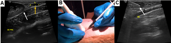
Ovaries were located by scanning caudomedially and caudolaterally to the caudal poles of the kidneys, appearing as hypoechoic, oval-shaped structures (Figure 1A). After initial visualization by the examiner, the procedure was standardized as follows: (1) measure ovarian length and width before injection (M1); (2) position a 1 mL syringe preloaded with 0.1 mL of 40% patent blue dye, attached to a needle, and orient it cranially at a 45° angle to the transducer (Figure 1B); (3) advance the needle into the ovarian parenchyma until reaching the medullary region (Figure 1C); (4) inject the dye; (5) record any ultrasonographic evidence of compound leakage into the abdominal cavity; and (6) measure ovarian length and width again immediately after injection (M2) (Table 1).
(A) Abdominal ultrasonographic image of a female cat showing the caudal pole of the kidney (white arrow) and the left ovary (yellow arrow, LO); (B) A 22-gauge needle attached to a 1 mL syringe positioned in a craniocaudal direction at a 45° angle relative to the transducer; (C) Moment of intraovarian injection, with the white arrow indicating the needle within the ovary.
Ovarian diameter was estimated as D = (length + width)/2, based on the ultrasonographic measurements (Table 1). Additionally, the total and individual procedure times were recorded, with a maximum duration of 1 hour for completion of both injections, beginning with the left ovary due to its more favorable position.
Ovariohysterectomy and macroscopic evaluation
After the ultrasound-guided procedure, an exploratory celiotomy was performed to inspect surrounding organs for perforations and check for staining in the abdominal cavity that might indicate leakage of the injected dye. Ovariohysterectomy was then performed, and the ovaries were collected, sectioned longitudinally, and examined macroscopically. Successful injection was confirmed when the ovarian parenchyma was visibly filled with dye (Figure 2).
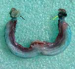
Macroscopic image of feline ovarian parenchyma following ultrasound-guided intraovarian injection of 0.1 mL of 40% patent blue, performed prior to ovariohysterectomy. The black arrow indicates a non-stained ovary, and the yellow arrow a stained ovary.
Statistical analysis
Data were analyzed using IBM SPSS Statistics software (version 20.0). Quantitative variables, such as total and individual procedure times, age, body weight, and ovarian measurements, were tested for normality using the Shapiro-Wilk test. The t-test was used to compare the time required for successful injection between left and right ovaries. The Wilcoxon test assessed pre- and post-injection differences in ovarian size. The mean percentage increase in ovarian dimensions was also calculated for successfully injected ovaries. Associations between injection success and age, body weight, parity, and estrous cycle phase were evaluated using Fisher’s exact test. Statistical significance was defined as P < 0.05.
RESULTS
Of the 42 ovaries, 24 (57.1%) were successfully injected, with 14 of these (58%) corresponding to the left ovary. The mean time required to correctly reach each ovary differed statistically (P < 0.05): 17.36 ± 12.53 minutes for the left ovary and 29.90 ± 12.13 minutes for the right ovary.
Ovaries that were successfully injected showed a significant post-injection increase in size when compared with pre-injection measurements (P < 0.001), representing a 44% increase in mean diameter (Table 1). By contrast, ovaries that were not reached showed no significant differences between the two time points (P > 0.05) (Table 1). After successful injections, ultrasound imaging also revealed changes in echogenicity, characterized by intraparenchymal hypoechoic areas and irregular ovarian contours (Figure 3).
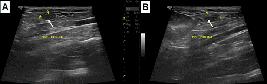
B-mode ultrasonographic images of feline ovaries in the longitudinal plane using a 12 MHz multifrequency transducer. (A) Left ovarian parenchyma (LO) before injection, appearing homogeneous, hypoechoic, and with a regular contour (arrow); (B) Left ovarian parenchyma (LO) after injection, showing intraparenchymal hypoechoic areas (arrow).
Evidence of dye leakage into the abdominal cavity was observed during all procedures and confirmed during exploratory celiotomy, which revealed free dye residues within the peritoneal cavity.
The success of the technique was significantly associated with the estrous cycle phase (P < 0.05). All unsuccessful attempts occurred in cats in diestrus, whereas successful injections, where one or both ovaries were stained, occurred in cats in estrus or interestrus (Table 2), as determined by vaginal cytology.
No significant associations were found between successful ovarian targeting and the animals’ body weight, age, or parity (P > 0.05).
DISCUSSION
The lower success rate observed for the right ovary appears to be primarily influenced by its anatomical position (LÉVY & FONTBONNE, 2007). The longer time required to perform the injection on this side reflects the greater difficulty in visualizing the right ovary in some animals. This ovary lies more cranially than the left and is partially covered by the costal arch (HOLUBIEV et al., 2023), which makes the procedure more technically demanding.
This limitation reduces the practicality of using the proposed technique for intraovarian administration of sclerosing agents for chemical sterilization, since effective bilateral injection is essential. As demonstrated by GOMES et al. (2021) in rats, ovarian stabilization is a key factor for successful targeting; however, despite the relative ease of palpating the abdominal organs, such stabilization was not feasible in this study. From a clinical standpoint, this finding is highly relevant when bilateral delivery is required, since in chemosterilization, the procedure should ideally begin on the right ovary. If this side is successfully targeted, the likelihood of achieving bilateral success increases. Despite the procedural challenge, no intraoperative complications, such as perforations or lacerations of adjacent organs, were observed during celiotomy.
The significant differences between pre- and post-injection ovarian measurements in successfully injected ovaries confirm that ultrasonography is an effective tool to verify correct needle placement and evaluate the practical feasibility of the technique. Similar findings were reported by GOMES et al. (2021) and DAI et al. (2024). In addition to the significant size increase of injected ovaries, post-injection changes in echogenicity, manifested as intraparenchymal hypoechoic areas and irregular ovarian contours, further supported successful intraovarian delivery. These observations are consistent with established ultrasonographic criteria for guided drug injections (KLUNKLIN et al., 2022).
Macroscopic examination of the correctly targeted ovaries corroborated the sonographic findings, reinforcing the conclusion that ultrasound-guided intraovarian injection, even without stabilization, is technically feasible in female cats. Sequential imaging and dimensional measurements can reliably indicate whether the injected compound reached the ovarian parenchyma.
Nevertheless, leakage of dye into the abdominal cavity occurred in all animals, evidenced by diffuse blue staining and peritoneal fluid discoloration. This underscores the need for caution when using sclerosing compounds (chemosterilants), since even minor leakage can cause adjacent tissue damage. The choice and concentration of the injected compound must account for this high risk of diffusion even when the ovaries are correctly targeted, as previously demonstrated by GOMES et al. (2022).
In this study, it was not possible to determine whether leakage resulted from the injection volume, the type of needle used, or excessive penetration beyond the ovarian tissue. The injected volume was considered appropriate, following the protocol proposed by SANTOS et al. (2022) for female cats. Conversely, DAI et al. (2024) reported no leakage or related complications in rats, using the same needle type but calculating injection volume based on organ weight rather than surface area. This suggested that more precise, individualized volume calculations are feasible and should be explored in future studies involving other species. In a pilot study conducted via celiotomy (unpublished data) using the same needle type, dye, and dilution, there were no signs of leakage during injection or needle withdrawal.
The highest rates of successful targeting occurred in female cats in interestrus, followed by estrus and diestrus. This may be related to ovarian enlargement, especially length and volume (CAMARGO et al., 2021), and increased blood flow during interestrus and estrus, which improve visualization and contrast of ovarian echotexture (VERCELLINI et al., 2018). Although, no statistically significant associations were found between success rate and body weight, age, or parity, 75% of successful cases occurred in nulliparous cats under one-year old and weighing more than 2.5 kg. While these findings require confirmation in larger populations, they suggested that selecting individuals with these characteristics may increase the chances of procedural success.
CONCLUSION
Ultrasound-guided intraovarian injection in female cats proved to be a feasible technique. A higher success rate was achieved in animals in the estrus and interestrus phases and when injections were performed in the left ovary. Ultrasonography effectively confirmed procedural success by detecting significant increases in ovarian dimensions, such as diameter, length, and width, and by identifying potential intracavitary leakage during injection. Despite the leakages observed, no perforations or lacerations of adjacent organs occurred, demonstrating that the technique is safe when the injected compound is appropriate.
Ethical considerations
This study was approved by the Animal Use Ethics Committee (CEUA, protocol no. 029/2022) and conducted in accordance with Normative Resolution No. 30/2016 of the National Council for the Control of Animal Experimentation (CONCEA).
ACKNOWLEDGMENTS
This study was partially funded by the Coordenação de Aperfeiçoamento de Pessoal de Nível Superior (CAPES) - Financial Code 001.
REFERENCES
-
ADIN, C. A. Complications of ovariohysterectomy and orchiectomy in companion animals. Veterinary Clinics: Small Animal Practice, v.41, n.5, p.1023-1039, 2011. Available from: <hAvailable from: ttps://www.vetsmall.theclinics.com/article/S0195-5616(11)00095-7/abstract >. Accessed: Feb. 10, 2024. doi: 10.1016/j.cvsm.2011.05.004.
» https://doi.org/10.1016/j.cvsm.2011.05.004.» ttps://www.vetsmall.theclinics.com/article/S0195-5616(11)00095-7/abstract -
BERGAMINO, C. et al. Developing a technique for ultrasound‐guided injection of the adult canine hip. Veterinary Radiology & Ultrasound, v.56, n.4, p.456-461, 2015. Available from: <Available from: https://onlinelibrary.wiley.com/doi/abs/10.1111/vru.12242 >. Accessed: Apr. 19, 2024. doi: 10.1111/vru.12242.
» https://doi.org/10.1111/vru.12242.» https://onlinelibrary.wiley.com/doi/abs/10.1111/vru.12242 -
CAKIROGLU, Y. et al. Effects of intraovarian injection of autologous platelet rich plasma on ovarian reserve and IVF outcome parameters in women with primary ovarian insufficiency. Aging (Albany NY), v.12, n.11, p.10211, 2020. Available from: <Available from: https://pmc.ncbi.nlm.nih.gov/articles/PMC7346073/ >. Accessed: Dec. 14, 2023. doi: 10.18632/aging.103403.
» https://doi.org/10.18632/aging.103403.» https://pmc.ncbi.nlm.nih.gov/articles/PMC7346073/ -
CAMARGO, L. S. et al. Ovarian and Oocyte Morphology During the Spring-Summer and Fall-Winter in Queens (Felis catus) Kept in a Tropical Climate. Topics in Companion Animal Medicine, v.43, p.100510, 2021. Available from: <Available from: https://www.sciencedirect.com/science/article/abs/pii/S1938973621000039 >. Accessed: May, 25, 2024. doi: 10.1016/j.tcam.2021.100510.
» https://doi.org/10.1016/j.tcam.2021.100510.» https://www.sciencedirect.com/science/article/abs/pii/S1938973621000039 -
CAVALIERI, J.; HAYES, L. Examination of the use of intraovarian administration of CaCl2 and zinc gluconate as potential chemosterilants in Bos indicus heifers. Australian veterinary journal, v.95, n.11, p.403-415, 2017. Available from: <Available from: https://onlinelibrary.wiley.com/doi/abs/10.1111/avj.12644 >. Accessed: May, 25, 2024. doi: 10.1111/avj.12644.
» https://doi.org/10.1111/avj.12644.» https://onlinelibrary.wiley.com/doi/abs/10.1111/avj.12644 -
DAI, F. et al. Model construction and drug therapy of primary ovarian insufficiency by ultrasound-guided injection. Stem Cell Research & Therapy, v.15, n.1, p.49, 2024. Available from: <Available from: https://link.springer.com/article/10.1186/s13287-024-03646- >. Accessed: Jun. 21, 2024. doi: 10.1186/s13287-024-03646-y.
» https://doi.org/10.1186/s13287-024-03646-y.» https://link.springer.com/article/10.1186/s13287-024-03646- -
DELANEY, L. J. et al. Making waves: how ultrasound-targeted drug delivery is changing pharmaceutical approaches. Materials Advances, v.3, n.7, p.3023-3040, 2022. Available from: <Available from: https://pubs.rsc.org/en/content/articlehtml/2022/ma/d1ma01197a >. Accessed: Apr. 19, 2024. doi: 10.1039/D1MA01197A.
» https://doi.org/10.1039/D1MA01197A.» https://pubs.rsc.org/en/content/articlehtml/2022/ma/d1ma01197a -
GRADY, S. T. et al. Effect of intra-ovarian injection of mesenchymal stem cells in aged mares. Journal of assisted reproduction and genetics, v.36, p.543-556, 2019. Available from: <Available from: https://link.springer.com/article/10.1007/s10815-018-1371-6 >. Accessed: Apr. 19, 2024. doi: 10.1007/s10815-018-1371-6.
» https://doi.org/10.1007/s10815-018-1371-6.» https://link.springer.com/article/10.1007/s10815-018-1371-6 -
GOMES, E. M. et al. Transcutaneous ultrasound guided intraovarian injection in rats (Rattus norvegicus). Journal of the American Association for Laboratory Animal Science, v.60, n.5, p.502-505, 2021. Available from: <Available from: https://www.ingentaconnect.com/content/aalas/jaalas/2021/00000060/00000005/art00002 >. Accessed: Oct. 13, 2023. doi: 10.30802/AALAS-JAALAS-20-000136.
» https://doi.org/10.30802/AALAS-JAALAS-20-000136.» https://www.ingentaconnect.com/content/aalas/jaalas/2021/00000060/00000005/art00002 -
GOMES, E. M. et al. Calcium chloride diluted in ethanol 95% as female sterilizing agent: effect of transcutaneous delivery in rats. BMC Veterinary Research, v.18, n.1, p.1-9, 2022. Available from: <Available from: https://link.springer.com/article/10.1186/s12917-022-03498-9 >. Accessed: Oct. 13, 2023. doi: 10.1186/s12917-022-03498-9.
» https://doi.org/10.1186/s12917-022-03498-9.» https://link.springer.com/article/10.1186/s12917-022-03498-9 -
HAAFTEN, K. V. A. et al. Effects of a single preappointment dose of gabapentin on signs of stress in cats during transportation and veterinary examination. Journal of the American Veterinary Medical Association, v.251, n.10, p.1175-1181, 2017. Available from: <Available from: https://avmajournals.avma.org/view/journals/javma/251/10/javma.251.10.1175.xml >. Accessed: Sept. 18, 2024. doi: 10.2460/javma.251.10.1175.
» https://doi.org/10.2460/javma.251.10.1175.» https://avmajournals.avma.org/view/journals/javma/251/10/javma.251.10.1175.xml -
HOEBER, S. et al. Ultrasound-guided hip joint injections are more accurate than landmark-guided injections: a systematic review and meta-analysis. British Journal of Sports Medicine, v.50, n.7, p.392-396, 2016. Available from: <Available from: https://bjsm.bmj.com/content/50/7/392.short >. Accessed: Nov. 15, 2023. doi: 10.1136/bjsports-2014-094570.
» https://doi.org/10.1136/bjsports-2014-094570.» https://bjsm.bmj.com/content/50/7/392.short -
HOLUBIEV, O. V. et al. Advantages and difficulties of ultrasound analysis to determine the fertile period in bitches. Theoretical and Applied Veterinary Medicine, v.11, n.2, p.14-20, 2023. Available from: <Available from: https://bulletin-biosafety.com/index.php/journal/article/view/361 >. Accessed: Nov. 19, 2023. doi: 10.32819/2023.11008.
» https://doi.org/10.32819/2023.11008.» https://bulletin-biosafety.com/index.php/journal/article/view/361 -
JANA, K.; SAMANTA, P. K. Sterilization of male stray dogs with a single intratesticular injection of calcium chloride: a dose-dependent study. Contraception, v.75, n.5, p.390-400, 2007. Available from: <Available from: https://www.sciencedirect.com/science/article/abs/pii/S0010782407000832 >. Accessed: Apr. 19, 2024. doi: 10.1016/j.contraception.2007.01.022.
» https://doi.org/10.1016/j.contraception.2007.01.022.» https://www.sciencedirect.com/science/article/abs/pii/S0010782407000832 -
JANA, K.; SAMANTA, P. K. Clinical evaluation of non-surgical sterilization of male cats with single intra-testicular injection of calcium chloride. BMC veterinary research, v.7, n.1, p.1-16, 2011. Available from: <Available from: https://link.springer.com/article/10.1186/1746-6148-7-39 >. Accessed: Apr. 19, 2024. doi: 10.32819/2023.11008.
» https://doi.org/10.32819/2023.11008.» https://link.springer.com/article/10.1186/1746-6148-7-39 -
JOHNSON, A. K. Normal feline reproduction: The queen. Journal of Feline Medicine and Surgery, v.24, n.3, p.204-211, 2022. Available from: <Available from: https://journals.sagepub.com/doi/full/10.1177/1098612X221079706 >. Accessed: Feb. 10, 2024. doi: 10.1177/1098612X221079706.
» https://doi.org/10.1177/1098612X221079706.» https://journals.sagepub.com/doi/full/10.1177/1098612X221079706 -
KARMAKAR, S. N.; DAS, S. K. Chemical and biological sciences chemosterilization induced by intratesticular injection of calcium chloride. Internacional Journal of Pharmaceutical, v.7, n.1, p.25-35, 2017. Available from: <Available from: https://openurl.ebsco.com/EPDB%3Agcd%3A10%3A14111642/detailv2?sid=ebsco%3Aplink%3Ascholar&id=ebsco%3Agcd%3A123219336&crl=c >. Accessed: Jun. 21, 2024.
» https://openurl.ebsco.com/EPDB%3Agcd%3A10%3A14111642/detailv2?sid=ebsco%3Aplink%3Ascholar&id=ebsco%3Agcd%3A123219336&crl=c -
KREISLER, R. E. et al. Use of nalbuphine as a substitute for butorphanol in combination with dexmedetomidine and tiletamine/zolazepam: A randomized non-inferiority trial. Journal of feline medicine and surgery, v.22, n.2, p.100-107, 2020. Available from: <Available from: https://journals.sagepub.com/doi/full/10.1177/1098612X19826715 >. Accessed: Oct. 13, 2023. doi: 10.1177/1098612X1982671.
» https://doi.org/10.1177/1098612X1982671.» https://journals.sagepub.com/doi/full/10.1177/1098612X19826715 -
KLUNKLIN, K. et al. Efficacy and safety of ultrasound-guided caudal epidural steroid injection in patients with low back pain and sciatica. Journal of Back and Musculoskeletal Rehabilitation, v.35, n.2, p.317-322, 2022. Available from: <Available from: https://content.iospress.com/articles/journal-of-back-and-musculoskeletal-rehabilitation/bmr200224 >. Accessed: Nov. 19, 2023. doi: 10.3233/BMR-200224.
» https://doi.org/10.3233/BMR-200224.» https://content.iospress.com/articles/journal-of-back-and-musculoskeletal-rehabilitation/bmr200224 -
LEOCI, R. et al. A dose-finding, long-term study on the use of calcium chloride in saline solution as a method of nonsurgical sterilization in dogs: evaluation of the most effective concentration with the lowest risk. Acta veterinary scandinavica, v.56, n.1, p.1-8, 2014. Available from: <Available from: https://link.springer.com/article/10.1186/s13028-014-0063-1 >. Accessed: Sept. 18, 2024. doi: 10.1186/s13028-014-0063-1.
» https://doi.org/10.1186/s13028-014-0063-1.» https://link.springer.com/article/10.1186/s13028-014-0063-1 -
LÉVY, X.; FONTBONNE, A. Determining the optimal time of mating in bitches: particularities. Rev Bras Reprod Anim, v.31, n.1, p.128-134, 2007. Available from: <Available from: https://citeseerx.ist.psu.edu/document?repid=rep1&type=pdf&doi=f1bb763ff5c473305c29af796e704b00019c22fa >. Accessed: Jun. 21, 2024.
» https://citeseerx.ist.psu.edu/document?repid=rep1&type=pdf&doi=f1bb763ff5c473305c29af796e704b00019c22fa -
MALARD, P. F. et al. Intraovarian injection of mesenchymal stem cells improves oocyte yield and in vitro embryo production in a bovine model of fertility loss. Scientific Reports, v.10, n.1, p.8018, 2020. Available from: <Available from: https://www.nature.com/articles/s41598-020-64810-x >. Accessed: Apr. 19, 2024. doi: 10.1038/s41598-020-64810-x.
» https://doi.org/10.1038/s41598-020-64810-x.» https://www.nature.com/articles/s41598-020-64810-x -
PANKRATZ, K. E. et al. Use of single-dose oral gabapentin to attenuate fear responses in cage-trap confined community cats: a double-blind, placebo-controlled field trial. Journal of feline medicine and surgery, v.20, n.6, p.535-543, 2018. Available from: <Available from: https://journals.sagepub.com/doi/full/10.1177/1098612X17719399 >. Accessed: Jun. 21, 2024. doi: 10.1177/1098612X17719399.
» https://doi.org/10.1177/1098612X17719399» https://journals.sagepub.com/doi/full/10.1177/1098612X17719399 -
ROBERTSON, S. A. et al. AAFP feline anesthesia guidelines. Journal of feline medicine and surgery, v.20, n.7, p.602-634, 2018. Available from: <Available from: https://journals.sagepub.com/doi/full/10.1177/1098612X18781391 >. Accessed: Apr. 19, 2024. doi: 10.1177/1098612X18781391.
» https://doi.org/10.1177/1098612X18781391.» https://journals.sagepub.com/doi/full/10.1177/1098612X18781391 -
SANTOS, B. G. et al. Ovarian morphometry applied to the use of chemosterilant in bitches and queens. Archives of veterinary science, v.27, p.1, 2022. Available from: <Available from: https://www.cabidigitallibrary.org/doi/full/10.5555/20220422786 >. Accessed: Jun. 21, 2024. doi: 10.5380/avs.v27i3.81753.
» https://doi.org/10.5380/avs.v27i3.81753.» https://www.cabidigitallibrary.org/doi/full/10.5555/20220422786 -
THIN, Z. M. et al. Stem cell delivery to kidney via minimally invasive ultrasound-guided renal artery injection in mice. Scientific reports, v.10, n.1, p.7514, 2020. Available from: <Available from: https://www.nature.com/articles/s41598-020-64417-2 >. Accessed: Apr. 19, 2024. doi: 10.1038/s41598-020-64417-2.
» https://doi.org/10.1038/s41598-020-64417-2.» https://www.nature.com/articles/s41598-020-64417-2 -
VERCELLINI, R. et al. Ultrasonographic evaluation of ovarian morphology and blood flow in prepubertal and pubertal cats. Reproduction in Domestic Animals, v.53, p.74-78, 2018. Available from: <Available from: https://onlinelibrary.wiley.com/doi/abs/10.1111/rda.13355 >. Accessed: Apr. 19, 2024. doi: 10.1111/rda.133.
» https://doi.org/10.1111/rda.133.» https://onlinelibrary.wiley.com/doi/abs/10.1111/rda.13355
-
CR-2024-0643.R1
-
DECLARATION OF CONFLICT OF INTEREST
The authors declare that there are no conflicts of interest. The funding agency was not involved in the study design; data collection, analysis, or interpretation; in manuscript preparation; or in the decision to publish the results.
-
BIOETHICS AND BIOSECURITY COMMITTEE APPROVAL
This study was carry out in strict accordance with the recommendations of the Ethics Committee on the Use of Animals of the Universidade Federal do Pampa - UNIPAMPA (CEUA-UNIPAMPA opinion no. 09/2022). And the guidelines of the Brazilian college of Animal Use on Experiments (COBEA).
-
DATA AVAILABILITY STATEMENT
Research data is only available upon request.
-
DECLARATION OF USE OF ARTIFICIAL INTELLIGENCE
We declare that no artificial intelligence resources were used in the writing or development of this manuscript.
Edited by
-
ASSOCIATE EDITOR:
Rudi Weiblen (0000-0002-1737-9817)
-
SCIENTIFIC EDITOR:
Alonso Guedes (0000-0002-7980-9018)
Research data is only available upon request.