 Descriptive skull morphology of Subulo gouazoubira and Mazama nana (Artiodactyla, Cervidae)
ABSTRACT:
Osteology plays an indispensable role in understanding the normal patterns of different species, serving as the foundation for zoological understanding. Mazama nana is well-known in Brazil; however, basic morphological descriptions of the species are scarce, while Subulo gouazoubira, currently revalidated as Mazama gouazoubira, is also prevalent in Brazil and has recently been the subject of various phylogenetic studies. In this respect, in the present study, 19 cervid heads (16 Subulo gouazoubira and three Mazama nana) were osteologically prepared. Next, computed tomographies (CTs) were performed to enhance the assessment of bone accidents, as well as internal structures and foramina through three-dimensional (3D) reconstruction techniques due to the difficult visibility in intact specimens. A comparison was then made between the reconstructions, digital photographs, and CT cross-sections of the skull, which enabled the visualization of anatomical peculiarities and exclusivities, such as nasolacrimal-maxillary fenestra, paranasal sinuses, and thin bone architecture, as well as the unique shape of cranial bones, compared to other species that exhibit a unique and genuine phenotype.
Key words:
Cervidae; description; osteology; skull; tomography
RESUMO:
A osteologia desempenha um papel indispensável para o conhecimento dos padrões de normalidade das diversas espécies, sendo a base para o entendimento zoológico. Mazama nana é bastante conhecido no Brasil, porém descrições morfológicas básicas acerca da espécie são escassas, enquanto Subulo gouazoubira, revalidação atual de Mazama gouazoubira, também é incidente no Brasil e, atualmente, tem sido alvo de diversos estudos filogenéticos. Nesse escopo, no presente estudo, foram preparadas, osteologicamente, 19 cabeças de cervídeos (16 Subulo gouazoubira e três Mazama nana) e, em seguida, tomografias computadorizadas (TCs) foram realizadas a fim de aprimorar as formas de avaliação de acidentes ósseos, além de estruturas internas e forames mediante técnicas de reconstrução em três dimensões (3D) devido a difícil visibilidade em peças intactas. Posteriormente, realizou-se um comparativo entre as reconstruções, fotografias digitais e secções tomográficas transversais do crânio, o qual possibilitou a visualização de peculiaridades e exclusividades anatômicas, como a presença da fenestra nasolacrimomaxilar, detalhamento dos seios paranasais e arquitetura de ossos finos, além do formato exclusivo dos ossos cranianos, em comparação a outras espécies, que apresentam um fenótipo único e genuíno.
Palavras-chave:
Cervidae; crânio; descrição; osteologia; tomografia
INTRODUCTION
Deer (family Cervidae) belong to the order Artiodactyla (BLACK-DECIMA & VOGLIOTTI, 2015), and eight of the known species occur in Brazil: Passalites nemorivagus (MORALES-DONOSO et al., 2023), Blastocerus dichotomus, Odocoileus virginiarus, Ozotoceros bezoarticus, Subulo gouazoubira, Mazama rufa, Mazama bororo and Mazama nana (DUARTE & REIS, 2012). The present study investigated M. nana and S. gouazoubira, both found throughout the country.
Considered the smallest species in the genus, M. nana is part of the biologically and ecologically least known Brazilian deer group (DUARTE & REIS, 2012), which hinders complete taxonomic agreement. This deer species displays nocturnal and crepuscular habits; however, DE ABREU et al. (2009) reported that there is no consistent information about the location and type of vegetation inhabited by this group in South America, but suggested the possibility of a significant presence in mixed ombrophilous forests, as do DUARTE et al. (2011) and DUARTE et al. (2015), besides, there are no studies that specifically address the species’ diet, which is currently in a vulnerable situation (IUCN, 2018).
Previously classified as Mazama gouazoubira, the current nomenclature Subulo gouazoubira, proposed in phylogenetic studies by BERNEGOSSI et al. (2022), validated the genus Subulo, resolving taxonomic uncertainties.
Subulo gouazoubira is also a small species, but larger than Mazama nana, and may be present in all Brazilian biomes (LACERDA et al., 2020), they are considered shy and elusive animals, with diurnal, solitary, and territorial habits, and their diet is based on fruits, flowers, and leaves (BLACK-DECIMA & VOGLIOTTI, 2015; DELLAFIORE & MACEIRA, 2001; DUARTE et al., 2012). Currently, the species is not considered endangered primarily due to its ecological plasticity (DUARTE et al., 2012).
Irrespective of the genus, the skeleton is the basis for vertebrate structure (FARIAS, 2015), calcium reservoirs (LIMA et al., 2021), and protection of the brain and sensory organs located in the head, including cranial (occipital, interparietal, basisphenoid, presphenoid, pterygoid, temporal, parietal, frontal, ethmoid, and vomer), and facial bones (nasal, lacrimal, maxillary, incisive, palatine, zygomatic, mandible, and hyoid) (GETTY et al., 1986; KÖNIG & LIEBICH, 2016).
The knowledge of normal patterns is essential for recognizing alterations that may be present in different diseases and species, as well as serving as the basis for studies related to morphology and phenotypes (POUGH et al., 2006), because the anatomy, like of the skull, demonstrated many interspecific, racial, sexual, and age differences (DYCE et al., 2011). Thus, to guide the imaging diagnosis of wild animals, it is often essential to compare baseline anatomical images contained in the scientific literature (BORTOLINI et al., 2013).
Computed tomography (CT) has been an excellent means of assessing the bony head due to its high image resolution, formulation of thin axial, sagittal, and coronal cross-sections, and excellent detailing of bone features (SCHWARZ et al., 2002), in addition to enabling three-dimensional (3D) reconstruction, which can be applied to bone, vascular, and soft tissue structures (SCHWARZ & SAUNDERS, 2011).
Studying the anatomical structures of fauna for scientific purposes and clinical practice is of utmost importance since it helps in clinical treatments (SHIMMING et al., 2015) and conservation policies, by understanding the adaptive and environmental aspects of species. Nevertheless, according to RIBEIRO et al. (2013), anatomical information regarding the skeleton of wild animals is scarce, which hinders the handling and use of related data. As such, this study conducted an osteological description of Mazama nana and Subulo gouazoubira skulls, using osteological preparations, 3D reconstructions, and cross-sectional CT, in order to achieve detailed and specific anatomical information for the agreed species, which is extremely relevant for zoological studies, since morphology is a well-consolidated basis in taxonomy and general biology, as shown by POUGH et al. (2006).
MATERIALS AND METHODS
In the present study, 19 deer skulls were used, including 16 S. gouazoubira (nine females, seven males) and three M. nana (two females, one male) (Table 1). The specimens used to prepare anatomical pieces were obtained from the Laboratory of Veterinary Anatomy (LANAVET) at the State University of the Midwest (UNICENTRO) and came from animals that had died due to reasons unrelated to the research, after attempts at treatment, or were sent to the CETRAS (Center for the Screening and Rehabilitation of Wild Animals) of UNICENTRO.
Age of the animals used in this study, divided into species, according to a scale of odontological events established from the first to sixteenth month of life.
Osteological preparation of the heads, carried out at LANAVET, involved disarticulation, decapitation, and desiccation of the different parts, as well as thermochemical maceration and general cleaning and aesthetics of the specimens. Finally, the skeletons were atmospherically dried to obtain the desired whiteness, since the skeleton should be both beautiful and functional.
With respect to imaging techniques, the 12-channel “Somatom Emotion Multislice Siemens” tomograph was used for imaging diagnosis, but was later replaced by the 60-channel “Somatom go.Now Siemens Healthineers” model. For this reason, some pieces reconstructed in 3D exhibited better quality, as did the cross-sections, which did not hinder the study in question. Imaging exams were initially performed on females and later on males; thus, the former were assessed by the old tomograph model, and the latter by the new model. The Syngo and Syngo fastView program was used to process and visualize the cross-sections.
For cross-sectional imaging, each skull was placed at the entrance of the device (gantry), producing images along the entire length of the skull and face, in a rostro-caudal direction. Technique adjustment was set at 130kV and 21/45/182mAs, with an acquisition time of approximately 3-5 seconds per piece. The device generated around 170/180 sections per skull, 10 cross-sections of which were analyzed to determine the shape of the bone features, structures, and foramina. Additionally, three-dimensional (3D) reconstructions of the anatomical pieces were compared with images captured by a 12 MP dual-camera with backside illuminated sensor (iPhone).
Thus, the normal morphology of the animals included in this study was presented in several cross-sectional series of each of the 19 osteologically prepared skulls. In the CTs, the marked structures were identified based on the selection of the best-prepared skulls, which were selected to ensure the best identification of the structures.
RESULTS AND DISCUSSION
Due to the morphological proximity of the skulls of both Subulo gouazoubira and Mazama nana species, previously classified as belonging to the same genus (BERNEGOSSI et al., 2022), the converging results obtained were described generally, and species-specific peculiarities were emphasized and correlated throughout the text.
Several comparisons were made with phylogenetically distant groups of deer, given that cranial tomographic studies in vertebrates are scarce (especially in deer) and are scattered on the phylogenetic tree, with the purpose of studying comparative anatomy.
The occipital. It forms the nuchal wall of the skull and is divided into three parts: squamous (dorsal), lateral, and basal (Figure 1), with the emission of condyles that surround the foramen magnum, which are wide, round, and separated, except ventrally (Figure 1), and present the body located cranially to the foramen magnum, joining the basisphenoid bone and forming the caudal segment at the base of the skull (Figure 1). The squamous part contains the external occipital protuberance, but no external occipital crest or internal occipital protuberance. The jugular processes are short, tapering ventrally and sloping medially. When connected to the basisphenoid bone, two discrete muscular tubercles are visualized (Figure 1), while the condylar fossa is quite prominent and houses the hypoglossal foramen (Figure 1). Uniquely, the bone extends cranially on the dorsum of the cranial vault, towards the parietals (Figure 1), denominated the supraoccipital bone (KIERDORF & KIERDORF, 1992).

Dorsal and ventral views of the Subulo gouazoubira skull, 14 months old, received at LANAVET, from the Guarapuava, Parana state (PR) region. (A, C) Digital photograph. (B, D) 3D reconstruction. (A, B) Foramen magnum (1), occipital condyle (2), jugular process (3), muscular tubercle (4), condylar fossa (5), basisphenoid (6), oval foramen (7), vomer (8), temporal process of the zygomatic bone (9), supraorbital foramen (10), zygomatic process of the temporal bone (11), external acoustic meatus (12), orbital part of the lacrimal bone (13), petrooccipital fissure (14), pterygoid hamulus (15), horizontal lamina of the palatine bone (16), third upper molar (17), tympanic bulla (18), major foramina in the pterygoid bone (19), sulcus of the vomer bone (20). (C, D) Nasal process of the incisive bone (1), nasal bone (2), nasolacrimal maxillary fenestra (3), lacrimal bone (4), maxilla (5), frontal bone (6), supraorbital foramen and sulcus (7), palatine fissure (8), palatine process of the incisive bone (9), parietal bone (10), interparietal bone forming a triangular protuberance (11), mandibular coronoid process (12), supraoccipital bone (13).
The sphenoid. Composed of rostral, presphenoid, and caudal components, discrete basisphenoid with slightly prominent wings, perforated rostrally by the optic foramina and caudally by the oval foramen, forming the rostral base of the cranial cavity (BUSSE et al., 2009; KÖNIG & LIEBICH, 2016) (Figure 1).
The parietal and interparietal. Located dorsally and bordered by the frontal and occipital bones rostrally and caudally, respectively, forming a discrete triangular prominence between the right and left parietal bones, constituting most of the dorsolateral portion of the cranial wall (Figure 1). Additionally, the parietal bone is more dorsorostrally projected because the frontal bone is less prominent in the caudal region.
The frontal. Is convex and divided into four parts: orbital, temporal, nasal, and frontal squama. It is located bilaterally between the parietal and nasal bones, housing the frontal sinus (Figure 2) (KÖNIG & LIEBICH, 2016), besides, the bone is thick and contains the zygomatic process of the short, delicate frontal bone (Figure 3). It forms the medial part of the orbital wall and the supraorbital foramen, located dorsomedially to the orbit (Figure 1) in the course of the orbital sulcus (Figure 3), delicate in Mazama nana and Subulo gouazoubira. The base of the antlers, present in M. nana and S. gouazoubira males, projects from the caudodorsal margin of the eye socket, where a bony projection starts from the frontal bone and continues through the parietal bone, occupying an extensive part of the cranial vault.

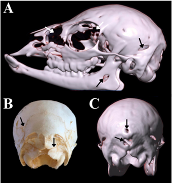
Subulo gouazoubira skull, two months old, received at LANAVET, from the Guarapuava-PR region. (A) Left lateral view through 3D reconstruction, showing incomplete ossification (arrows). (B) Caudal view obtained through digital photography, with evident sutures (arrows). (C) Caudal view through 3D reconstruction, demonstrating areas with fibrous connective tissue and cartilage (arrows).

Left lateral view of the Subulo gouazoubira skull, 14 months old, received at LANAVET, from the Guarapuava-PR region. (A) Image obtained through digital photography. (B) Image obtained through 3D reconstruction. Frontal bone (1), frontal process of the zygomatic bone (2), orbital sulcus (3), squamous part of the temporal bone (4), tympanic part of the temporal bone (4’), temporal crest (5), temporal fossa (6), zygomatic process of the temporal bone (7), articular tubercle (8), retroarticular process (9), jugular process (10), external acoustic meatus (11), parietal bone (12), zygomatic bone (13), frontal process of the zygomatic bone (13’), temporal process of the zygomatic bone (13”), infraorbital foramen (14), molar teeth (15), premolar teeth (16), maxilla (17), lacrimal bone (18), preorbital fossa (19), nasolacrimal maxillary fenestra (20).
The temporal. Divided into three parts (petrous, squamous, and tympanic) that join as the animal develops, it constitutes a part of the ventrolateral wall of the skull, with the squamous part more extensive (Figure 3). It has a slightly prominent temporal crest, and the area rostral to the temporal crest (Figure 3) is underdeveloped, continuing as the temporal fossa (Figure 3), which is also shallow. The zygomatic process of the temporal bone is delicate and thin, and due to the smaller fossa, the articular tubercle, which is continuous with the temporal crest, is almost imperceptible (Figure 3). The retroarticular process is discrete (Figure 3); the acoustic meatus thin and projected (Figure 3); the stylomastoid foramen easily visible in the intact piece, but identification was not possible with the images obtained. The tympanic bulla is round and small (Figure 1) and separated from the occipital bone by the petro-occipital fissure (Figure 1), while the temporal meatus is formed in the temporal bone and located near the articular tubercle.
Maxilla. Flattened dorsoventrally, it does not exhibit facial tuberosity but a discrete facial crest (Figure 4). The opening of the infraorbital foramen is located dorsally to the first molar, and there are alveoli for the insertion of the deciduous upper canine in juveniles (Figure 4), with the alveoli for the molars becoming gradually larger rostrocaudally. The palatine process is thin rostrally (Figure 4).

Cross-sectional CT of the Subulo gouazoubira skull 14 months old, received at LANAVET, from the Guarapuava-PR region. (A) 1/3 of the orbital region, with visualization of the frontal bone (1), zygomatic bone (2), internal orbital part (3), horizontal lamina of the palatine bone (4), mandibular canal (5), frontal sinus (6), caudal portion of the maxillary recess (7), vomer (8), choana (9), ethmoid bone (10), septal process of the frontal bone (11), second molar (12), endoturbinates (13). (B) Diastema region, with visualization of the nasal bone (1), maxilla (2), palatine process of the maxilla (3), cartilaginous nasal septum (4), vomer (5), maxillary sinus (6), mandibular canal (7), palatine sinus (8), dorsal nasal meatus (9), middle nasal meatus (10), ventral nasal meatus (11), dorsal part of the middle nasal concha (12), ventral part of the ventral nasal concha (13). (C) Region of the second premolar and beginning of the nasolacrimal maxillary fenestra, with visualization of the nasal bone (1), opening of the nasolacrimal maxillary fenestra (2), lacrimal bone (3), maxilla (4), nasolacrimal canal (5), infraorbital canal (6), palatine bone (7), vomer (8), perpendicular lamina of the ethmoid bone (9), palatine sinus (10), maxillary recess (11), second premolar (12), mandibular canal (13), mandibular body (14), middle nasal concha (15).
The incisive. This slender bone is in the rostrolateral portion of the skull (GETTY et al., 1986). The nasal process is quite small and does not reach the nasal bone; it is also convex laterally, while the palatine processes are thin and join on both sides (Figure 1).
The ethmoid. The bone is located internally in the skull, making it impossible to visualize in the intact skeleton; however, identification was possible in some CT cross-sections (Figure 4). The endoturbinate bones, one of its components, are projected rostrally up to the margin of the incisive bone, and are more pronounced when compared to other species (GETTY et al., 1986) (Figure 4).
The palatine. Exhibits an extensive and thick horizontal part (Figure 4), occupying about one-third of the hard palate, and does not contain the greater and lesser palatine foramina (Figure 1), because these foramina are usually in the caudal portion of the bone, in the horizontal plate, which is not visible in the deer species under study. However, some foramina are located in the region of the third molar.
The pterygoid. Located caudally to the palatine bone, this delicate bone forms the lateral contour of the choanae, and the pterygoid hamulus has a round and discrete shape (Figure 1).
The nasal. Is a thick, rectangular paired bone, located rostrodorsally (Figure 4), it connects caudally to the frontal bone and is wide mediolaterally (Figure 1). In the deer species assessed, it has a larger diameter in relation to the head size when compared to other ruminants, such as camels (ALSAFY et al., 2014).
The lacrimals. Located near the medial angle of the eye, articulate with the frontal bone (dorsally), zygomatic and maxilla bones (ventrally), and nasal bone (rostrally) (Figure 4) (KÖNIG & LIEBICH, 2016). At the cranial margin of the eye socket, they exhibit two lacrimal foramina, an intrinsic characteristic of the cranial anatomy of deer (HECKEBERG & WORHEIDE, 2019). At the connection to the nasal bone, there is thinning with less ossification, exhibiting cartilage, and between the lacrimal bone and the maxilla is a ventral recession to the nasolacrimal maxillary fenestra (suggested nomenclature of a characteristic structure of Cervidae according to the INTERNATIONAL COMMITTEE ON VETERINARY GROSS ANATOMICAL NOMENCLATURE (2017)) (Figure 3), forming a notch towards the facial crest on the maxilla. At the rostral margin of the lacrimal bone is a fossa that houses the preorbital fossa, the body of the infraorbital gland (Figure 3).
The zygomatic. Long and flattened dorsoventrally, this delicate bone projects laterally (Figure 4) and it connects with the frontal bone (dorsally), maxilla (rostrally), and temporal bone (caudally), forming a considerable part of the bony eye socket. It has some processes that project towards the bone to which they connect, such as the temporal and frontal processes of the zygomatic bone, which are quite discrete and delicate (Figure 3).
The vomer. Located and arranged longitudinally in the skull, it is an unpaired bone (Figures 1 and 4) that extends from the region of the choanae to the nasal cavity. The ventral surface of the nasal septum is connected to the septal grooves of the vomer (HARVEY, 1979), and its caudal end is narrow and forms a wide and distinctive groove on both sides (Figure 1).
The mandible. Exhibits a thin narrow body, and is tapered in its rostral part, as depicted in figure 3. The mental foramen, located in the middle part of the diastema, is easily visible and the coronoid process, located on the ramus of the mandible, is curved, and the condylar process head less pronounced. The mandibular foramen is quite wide, and the mandibular angle is circumscribed, which is more pronounced in the species M. nana, when compared to S. gouazoubira.
The hyoid apparatus (hyoideum). Exhibits a vestigial lingual process, hyoid and ceratohyoid of relatively the same size, while the thyrohyoid is smaller, but the stylohyoid angle is marked.
Cross-Sectional CT. Cross-section assessment provided a better understanding of bone thickness. Thus, the nasal bone exhibits average thickness (Figure 4), extending lateroventrally from the craniorostral portion, connecting to the maxilla (Figure 4), it also contains the nasal sinus, which has a small diameter but is easily visible in the cross-sections. Moreover, internally to the location of this bone is the dorsal nasal meatus (Figure 4), the dorsal nasal concha, and the cartilaginous nasal septum, separating both sides of the rostral portion of the nasal cavity (Figure 4).
The maxilla extends lateroventrally from the nasal bone until it reaches the ventromedial palatine bone (Figure 4) and exhibits average thickness. The presence of the maxillary sinus (Figure 4), maxillary recess (Figure 4), and infraorbital canal, which runs medially through this bone, migrating even more medially as it progresses caudally (Figure 4), can be observed. Internally, it contains the ventral and middle nasal meatuses (Figure 4) and connects with the palatine bone at the palatine sinus. This connects medially to the vomer bone (Figure 4), projecting the perpendicular plate of the ethmoid bone dorsomedially (Figure 4), which is an unpaired bony plate that divides the nasal meatuses. Following caudally, after the maxilla, between the lacrimal and nasal bones, is the nasolacrimal-maxillary fenestra (Figure 4), which does not have a bony connection due to the presence of cartilage and connective tissue in its composition.
The frontal bone follows caudal to the nasal bone (Figure 4), which internally contains the frontal sinus, making it thinner (Figure 4). In the region of the eye socket, medially to the endoturbinate bones (Figure 4), is the septal process of the frontal bone, located in the dorsal internal region (Figure 4), and part of the ethmoid bone in the ventral internal region. Ventral to the ethmoid bone, laterally on both sides of the vomer, is the nasal choanae, and more caudally, the caudal recess of the nasal cavity (Figure 4), the horizontal plate of the palatine bone (Figure 4), which forms its floor, and caudally, in the region of the last molar, the pterygoid bone forms the floor.
The zygomatic bone is in the region of the eye socket, forming it (Figure 4) along with the internal bony part of the orbit and, medially, to this bone is the caudal portion of the maxillary recess, which has a small diameter. At the caudal portion of the frontal sinus, at the level of the pterygoid hamulus, is the internal cranial cavity, with a large diameter, besides, the cranial portion of the basisphenoid bone is prominent, forming part of the cranial floor. From there, the vomer bone reaches its caudal and final region, becoming thinner compared to its previous shape.
In the region of the parietal bone, which is one of the thickest in M. nana and S. gouazoubira, the cranial vault reaches its greatest diameter. In the region where the oval foramen is located, the prevalence of the bone on the cranial vault floor and its composition, medially by the basisphenoid bone, and laterally by the wings of the basisphenoid, can be observed. The temporal bone is also evident in this region, where part of the zygomatic process of the temporal bone and the junction of the mandibular condylar process with the articular tubercle can be seen, as the apex of the mandibular coronoid process is visible in this region.
In the region of the tympanic bulla, the parietal bone forms the largest part of the cranial vault, with the lateral walls formed by the parietal bones. The large extension of the parietal bones in the described species is evident, since they are still prominent in the caudal cranial portion. In the temporal bone, the external opening of the temporal meatus occurs at the level of the bulla, which shows a pronounced apex, the tympanic cavity and the external acoustic meatus, easily identified in cross-sectional views.
The thick occipital bone forms the base of the cranial vault, medially to the petrooccipital fissure and in the most caudal region, passing transversely through the jugular process, one can observe that the occipital bone is curved and medially tilted. Additionally, the consolidation of the tympanic bulla, which becomes more solid, making up the petrous part of the temporal bone, is analyzed. Finally, the occipital condyles are evident and demonstrate the opening of the foramen magnum.
In the anatomical description of the occipital bone for the species assessed, the conformation of the foramen magnum is similar to that of cattle (GETTY et al., 1986), while in carnivores, it is oval-shaped (KARAN et al., 2006). Other characteristics identical to cattle are the widely separated occipital condyles (except ventrally) and the absence of the external occipital crest (GETTY et al., 1986), which differs in cats, otters (Lutra lutra), and badgers (Meles meles), which have an indistinct crest, and dogs, whose crest is prominent. The southern pudu (Pudu puda), a South American deer, exhibits an external occipital protuberance in the squamous part of the occipital bone (WORLD DEER, 2024), as in S. gouazoubira and M. nana, while cats also do not have an internal occipital protuberance. The bone extends dorsorostrally in the cranial vault, as in the capybara; however, the jugular process is short and thin, differing completely from that of the capybara (PEREIRA et al., 2020).
The sphenoid bones are proportionally larger than those of cats and cattle, and shallower compared to cattle (GETTY et al., 1986) and African buffalo (Syncerus caffer caffer). However, the basisphenoid bone is similar to that of capybaras (PEREIRA et al., 2020) and the presphenoid bone is not visible ventrally in the intact skull.
The parietal bones encompass a significant portion of the cranial wall, while in cattle, they are smaller and less evident, and in capybaras occupy a smaller part of the cranial vault (PEREIRA et al., 2020), and there is no interparietal process of the occipital bone.
In cattle and the southern pudu (Pudu puda), the frontal bone is quite broad (KARAN et al., 2005) and prominent (WORLD DEER, 2024), while in M. nana and S. gouzoubira, the bone is less prominent, similar to capybaras (PEREIRA et al., 2020). The zygomatic process of the frontal bone resembles that of otters (Lutra lutra) because they are less developed (KARAN et al., 2005), it is also a convex bone, similar to the frontal bone of a canine. There are lacrimal foramina at the cranial margin of the eye socket, which do not occur in horses and capybaras (PEREIRA et al., 2020), but were described by HECKEBERG & WORHEIDE (2019) in several deer species, as well assupraorbital foramina, similar to Bardhoka sheep (Ovis aries) (GÜNDEMIR et al., 2020), and the southern pudu (Pudu puda) (WORLD DEER, 2024). The number and size of supraorbital foramina vary among deer species and can be used to distinguish groups (HECKEBERG & WORHEIDE, 2019). In cattle, there is the lacrimal sac fossa (GETTY et al., 1986).
With respect to the temporal bone, the temporal crest is poorly developed, as is its rostral area and the temporal fossa. The tympanic bulla is round and small, similar to dogs and cats, but unlike otters, which are dorsoventrally compressed (KARAN et al., 2005), and Axis deer, where it is large and inflated (HECKEBERG & WORHEIDE, 2019). The pampas deer (Ozotoceros bezoarticus) also has small but flattened tympanic bullae (HECKEBERG & WORHEIDE, 2019).
The infraorbital foramen is located dorsally to the first molar, while the configuration of the dental alveoli is the same as that of cattle (GETTY et al., 1986), since both are ruminants. The configuration of the maxilla in the capybara is significantly different from deer assessed, given that it has the maxillary foramen instead of its infraorbital counterpart, and the maxilla is also an evidently irregular bone in this species (PEREIRA et al., 2020). The nasolacrimal-maxillary fenestra (nomenclature suggestion) in this study differs from the nasolacrimal and nasomaxillary fissures present in the skull of the Korean native goat (Capra hircus), since they belong to different families and due to their bone structure (YI et al., 2001), but converges with the preorbital void, a structure located between the lacrimal, maxillary, and nasal bones, described by HECKEBERG & WÖRHEIDE (2019) as an intrinsic characteristic of deer skulls. KENEISENUO et al. (2021) reported the presence of a nasolacrimal fissure for Muntiacus muntjak and Rusa unicolor.
The incisive bone exhibits thin palatine processes that join on both sides, which does not occur in camels (Camelus dromedarius) (EL ALLALI et al., 2017), pigs and ruminants (GETTY et al., 1986). The palatine foramina, located in the region of the 3rd molar of the evaluated deer, also occur in dromedaries. However, the greater palatine foramina are discrete and located on the pterygoid bone (Figure 1), while in dromedaries, they are located between the 2nd and 3rd molar (EL ALLALI et al., 2017).
With a round and discreet shape, the pterygoid hamulus (Figure 1) is different in cattle, where it has a hook shape and is thin and sharp (GETTY et al., 1986), and in capybaras, where it also has this characteristic shape (PEREIRA et al., 2020). M. nana and S. gouazoubira have a larger nasal bone diameter when compared to camels (ALSAFY et al., 2014).The zygomatic bone is long and dorsoventrally flattened, delicate, and thin, as are its processes, unlike capybaras, where it is quite prominent, thick, and broad (PEREIRA et al., 2020). Bardhoka sheep and the southern pudu also have thin delicate bones (GÜNDEMIR et al., 2020; WORLD DEER, 2024). The vomer bone forms the caudoventral bony part of the nasal septum. However, according to GETTY et al. (1986), in cattle, this bone is not directly in contact with the caudal part of the nasal cavity floor and does not divide the choanae because the two blades curve laterally proximally to the choanae, joining the palatine bones, in addition to helping form the basal lamina, which is also observed in S. gouazoubira and M. nana.
In young animals, the bones are separated by thin fibers of fibrous connective tissue or cartilage (Figure 2), providing enough mobility to allow growth (DYCE et al., 2011), including in deer. At the end of growth, ossification extends to the connective tissue and cartilage, making the joints located between most of the bones of the bony head immobile. These are called sutures when associated with connective tissue or synchondroses when associated with cartilaginous tissue (GETTY et al., 1986). This ossification is often incomplete, allowing the contours of the different cranial bones to be distinguished, even in geriatric animals (DYCE et al., 2011).
The CT sections of the present study were obtained from osteologically prepared heads, with no reference to soft tissues, muscles, and structures other than the bones that make up the head. Diagnostic imaging in wild and captive animals provides biologists, zoologists, anatomists, and veterinarians with a better understanding of their anatomy (FARROW, 2009), since CT advances allow for a better anatomical description and clinical assessment of healthy animals, given that some species of wild animals exhibit regions that are not fully interpreted through conventional radiographic techniques.
The results obtained in the present study add to knowledge about the species involved since all necessary techniques and procedures are developed through anatomy. The shape and structure of organisms are unique in every species. In M. nana and S. gouazoubira, the results obtained here showed distinct bone features, shapes, and sizes that will help professionals develop specific methods based on the clinical and anatomical aspects of the animals potentially involved.
CONCLUSION
Based on the results obtained through 3D reconstruction and cross-sectional analysis, it is concluded that computed tomography is an excellent tool in the osteological description of cervid skulls. The anatomical description provided demonstrated that Mazama nana and Subulo gouazoubira skulls have unique characteristics, such as the nasolacrimal-maxillary fenestra, as well as exclusive bone shape when compared to other species, characterizing a distinctive and genuine phenotype.
The results achieved were satisfactory. The cross-sectional images showed very clear details of thin bone, paranasal sinus, and nasal cavity architecture, particularly displaying structures that are not externally visible, as described earlier. As such, this study provides additional information on species of the Cervidae family. Furthermore, research using different imaging diagnostic methods, such as magnetic resonance imaging, combined with the use of intact animal heads, are suggested for more descriptive results for adjacent structures and soft tissues.
ACKNOWLEDGMENTS
To Tomocenter - Guarapuava, which provided time slots for cranial tomographies.
REFERENCES
-
ALSAFY, M. A. M. et al. Computed Tomography and Gross Anatomical Studies on the Head of One-Humped Camel (Camelus dromedarius). The Anatomical Record, v.297, n.4, p.630-642, 2014. Available from: <Available from: https://www.researchgate.net/publication/260876241_Computed_Tomography_and_Gross_Anatomical_Studies_on_the_Head_of_One-Humped_Camel_Camelus_dromedarius >. Accessed: Oct. 27, 2023. doi: 10.1002/ar.22865.
» https://doi.org/10.1002/ar.22865.» https://www.researchgate.net/publication/260876241_Computed_Tomography_and_Gross_Anatomical_Studies_on_the_Head_of_One-Humped_Camel_Camelus_dromedarius -
BERNEGOSSI, A. M. et al. Resurrection of the genus Subulo Smith, 1827 for the gray brocket deer, with designation of a neotype. Journal of Mammalogy, v.104, n.3, p.619-633, 2022. Available from: <Available from: https://academic.oup.com/jmammal/articleabstract/104/3/619/6702645?redirectedFrom=fulltext >. Accessed: Oct. 27, 2023. doi: 10.1093/jmammal/gyac068.
» https://doi.org/10.1093/jmammal/gyac068.» https://academic.oup.com/jmammal/articleabstract/104/3/619/6702645?redirectedFrom=fulltext -
BLACK-DECIMA, P. A.; VOGLIOTTI, A. Mazama gouazoubira The IUCNRed List of threatened species, 15 Jun. 29, 2015. Online. Available from: <Available from: https://www.iucnredlist.org/species/29620/22154584#assessment-information >. Accessed: Oct. 27, 2023.
» https://www.iucnredlist.org/species/29620/22154584#assessment-information -
BORTOLINI, Z. et al. Casuistics of diagnostic imaging in wildlife medicine - from 2009 to 2010. Arquivo Brasileiro de Medicina Veterinária e Zootecnia, v.65, n.4, p.1247-1252, 2013. Available from: <Available from: https://www.scielo.br/j/abmvz/a/gyHrkQh49fpsn4DwxLw4wzt/?lang=pt >. Accessed: Oct. 27, 2023. doi: 10.1590/S0102-09352013000400042.
» https://doi.org/10.1590/S0102-09352013000400042.» https://www.scielo.br/j/abmvz/a/gyHrkQh49fpsn4DwxLw4wzt/?lang=pt -
BUSSE, C. et al. Suspected sphenoid bone osteomyelitis causing visual impairment in two dogs and one cat. Veterinary Ophthalmology, v.12, n.2, p.71-77, 2009. Available from: <Available from: https://onlinelibrary.wiley.com/doi/full/10.1111/j.1463-5224.2008.00678.x. >. Accessed: Oct. 27, 2023. doi: 10.1111/j.1463-5224.2008.00678.x.
» https://doi.org/10.1111/j.1463-5224.2008.00678.x.» https://onlinelibrary.wiley.com/doi/full/10.1111/j.1463-5224.2008.00678.x. -
DE ABREU, C. O. et al. Reproductive characteristics of Brazilian dwarf brocket deer (Mazama nana). Pesquisa Veterinária Brasileira, v.29, n.12, p.993-998, 2009. Available from: <Available from: https://www.scielo.br/j/pvb/a/gMn4MKt7FLNY3VjsrFCjCDx/?lang=pt >. Accessed: Oct. 27, 2023. doi: 10.1590/S0100-736X2009001200007.
» https://doi.org/10.1590/S0100-736X2009001200007.» https://www.scielo.br/j/pvb/a/gMn4MKt7FLNY3VjsrFCjCDx/?lang=pt - DELLAFIORE, C. M.; MACEIRA, N. Los ciervos autóctonos de la Argentina y la acción del hombre. Buenos Aires: Grupo abierto comunicaciones, 2001. 1 ed.
-
DUARTE, J. M. B. et al. Avaliação do risco de extinção do Veado-bororó Mazama nana Hensel, 1872, no Brasil. Biodiversidade Brasileira, v.2, n.3, p.59-69, 2011. Available from: <Available from: https://revistaeletronica.icmbio.gov.br/BioBR/article/view/239/148 >. Accessed: Oct. 27, 2023.
» https://revistaeletronica.icmbio.gov.br/BioBR/article/view/239/148 -
DUARTE, J. M. B. et al. Avaliação do Risco de Extinção do Veado-catingueiro Mazama gouazoubira G. Fischer [Von Waldheim], 1814, no Brasil. Biodiversidade Brasileira, v.2, n.3, p.50-58, 2012. Available from: <Available from: https://www.researchgate.net/publication/259685533_Avaliacao_do_risco_de_extincao_de_Mazama_gouazoubira >. Accessed: Oct. 27, 2023.
» https://www.researchgate.net/publication/259685533_Avaliacao_do_risco_de_extincao_de_Mazama_gouazoubira -
DUARTE, J. M. B. et al. Mazama nana. The IUCN Red List of Threatened Species. 2015. Available from: <Available from: https://www.iucnredlist.org/species/29621/22154379 >. Accessed: Aug. 09, 2024.
» https://www.iucnredlist.org/species/29621/22154379 - DUARTE, J. M. B.; REIS, M. L. Plano de Ação Nacional Para a Conservação dos Cervídeos Ameaçados de Extinção. Brasília: Instituto Chico Mendes de Conservação da Biodiversidade, 2012. n.22.
- DYCE, K. M. et al. Tratado de Anatomia Veterinária. Rio de Janeiro: Elsevier, 2011. 4 ed.
-
EL ALLALI, K. et al. Anatomy of the dromedary head skeleton revisited. Journal of Morphological Sciences, v.34, n.2, p.73-88, 2017. Available from: <Available from: https://www.thieme-connect.de/products/ejournals/abstract/10.4322/jms.100916 >. Accessed: Oct. 27, 2023. doi: 10.4322/jms.100916.
» https://doi.org/10.4322/jms.100916.» https://www.thieme-connect.de/products/ejournals/abstract/10.4322/jms.100916 - FARROW, C. S. Veterinary diagnostic imaging: birds, exotic pets and wildlife. St. Louis: Mosby, 2009. 1 ed.
- GETTY, R. et al. Anatomia dos Animais Domésticos. Rio de Janeiro:Interamericana,1986.
-
GÜNDEMIR, O. et al. A study on morphology and morphometric parameters on skull of the Bardhoka autochthonous sheep breed in Kosovo. Anatomia, Histologia, Embryologia, v.49, n.3, p.365-371, 2020. Available from: <Available from: https://onlinelibrary.wiley.com/doi/10.1111/ahe.12538 >. Accessed: Oct. 27, 2023. doi: 10.1111/ahe.12538.
» https://doi.org/10.1111/ahe.12538.» https://onlinelibrary.wiley.com/doi/10.1111/ahe.12538 -
HARVEY, C. E. The Nasal Septum of the Dog: Is It Visible RadiographicaIly?. Veterinary Radiology & Ultrasound, v.20, p.88-90, 1979. Available from: <Available from: https://onlinelibrary.wiley.com/doi/10.1111/j.1740-8261.1979.tb01185.x >. Accessed: Oct. 27, 2023. doi: 10.1111/j.1740-8261.1979.tb01185.x.
» https://doi.org/10.1111/j.1740-8261.1979.tb01185.x.» https://onlinelibrary.wiley.com/doi/10.1111/j.1740-8261.1979.tb01185.x -
HECKEBERG, N. S.; WÖRHEIDE, G. A. comprehensive approach towards the systematics of Cervidae. PeerJ Preprints, 7:e27618v1, 2019. Available from: <Available from: https://peerj.com/preprints/27618/ >. Accessed: Apr. 01, 2024. doi: 10.7287/peerj.preprints.27618v1.
» https://doi.org/10.7287/peerj.preprints.27618v1.» https://peerj.com/preprints/27618/ - INTERNATIONAL COMMITTEE ON VETERINARY GROSS ANATOMICAL NOMENCLATURE. Nomina Anatomica Veterinaria. Hannover: World Association of Veterinary Anatomists, 2017. 6 ed.
-
IUCN. 2018 IUCN Red List of Threatened Species. 2018. Available from: <Available from: https://www.iucnredlist.org/species/29621/22154379 >. Accessed: Apr. 02, 2024.
» https://www.iucnredlist.org/species/29621/22154379 -
KARAN, M. et al. Comparative Macroanatomical Study of the Neurocranium in some Carnivora. Anatomia, Histologia, Embryologia, v.35, n.1, p.53-56, 2006. Available from: <Available from: https://onlinelibrary.wiley.com/doi/10.1111/j.1439-0264.2005.00659.x >. Accessed: Oct. 27, 2023. doi:10.1111/j.1439-0264.2005.00659.x.
» https://doi.org/10.1111/j.1439-0264.2005.00659.x.» https://onlinelibrary.wiley.com/doi/10.1111/j.1439-0264.2005.00659.x -
KENEISENUO, K. et al. A comparative study on the morphology, radiography and computed tomography of the skull bones of barking deer (Muntiacus muntjak) and sambar deer (Rusa unicolor). Folia Morphologica, v.81, n.1, p.164-174, 2021. Available from: <Available from: https://journals.viamedica.pl/folia_morphologica/article/view/FM.a2021.0015/54796 >. Accessed: Apr. 02, 2024. doi: 10.5603/FM.a2021.0015.
» https://doi.org/10.5603/FM.a2021.0015.» https://journals.viamedica.pl/folia_morphologica/article/view/FM.a2021.0015/54796 -
KIERDORF, U.; KIERDORF, H. Studies on the Parietal Region of the Cervid Skull. Anatomia, Histologia, Embryologia, v.21, n.4, p.373-379, 1992. Available from:<Available from:https://onlinelibrary.wiley.com/doi/10.1111/j.1439-0264.1992.tb00467.x >. Accessed: Oct. 27, 2023. doi: 10.1111/j.1439-0264.1992.tb00467.x.
» https://doi.org/10.1111/j.1439-0264.1992.tb00467.x.» https://onlinelibrary.wiley.com/doi/10.1111/j.1439-0264.1992.tb00467.x - KÖNIG, H. E.; LIEBICH, H. Anatomia dos Animais Domésticos. Porto Alegre: Artmed, 2016. 6 ed.
-
LACERDA, R. G. et al. Trauma in Mazama gouazoubira: aspects of rehabilitation in Ceptas of Cubatão, SP. Brazilian Journal of Animal and Environmental Research, v.3, n.4, p.4171-4178, 2020. Available from: <Available from: https://ojs.brazilianjournals.com.br/ojs/index.php/BJAER/article/view/21722/17320 >. Accessed: Oct. 27, 2023. doi: 10.34188/bjaerv3n4-112.
» https://doi.org/10.34188/bjaerv3n4-112.» https://ojs.brazilianjournals.com.br/ojs/index.php/BJAER/article/view/21722/17320 -
LIMA, L. A. et al. Skeletal anatomical, osteotechnical and osteomontage description of a ocelot jaguar (Leopardus pardalis) Mountain ranged the Sea-Bertioga-São Paulo-SP. Brazilian Journal Of Animal And Environmental Research, v.4, n.4, p.5373-5386, 2021. Available from: <Available from: https://ojs.brazilianjournals.com.br/ojs/index.php/BJAER/article/view/38271 >. Accessed: Oct. 27, 2023. doi: 10.34188/bjaerv4n4-040.
» https://doi.org/10.34188/bjaerv4n4-040.» https://ojs.brazilianjournals.com.br/ojs/index.php/BJAER/article/view/38271 -
MORALES-DONOSO, J. A. et al. Revalidation of Passalites Gloger, 1841 for the Amazon brown brocket deer P. nemorivagus (Cuvier, 1817) (Mammalia, Artiodactyla, Cervidae). Zookeys, v.1167, p.241-264, 2013. Available from: <Available from: https://zookeys.pensoft.net/article/100577/ >. Accessed: Oct. 27, 2023. doi: 10.3897/zookeys.1167.100577.
» https://doi.org/10.3897/zookeys.1167.100577.» https://zookeys.pensoft.net/article/100577/ -
PEREIRA, F. M. A. M. et al. Anatomy of the skull in the capybara (Hydrochoerus hydrochaeris) using radiography and 3D computed tomography. Anatomia, Histologia, Embryologia, v.49, n.3, p.317-324, 2020. Available from: <Available from: https://onlinelibrary.wiley.com/doi/10.1111/ahe.12531 >. Accessed: Oct. 27, 2023. doi: 10.1111/ahe.12531.
» https://doi.org/10.1111/ahe.12531.» https://onlinelibrary.wiley.com/doi/10.1111/ahe.12531 - POUGH, F. H. et al. A vida dos Vertebrados. São Paulo: Atheneu, 2006. 4 ed.
-
RIBEIRO, P. R. Q. et al. Bone anatomy of the pelvic girdle, the thigh and the leg of Myrmecophaga tridactyla (Myrmecophagidae: Pilosa). Biotemas, v.26, n.4, p.153-160, 2013. Available from: <Available from: https://periodicos.ufsc.br/index.php/biotemas/article/view/2175-7925.2013v26n4p153 >. Accessed: Oct. 27, 2023. doi: 10.5007/2175-7925.2013v26n4p153.
» https://periodicos.ufsc.br/index.php/biotemas/article/view/2175-7925.2013v26n4p153 -
SCHWARZ, T. et al. Imaging of the canine and feline temporomandibular joint: A Review. Veterinary Radiology & Ultrasound, v.43, n.2, p.85-97, 2002. Available from: <Available from: https://onlinelibrary.wiley.com/doi/10.1111/j.1740-8261.2002.tb01656.x >. Accessed: Oct. 27, 2023. doi: 10.1111/j.1740-8261.2002.tb01656.x.
» https://doi.org/10.1111/j.1740-8261.2002.tb01656.x.» https://onlinelibrary.wiley.com/doi/10.1111/j.1740-8261.2002.tb01656.x - SCHWARZ, T.; SAUNDERS, J. Special software applications. In: KINNS, J. et al. FROM Veterinary computed tomography. West Sussex: Wiley-Blackwell, 2011, p.67-75.
-
SHIMMING, J. L. et al. Osteology and radiographic anatomy of the hind limbs in Marshdeer (Blastocerus dichotomus). Pesquisa Veterinária Brasileira, v.35, n.12, p.997-1001, 2015. Available from: <Available from: https://www.scielo.br/j/pvb/a/LrFKWZJm8P4K5gbd59BD7cs/?lang=en >. Accessed: Oct. 27, 2023. doi: 10.1590/S0100-736X2015001200009.
» https://doi.org/10.1590/S0100-736X2015001200009.» https://www.scielo.br/j/pvb/a/LrFKWZJm8P4K5gbd59BD7cs/?lang=en -
WORLD DEER. Deer Skull Anatomy. Available from: <Available from: https://worlddeer.org/deer-skull-anatomy/ >. Accessed: Apr. 02, 2024.
» https://worlddeer.org/deer-skull-anatomy/ -
YI, S. J. et al. Comparative Anatomical Study on Fissura Nasolacrimalis and Nasomaxillaris in Skull of the Korean Native Goat (Capra hircus). Anatomia, Histologia, Embryologia, v.30, n.3, p.133-134, 2001. Available from: <Available from: https://onlinelibrary.wiley.com/doi/10.1111/j.1439-0264.2001.t01-1-0282.x >. Accessed: Nov. 01, 2023. doi: 10.1111/j.1439-0264.2001.t01-1-0282.x.
» https://doi.org/10.1111/j.1439-0264.2001.t01-1-0282.x.» https://onlinelibrary.wiley.com/doi/10.1111/j.1439-0264.2001.t01-1-0282.x
BIOETHICS AND BIOSECURITY COMMITTEE APPROVAL
Edited by
-
Editors: Rudi Weiblen (0000-0002-1737-9817) Adriano Carregaro (0000-0002-0580-0467)