 Cone degeneration in two Shih Tzu dogs: clinical and genetic investigation
ABSTRACT:
Two cases of cone degeneration in adult Shih Tzu canine patients are reported herein. Definitive diagnosis was made based on the ophthalmic examination finding, typical clinical signs, and characteristic electroretinography results that demonstrated normal rod function and absence of recordable cone function. Sanger sequencing of the entire coding sequence of the cyclic nucleotide gated channel β3 (CNGB3) gene did not reveal disease causing variants in these dogs. To the authors’ knowledge, this is the first report of clinical, electroretinographic and genetic investigation of cone degeneration in Shih Tzu.
Key words:
cone degeneration; achromatopsia; hemeralopia; day blindness; Shih Tzu; electroretinogram
RESUMO:
O presente trabalho descreve dois casos de degeneração de cones, diagnosticados em cães da raça Shih Tzu. O diagnóstico definitivo foi alcançado com base nos achados oftalmológicos, nos sinais clínicos sugestivos da doença e na eletrorretinografia, cujos resultados demonstraram função normal dos bastonetes e ausência de respostas detectáveis de cones. O sequenciamento da codificação do gene CNGB3 não revelou variantes que sugerissem ser a causa da enfermidade nesses cães. No entendimento dos autores, este é o primeiro relato de caso da degeneração de cones em cães da raça Shih Tzu, descrevendo os aspectos cínicos e eletrorretinográficos, associados à investigação genética.
Palavras-chave:
degeneração de cones. acromatopsia; hemeralopia; cegueira diurna; shih tzu; eletrorretinograma.
INTRODUCTION
Cone degeneration (CD), also known as achromatopsia, hemeralopia or day-blindness, is a genetic autosomal recessive retinal disease characterized by progressive degeneration of cone photoreceptors. In dogs, CD was first described in the Alaskan Malamutes (RUBIN et al., 1967; RUBIN, 1971) and more recently in the Miniature Poodle (RUBIN, 1989), Rhodesian Ridgeback-cross, Chihuahua, Australian Cattledog (HURN et al., 2003), German Wirehaired Pointer (MCELROY, 2006), German Short-Haired Pointer (SIDJANIN et al., 2002) and Labrador Retriever (DIXON, 2016). Clinical signs of CD may be seen as early as 6-8 weeks postnatally and include sudden blindness in bright light followed by gradual vision improvement when returning to dim light. Affected dogs can navigate effectively around obstacles while indoors or under low illumination conditions. No fundic or pupillary light reflex (PLR) abnormalities were seen in these cases (RUBIN, 1971a; RUBIN, 1971b) except for one case reported to have pinpoint-sized pupils in bright day light (HURN et al., 2003). Electroretinographic (ERG) evaluation of affected dogs demonstrates normal rod function and absence of the cone response (RUBIN, 1971).
In the early stage (7 weeks of age), the retina of affected Alaskan Malamutes showed abnormalities in the lamellar discs and inner segments of some cones, though most cones show no abnormalities. With aging, progressive cone degeneration occurs until cones can no longer be identified and the retina is morphologically a pure rod retina. The rods remain completely normal throughout the life of these dogs (AGUIRRE & RUBIN, 1974; AGUIRRE & RUBIN, 1975).
Clinical diagnosis of CD relies on typical clinical signs, i.e., acute blindness in bright light and gradual return of vision in dim light, with the absence of visual changes to the fundus, as well as characteristic ERG changes. An obstacle avoidance course can also be used, under dim and bright light conditions, to aid with the diagnosis of hemeralopia in dogs (GARCIA et al., 2010).
In human patients, the predominant cause of achromatopsia is a mutation in the cyclic nucleotide gated channel β3 (CNGB3) gene (KOHL et al., 2004). SEDDON et al. (2006) found that CD in the Alaskan Malamute presents genetic heterogeneity and that the deletion of CNGB3 is not sufficient to explain all cases of day blindness. It was suggested that the disease is more common than appreciated and may affect additional breeds than is currently known (SEDDON et al., 2006). The purpose of this report is to describe CD in two adult Shih Tzu dogs, a breed in which it has not been previously reported with genetic investigation, and to determine if previously reported mutations in CNGB3 are responsible for the disease in these dogs.
MATERIALS AND METHODS
Case One
A 9-year-old female spayed Shih Tzu dog was presented for evaluation of blindness. The dog was deemed blind by the referring veterinarian during a routine appointment, following ophthalmic examination that revealed negative menace response in both eyes (OU) and a poor performance on obstacle course testing in bright light conditions. Based on the physical examination and the lack of previous report of visual impairment, the referring veterinarian suspected sudden acquired retinal degeneration syndrome as the cause of blindness and referred the patient for further evaluation.
A complete ophthalmic examination including slit lamp biomicroscopy and indirect ophthalmoscopy was performed. Ophthalmic examination under bright room light conditions revealed moderately dilated (~80%), unresponsive pupils and the absence of menace response OU. Dazzle response was also negative under bright light conditions. Chromatic pupillary light reflexes (CPLR) were not tested, once the equipment was unavailable. Both fundi appeared normal and no other abnormalities were noted OU.
Electroretinography was then performed, using the BPM 200 ERG machine (Retinographics Inc, Norwalk, CT, USA). Prior to ERG testing both pupils were dilated using one drop of 0.5% tropicamide and the dog was dark adapted for 20 minutes. The patient was lightly sedated with acepromazine (0.02mg/Kg, subcutaneously) and the electrodes were routinely placed. Briefly, the recording electrode (ERG-jetTM, Universo Plastique SA, Le Cret-du-Locle, Switzerland) was positioned on the cornea following the application of topical anesthesia (Proxymetacaine hydrochloride 0.5%) and 0.5% methylcellulose eye drop was used for conductivity and stabilization of the contact lens electrode. The reference and ground needle electrodes were placed subcutaneously, 3 cm caudal to the lateral canthus and behind the ear, respectively.
Following dark adaptation, rod function was recorded in response to low intensity stimulus (0.0087 cd·s/m2). Combined rod-cone function was then recorded in response to high intensity stimulus (2.75 cd·s/m2). Subsequently, the patient was light adapted for 10 minutes, and cone function was evaluated in response to bright light stimulus (2.75 cd·s/m2) and a flicker stimuli of 30Hz (8.7 cd·s/m2).
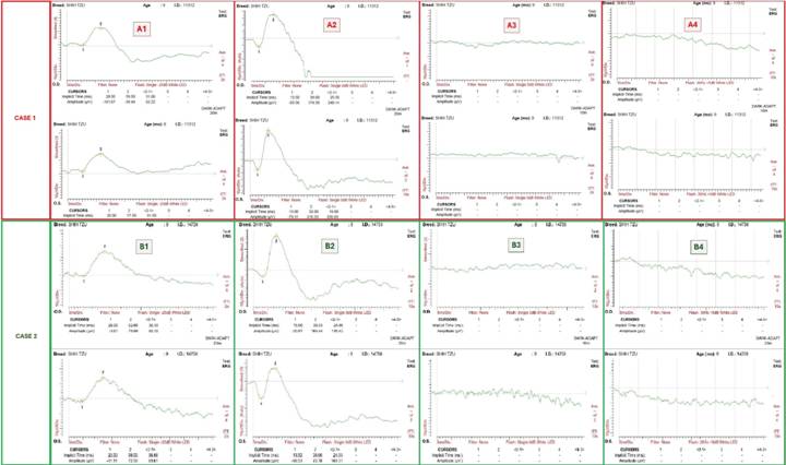
Normal retinal function was recorded OU under scotopic conditions (Figure 1 A1, A2). In contrast, under photopic conditions, no cone activity was recorded (Figure 1 A3, A4). Considering the ERG results, further ophthalmic evaluation was requested, including photopic and scotopic tests. Following dark adaptation (3 minutes), a positive menace response, and dazzle reflex were noted under dim light, OU. In addition, scotopic maze test using 20 cm white plastic obstacles showed positive results. Once the patient was light adapted (3 minutes), the menace response and dazzle reflex were once again negative OU, as occurred in the photopic maze test. Based on the typical clinical signs, the ophthalmic examination findings, and ERG results, a diagnosis of CD was made. When the diagnosis was discussed with the owner, the owner indicated that the dog, throughout his life, was indeed more active at night and slept throughout the day, supporting the diagnosis of day blindness.
Bilateral electroretinographic findings of two shih tzu dogs affected by Cone Degeneration. Case 1: 9-year-old female. Case 2: 9-year-old male shih tzu. Normal rod function as recorded after 20 minutes of dark adaptation, in response to a low (A1, B1) and high (A2, B2) intensity light stimuli from Case 1 and Case 2, respectively. No cone function was evident in response to a bright single flash stimulus (A3, B3) and a flicker (A4, B4), recorded after 10 minutes of light adaptation, from Cases 1 and 2, respectively.
Case Two
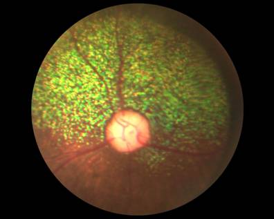
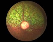
A 9-year-old intact male Shih Tzu dog, with no known relation to the dog presented in case 1, was presented for the evaluation of visual impairment. The owner indicated that the visual impairment was recently noted after their move to a new house. Ophthalmic examination and ERG testing were performed similarly to Case One. The ophthalmic examination revealed negative menace response under photopic conditions OU, and positive under scotopic conditions in the left eye (OS), but negative in the right eye (OD). Pupils were symmetric and revealed mild dilation (~50%). The pupillary light reflex under bright room light was absent OD and partial OS, but present OU under dim room light. CPLR was also not performed for unavailability reasons likewise case 1. Both fundi appeared normal (Figure 2) and no other abnormalities were noted OU. Under bright room light, this dog failed to complete an obstacle course, while under dim light the obstacle course was completed successfully. The obstacles were the same used for case one. Unlike the first case, the photopic and scotopic tests were performed a few days before the ERG. The ERG testing revealed similar results to Case One, with normal retinal function recorded under scotopic condition, but no recordable retinal activity was observed under photopic conditions following 10 minutes of light adaptation.
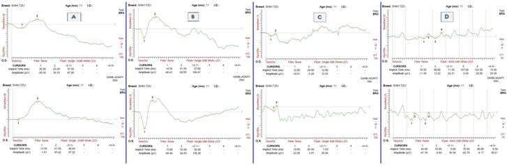
An 11-year-old female shih tzu without visual impairment was used as a control. In the ophthalmic evaluation, both photopic and scotopic tests were positive and there were no signs of any ocular disease. ERG was performed following the same protocol used for cases one and two, revealing normal retinal function under scotopic and photopic conditions (Figure 3), differently from the cases previously presented.

Bilateral electroretinographic findings of a control 11-year-old female shih tzu. Normal rod function as recorded in response to a low (A) and high (B) intensity light stimuli. Normal cone response to a bright single flash stimulus (C) and a flicker (D), recorded after 10 minutes of light adaptation.
Genetic investigation
To determine if previously reported disease-causing mutations in CNGB3 were responsible for CD in the animals reported, genomic DNA was subjected to PCR analysis using overlapping primer pairs spanning the CNGB3 protein coding sequence (Table 1). All exons amplified, indicating that unlike the common Alaskan Malamute allele, CNGB3 was found to be intact (i.e., the previously reported large deletion removing all exons of CNGB3 was not detected) (SIDJANIN et al., 2002). PCR products were subsequently TA cloned using the TOPO PCR2.1 TA cloning kit (Thermo Fisher Scientific, Waltham, MA, USA) via the manufacturer’s protocol. A minimum of 4 colonies were picked for each amplicon and subjected to bidirectional Sanger Sequencing as described previously (TUCKER et al., 2011; TUCKER et al., 2013; BURNIGHT et al., 2017). Sequences were aligned and read using the LaserGene SeqMan Pro alignment software (DNASTAR). A heterozygous variant of unknown significance was identified in exon 4 (c.466 A > G; N149D) of case 2. No other coding sequence variants were identified using the approach described. That said, is important to note that as the genome of the dog is not well annotated, we were unable to develop reliable primer pairs within intronic space that would cover the 3’ and 5’ end of each exon along with their adjacent splice sequences. Because of this inherent limitation, we cannot exclude the fact that disease causing mutations may exist within these regions.
DISCUSSION
Cone degeneration in canine patients was initially described in the Alaskan Malamute a few decades ago (RUBIN, 1967). The cone photoreceptors develop normally in these dogs but show structural abnormalities as early as 7 weeks postnatally. Consequently, massive death of cone photoreceptors occurs, and at 4 years of age the retina is cone free (AGUIRRE & RUBIN, 1974; KOCH & RUBIN, 1971). Nonetheless, clinical signs of moderate-severe visual impairment (“clumsiness”) or blindness in bright light, as well as characteristic changes to the ERG, are evident within the first 8-10 weeks of life (HURN et al., 2003; AGUIRRE & RUBIN, 1975). The mode of inheritance of CD is autosomal recessive and was shown to affect the CNGB3 gene in the Alaskan Malamute (SIDJANIN et al., 2002). In recent years, CD was reported also in the Miniature Poodle (RUBIN, 1989), Rhodesian Ridgeback-cross, Chihuahua, Australian Cattledog (HURN et al., 2003), German Wirehaired Pointer (MCELROY, 2006), German Short-Haired Pointer (SIDJANIN et al., 2002) and Labrador Retriever (DIXON, 2016). Recent study in human patients showed that only 50% of all cases with autosomal recessive achromatopsia are due to a mutation in the CNGB3 gene (KOHL et al., 2004). TANAKA et al. (2015) discovered two novel canine mutations in another gene related to CNG channels (CNGA3), encoding a subunit essential for the generation of light-evoked electrical responses in cone outer segment membranes. Genetic heterogeneity within the Alaskan Malamute populations in Australia was also demonstrated in a study by Seddon and colleagues. They further suggested that CD is more common in dogs than generally appreciated, in part due to the behavioral adaptability of affected dogs, and the need for advanced diagnostic techniques (i.e., ERG) for definitive diagnosis (SEDDON et al., 2006). Genetic heterogeneity also supports the possibility that the prevalence of CD among dog breeds is higher than currently acknowledged. Variability in the severity of achromatopsia was recognized in human patients, and some may show slightly better visual acuity than others, which has been suggested to be phenotypical variations of a single genetic defect (SIMUNOVIC & MOORE, 1998). Similarly, moderate manifestation of clinical signs, as seen in the two cases reported herein and in a previously published case (HURN et al., 2003), together with indoor living and oversight by owners, may also contribute to the underdiagnoses of this disease in dogs.
Clinical signs in these cases included visual impairment and clumsiness in bright light, sleeping during the day, and increased activity during nighttime. Electroretinographic testing demonstrated normal function of rods and abolished cone function, similar to other reports (RUBIN, 1971; HURN et al., 2003; AGUIRRE & RUBIN., 1975; GARCIA et al., 2010). DRAZEK-KUBIAK et al. (2018) reported mild cone activity in CD 3 dogs younger than 6 months. The remaining 9 affected dogs examined had undetectable photopic response, in accordance with our findings. The ocular examination findings were consistent with previous reports and showed no abnormalities to the fundus and other ocular structures, except for moderately dilated pupils and the lack of PLRs in bright light in Case One, and unilaterally absent PLRs in bright light in Case Two. Previously, positive PLRs were reported in CD dogs, and pupil size was normal, or miotic (HURN et al., 2003; DRAZEK-KUBIAK et al., 2018). The reason for the relative mydriasis in our cases and PLR abnormality in case 2 is unclear and could not be explained by our ophthalmic examination findings. It is possible that relative mydriasis or slow PLR are another phenotypic variation of clinical signs in this breed.
The referring veterinarian of the dog in Case One recognized the patient’s visual impairment, and suspected sudden acquired retinal degeneration syndrome as the underlaying cause, as previous visual impairment was not reported by the owner. In Case Two, no visual impairment was recognized by the owner prior to a move to a new house. The definitive diagnosis in these two cases was based on the typical ERG results. The importance of ERG evaluation of patients with visual impairment is well demonstrated in this case and cannot be overstated. Increased awareness for this disease by veterinarians may lead to diagnosis of CD in additional breeds and patients. To the authors’ knowledge, this is the first report of CD in the Shih Tzu associated with genetic investigation. Further studies are needed to identify the genetic mutation leading to achromatopsia in this breed.
ACKNOWLEDGEMENTS
The authors would like to thank Drs. Paulo Lasmar, Luciana Amaral and Fábio Gondim for their support with the clinical management of these cases. The present paper is dedicated to the memory of Dr. Gil Ben-Shlomo, who was essential for the development of this study and the enhancement of veterinary ophthalmology worldwide.
REFERENCES
-
AGUIRRE, G. D.; RUBIN, L. F. Pathology of hemeralopia in the Alaskan malamute dog. Investigative Ophthalmology & Visual Science, v.13, p.231-235, 1974. Available from: <Available from: https://iovs.arvojournals.org >. Accessed: May, 08, 2024. doi: 10.1167/iovs.13-3-231.
» https://doi.org/10.1167/iovs.13-3-231.» https://iovs.arvojournals.org -
AGUIRRE, G. D.; RUBIN, L. F. The electroretinogram in dogs with inherited cone degeneration. Investigative Ophthalmology & Visual Science, v.14, p.840-847, 1975. Available from: <Available from: https://iovs.arvojournals.org >. Accessed: May, 08, 2024. doi: 10.1167/iovs.14-11-840.
» https://doi.org/10.1167/iovs.14-11-840.» https://iovs.arvojournals.org -
BURNIGHT, E. R. et al. Using CRISPR-Cas9 to Generate Gene-Corrected Autologous iPSCs for the Treatment of Inherited Retinal Degeneration. Molecular Therapy, v.25, p.1999-2013, 2017. Available from: <Available from: https://pubmed.ncbi.nlm.nih.gov/28619647/ >. Accessed: May, 08, 2024. doi: 10.1016/j.ymthe.2017.05.015.
» https://doi.org/10.1016/j.ymthe.2017.05.015.» https://pubmed.ncbi.nlm.nih.gov/28619647/ -
DIXON, C. J. Achromatopsia in three sibling Labrador Retrievers in the UK. Veterinary Ophthalmology, v.19, n.1, p.68-72, 2016. Available from: <Available from: https://onlinelibrary.wiley.com/doi/10.1111/vop.12265 >. Accessed: May, 08, 2024. doi: 10.1111/vop.12265.
» https://doi.org/10.1111/vop.12265.» https://onlinelibrary.wiley.com/doi/10.1111/vop.12265 -
DRAZEK-KUBIAK, M. et al. Electroretinographic findings in day-blind dogs. Acta Veterinaria Hungarica, v.66, n.2, p.204-214, 2018. Available from: <Available from: https://pubmed.ncbi.nlm.nih.gov/29958531/ >. Accessed: Oct, 30, 2024. doi: 10.1556/004.2018.019.
» https://doi.org/10.1556/004.2018.019.» https://pubmed.ncbi.nlm.nih.gov/29958531/ -
GARCIA, M. M. Y. et al. Evaluation of a behavioral method for objective vision testing and identification of achromatopsia in dogs. American Journal of Veterinary Research, v.71 p.97-102, 2010. Available from: <Available from: https://avmajournals.avma.org/view/journals/ajvr/71/1/ajvr.71.1.97.xml >. Accessed: May, 08, 2024 doi: 10.2460/ajvr.71.1.97.
» https://doi.org/10.2460/ajvr.71.1.97.» https://avmajournals.avma.org/view/journals/ajvr/71/1/ajvr.71.1.97.xml -
HURN, S. D. et al. Day-blindness in three dogs: clinical and electroretinographic findings. Veterinary Ophthalmology, v.6, p.127-130, 2003; Available from: <Available from: https://onlinelibrary.wiley.com/doi/10.1046/j.1463-5224.2003.00278.x >. Accessed: May, 08, 2024.
» https://onlinelibrary.wiley.com/doi/10.1046/j.1463-5224.2003.00278.x -
KOCH, S. A.; RUBIN L. F. Distribution of cones in the hemeralopic dog. Journal of the American Veterinay Medical Association. v.159, p.1257-1259, 1971. Available from: <Available from: https://real.mtak.hu/81529/1/004.2018.019.pdf >. Accessed: May, 08, 2024.
» https://real.mtak.hu/81529/1/004.2018.019.pdf -
KOHL, S. et al. CNGB3 mutations account for 50% of all cases with autosomal recessive achromatopsia. European Journal of Human Genetics, v.13, p.302-308, 2004. Available from: <Available from: https://www.nature.com/articles/5201269 >. Accessed: Oct, 30, 2024. doi: 10.1038/sj.ejhg.5201269.
» https://doi.org/10.1038/sj.ejhg.5201269.» https://www.nature.com/articles/5201269 -
MCELROY, R. P. Day blindness in a German wirehaired pointer. Veterinary Record, v.158, p.175, 2006. Available from: <Available from: https://sci-hub.se/10.1136/vr.158.5.175-a >. Accessed: May, 08, 2024 doi: 10.1136/vr.158.5.175-a.
» https://doi.org/10.1136/vr.158.5.175-a.» https://sci-hub.se/10.1136/vr.158.5.175-a - RUBIN, L. F. Clinical features of hemeralopia in the adult Alaskan malamute. Journal of the American Veterinary Medical Association, v.158, p.1696-1698, 1971a. PMID: 5314319. Accessed: May, 08, 2024.
- RUBIN, L. F. et al. Hemeralopia in dogs: Heredity of hemeralopia in Alaskan malamutes. American Journal of Veterinary Research, v.28, p.355-357, 1967. Accessed: Oct, 30, 2024.
- RUBIN, L. F. Inherited Eye Diseases in Purebred Dogs. Williams & Wilkins, Baltimore, 1989. ISBN: 0683074520.
-
RUBIN, L. F. Hemeralopia in Alaskan Malamute pups. Journal of the American Veterinary Medical Association, v.158, p.1699-1701, 1971b. Accessed: May, 08, 2024. doi: 10.1353/dog.1971.0035.
» https://doi.org/10.1353/dog.1971.0035. -
SIDJANIN, D. J. et al. Canine CNGB3 mutations establish cone degeneration as orthologous to the human achromatopsia locus ACHM3. Human Molecular Genetics, v.11, p.1823-1833, 2002. Available from: <Available from: https://academic.oup.com/hmg/article/11/16/1823/657255 >. Accessed: May, 08, 2024. doi: 10.1093/hmg/11.16.1823.
» https://doi.org/10.1093/hmg/11.16.1823.» https://academic.oup.com/hmg/article/11/16/1823/657255 -
SEDDON, J. M et al. Genetic heterogeneity of day blindness in Alaskan Malamutes. Animal Genetics, v.37, p.407-410, 2006. Available from: <Available from: https://onlinelibrary.wiley.com/doi/abs/10.1111/j.1365-2052.2006.01484.x >. Accessed: May, 08, 2024. doi: 10.1111/j.1365-2052.2006.01484.x.
» https://doi.org/10.1111/j.1365-2052.2006.01484.x.» https://onlinelibrary.wiley.com/doi/abs/10.1111/j.1365-2052.2006.01484.x -
SIMUNOVIC, M. P.; MOORE, A. T. The cone dystrophies. Eye (London), v.12(Pt 3b), p.553-565, 1998. Available from: <Available from: https://www.nature.com/articles/eye1998145# >. Accessed: May, 08, 2024. doi: 10.1038/eye.1998.145.
» https://doi.org/10.1038/eye.1998.145.» https://www.nature.com/articles/eye1998145# -
TANAKA, N. et al. Canine CNGA3 gene mutations provide novel insights into human achromatopsia-associated channelopathies and treatment. PLoS One, v.10. 2015. Available from: <Available from: https://pmc.ncbi.nlm.nih.gov/articles/PMC4583268/ > Accessed: May, 08, 2024 doi: 10.1371/journal.pone.0138943.
» https://doi.org/10.1371/journal.pone.0138943.» https://pmc.ncbi.nlm.nih.gov/articles/PMC4583268/ -
TUCKER, B. A. et al. Exome sequencing and analysis of induced pluripotent stem cells identify the cilia-related gene male germ cell-associated kinase (MAK) as a cause of retinitis pigmentosa. Proceedings of the National Academy of Sciences of the United States of America, v.108, p.E569-576, 2011. Available from <Available from https://pubmed.ncbi.nlm.nih.gov/21825139/ >. Accessed: May, 08, 2024. doi: 10.1073/pnas.1108918108.
» https://doi.org/10.1073/pnas.1108918108.» https://pubmed.ncbi.nlm.nih.gov/21825139/ -
TUCKER, B. A. Patient-specific iPSC-derived photoreceptor precursor cells as a means to investigate retinitis pigmentosa. Elife, v.2, e00824, 2013. Available from: <Available from: https://pubmed.ncbi.nlm.nih.gov/23991284/ >. Accessed: May, 08, 2024. doi: 10.7554/elife.00824.
» https://doi.org/10.7554/elife.00824.» https://pubmed.ncbi.nlm.nih.gov/23991284/
Edited by
-
Editors
Rudi Weiblen (0000-0002-1737-9817)Alexandre Pinto Ribeiro (0000-0002-8922-2886)