 SPINAL OSTEOCHONDROMAS IN PEDIATRIC PATIENTS: CASE SERIES AND LITERATURE REVIEW
ABSTRACT
Objective: To evaluate a pediatric series of spinal osteochondromas (OCs), with a literature review and emphasis on management.
Methods: Multicentric retrospective review of growing patients with spinal OCs. In statistical analysis non-parametric Wilcoxon Rank tests and multivariable Pearson correlation were used.
Results: We collected 13 patients (14 OCs), 8 males/5 females, with a mean age at diagnosis of 11.5 years (2-17 years); cervical tumors predominated. Three cases were associated with Multiple Osteochondromatosis (MO); 7 were intracanal (IC) and 7 with extracanal exophytic growth (EC). Four patients (5 OCs) were asymptomatic at diagnosis; the others were predominantly characterized by mass and/or pain; 3 presented with neurological deficit. Mean tumor volume: 31.50 cm3, with a difference between EC (37.75 cm3) and IC (19 cm3). The IC showed significant percentages of canal occupation (mean 53.9%), being higher in cases with deficit (57.4%). Eleven of the 13 patients underwent surgery, mostly with marginal resections; one had spontaneous disappearance. Mean follow-up: 10.5 years (6 months-26 years). Two cases with neurological deficits showed recovery. Two spinal deformities and one hip subluxation due to neurological impairment appeared during follow-up. There were no recurrences, only one case of residual tissue due to incomplete resection.
Conclusions: 1) Consider resecting bulky asymptomatic EC OCs due to possible malignancy. 2) IC OCs that enlarge or become symptomatic should be resected regardless of their volume. 3) Closely control patients with OM due to the possibility of developing symptomatic IC OCs. 4) Avoid intralesional ablation due to increased risk of recurrence or residue. Marginal resection is adequate, but a wide resection may be necessary. Level of Evidence IV; Case Series.
Keywords:
Osteochondromas; Spine; Children; Surgery.
RESUMO
Objetivo: Avaliar uma série pediátrica de osteocondromas espinhais (OCs) com revisão de literatura e ênfase no tratamento.
Métodos: Revisão retrospectiva multicêntrica de pacientes em crescimento com OCs espinhais. Nas avaliações estatísticas utilizaram-se testes não paramétricos de Wilcoxon Rank e a correlação multivariada de Pearson.
Resultados: Foram coletados 13 pacientes (14 OCs), 8 homens/5 mulheres, com idade média ao diagnóstico de 11.5 anos (2 - 17 anos); predominaram as cervicais. Três casos associados à Osteocondromatose Múltipla (OM); 7 eram com crescimento intracanal (IC) e 7 eram com crescimento exofítico extracanal (EC). Quatro pacientes (5 OCs) assintomáticos ao diagnóstico; nos demais predominou a presença de massa e/ou dor; 3 estreou com déficit neurológico. Volume tumoral médio: 31,50 cm3, com diferença entre EC (37,75 cm3) e IC (19 cm3). Os IC apresentaram percentuais significativos de ocupação do canal (média 53,9%), sendo maior nos casos com déficit (57,4%). Onze dos 13 pacientes foram submetidos à cirurgia, a maioria com ressecções marginais; um teve desaparecimento espontâneo. Acompanhamento médio: 10,5 anos (6 meses - 26 anos). Dois casos com déficit neurológico apresentaram recuperação. No início tardio, houve duas deformidades na coluna e uma subluxação neurológica do quadril. Não houve recidivas, apenas um caso de resíduo devido à ressecção incompleta.
Conclusões: 1) Considerar a ressecção de OCs EC assintomáticos volumosos devido à possível malignidade. 2) Os OCs IC que crescem ou apresentam sintomas devem ser ressecados independentemente do seu volume. 3) Monitoramento rigoroso dos pacientes com OM para verificar a possibilidade de apresentar OCs IC sintomáticos. 4) Evitar ablação intralesional devido ao risco de recorrência ou resíduo. A ressecção marginal é adequada, mas uma ressecção ampla pode ser necessária. Nível de Evidência IV; Série de Casos.
Descritores:
Osteocondromas; Coluna; Crianças; Cirurgia.
RESUMEN
Objetivo: Evaluar una serie pediátrica propia de Osteocondromas (OCs) espinales con revisión de literatura y énfasis en el manejo.
Métodos: Revisión retrospectiva multicéntrica de pacientes en crecimiento con OCs raquídeos. Análisis estadístico con tests no paramétricos de Wilcoxon Rank y correlación multivariable de Pearson.
Resultados: Recolectamos 13 pacientes (14 OCs), 8 varones/5 mujeres, con edad promedio al diagnóstico de 11,5 años (2 - 17 años); predominaron los cervicales. Tres casos asociados a Osteocondromatosis Múltiple (OM); 7 eran intracanal (IC) y 7 de crecimiento exofítico extracanal (EC). Cuatro pacientes (5 OCs) asintomáticos al diagnóstico; en los otros predominó la presencia de masa y/o dolor; 3 debutaron con déficit neurológico. Volumen tumoral promedio: 31,50 cm3, con diferencia entre EC (37,75 cm3) e IC (19 cm3). Los IC mostraron porcentajes importantes de ocupación del conducto (promedio 53,9%), siendo mayor en los casos con déficit (57,4%). Once de 13 pacientes fueron operados, mayoritariamente con resecciones marginales; uno tuvo desaparición espontánea. Seguimiento promedio: 10,5 años (6 meses - 26 años). Dos casos con déficit neurológico mostraron recuperación. Tardíamente hubo 2 deformidades espinales y una subluxación neurológica de cadera. No hubo recidivas, solo un caso de residuo por resección incompleta.
Conclusiones: 1) Considerar resecar OCs voluminosos EC asintomáticos por la posible malignización. 2) Los OCs IC que crecen o dan síntomas deben resecarse sin importar su volumen. 3) Seguir los pacientes con OM estrechamente por la posibilidad de desarrollar OCs IC sintomáticos. 4) Evitar la ablación intralesional por riesgo de recidiva o residuo. La resección marginal es adecuada, pero una resección amplia puede necesitarse. Nivel de Evidencia IV; Serie de Casos.
Descriptores:
Osteocondromas; Columna; Niños; Cirugía.
INTRODUCTION
Osteochondromas (OCs) are the most common benign bone tumors of the long bones. Only 3-4% are located in the spine, although this rate may increase significantly in cases of Multiple Osteochondromatosis (MO).1,2 Most spinal OCs are located in the cervical spine, accounting for approximately 50%.2
The aim of this study is to present a case series of spinal OCs in a pediatric population, analyze the most relevant characteristics, assess the treatments performed and their medium-term outcomes, and conduct a literature review.
MATERIALS AND METHODS
This is a retrospective, multicenter cohort study. It included patients under 18 years of age or skeletally immature (based on bone age) at the time of diagnosis, with spinal OCs, covering the period from January 1996 to February 2025 (29 years and 2 months). The variables collected and analyzed for each case are listed in Chart 1.
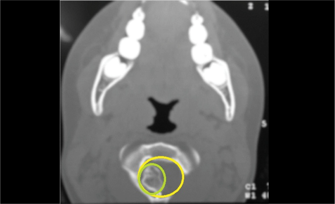
For preoperative volumetric measurement based on imaging studies, an approximation to the actual volume was calculated using the best available imaging modality, either computed tomography (CT) or magnetic resonance imaging (MRI). The tumor shape was assimilated to the geometric solid most closely resembling the best image, and the volume was estimated using mathematical formulas (Figure 1). To calculate the percentage of canal occupation, the canal area was measured similarly, and the respective ratio was computed. (Figure 2)
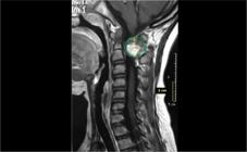
Case 3 in the series (Table 1); intracanal spinal osteochondroma (originating from the posterior arch of C1). T2 sagittal MRI using the approximate volumetric measurement method (6.28 cm3 volume).
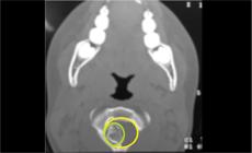
Axial CT scan of the same case as in the previous figure, showing the osteochondroma originating from the posterior arch of C1, and how to perform comparative measurements of the tumor and the canal.
Statistical analyses were performed using non-parametric Wilcoxon Rank tests and multivariable Pearson correlation; alpha was set at ≤ 0.05. Statistical processing was conducted using SPSS version 17.
As a multicenter observational study, each Ethics Committee of the participating institutions confirmed that no formal approval was required. However, all parents, guardians, or the patients themselves (depending on age, context, and applicable legislation) signed informed consent forms authorizing participation in the study and publication of their data and photographs, provided patient privacy was protected.
RESULTS
A total of 13 patients from three centers were included, with 14 spinal osteochondromas (OCs) - one patient had two tumors in different locations. The cohort comprised 8 males and 5 females, with a mean age at diagnosis of 11 years and 5 months (range: 2-17 years) (Table 1). Three patients had an associated condition (Multiple Osteochondromatosis - MO).
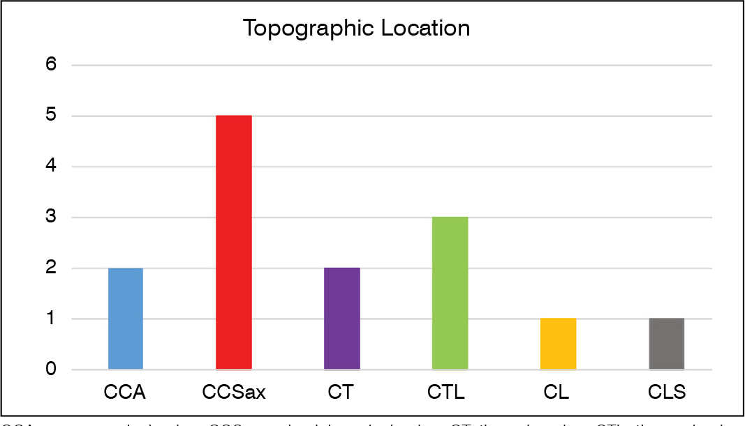
The cervical spine was the most common anatomical site. (Figure 3)

Seven OCs were intracanalar, affecting six patients; the remaining seven lesions showed extracanal exophytic growth. No statistically significant association was observed between age and intracanalar location (p = 0.34); however, there was a significant association with the female sex (p = 0.05). Furthermore, in this series, intracanalar OCs were associated with MO with a probability exceeding 95% (between 95% and 99%).
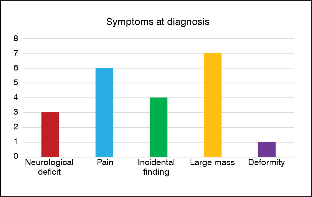
Five OCs in four patients were asymptomatic at diagnosis. At presentation (Figure 4), the most frequent findings were palpable mass or pain, but three patients exhibited neurological deficits (one of them only with neurogenic bladder - Case 4, Table 1). Extracanal OCs commonly presented as palpable and painful masses, especially when large in size (p = 0.005). One patient presented with systemic symptoms (weight loss).
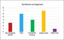
All patients underwent MRI, and 12 also had plain radiographs and CT; in two cases, bone scintigraphy was performed. MRI was the primary imaging modality used for tumor volume estimation. The overall mean tumor volume was 31.50 cm3 (range: 1.49-103 cm3). Extracanal tumors had a mean volume of 37.75 cm3, while intracanalar tumors averaged 19 cm3; this difference was not statistically significant (p = 0.24). However, intracanalar lesions demonstrated a high mean neural canal occupation (53.9%; range: 1.49%-75%). The difference in canal occupation between patients with and without neurological deficits (57.4% vs. 14.72%) was statistically significant in this subgroup (p = 0.029).
Only two patients underwent image-guided biopsy before definitive surgery, with histopathological findings always consistent with the final surgical specimens.
Eight (8) patients (with 9 OCs) were staged as Enneking Stage S1 (latent), and five as Stage S3 (aggressive). The Weinstein-Boriani-Biagini (WBB) classification for each case is presented in Table 1.
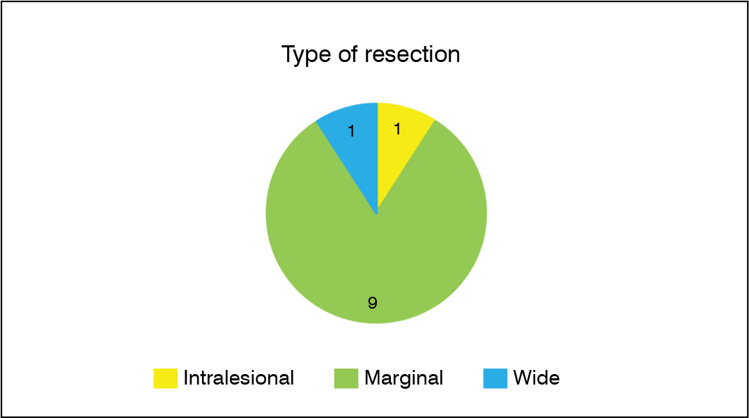
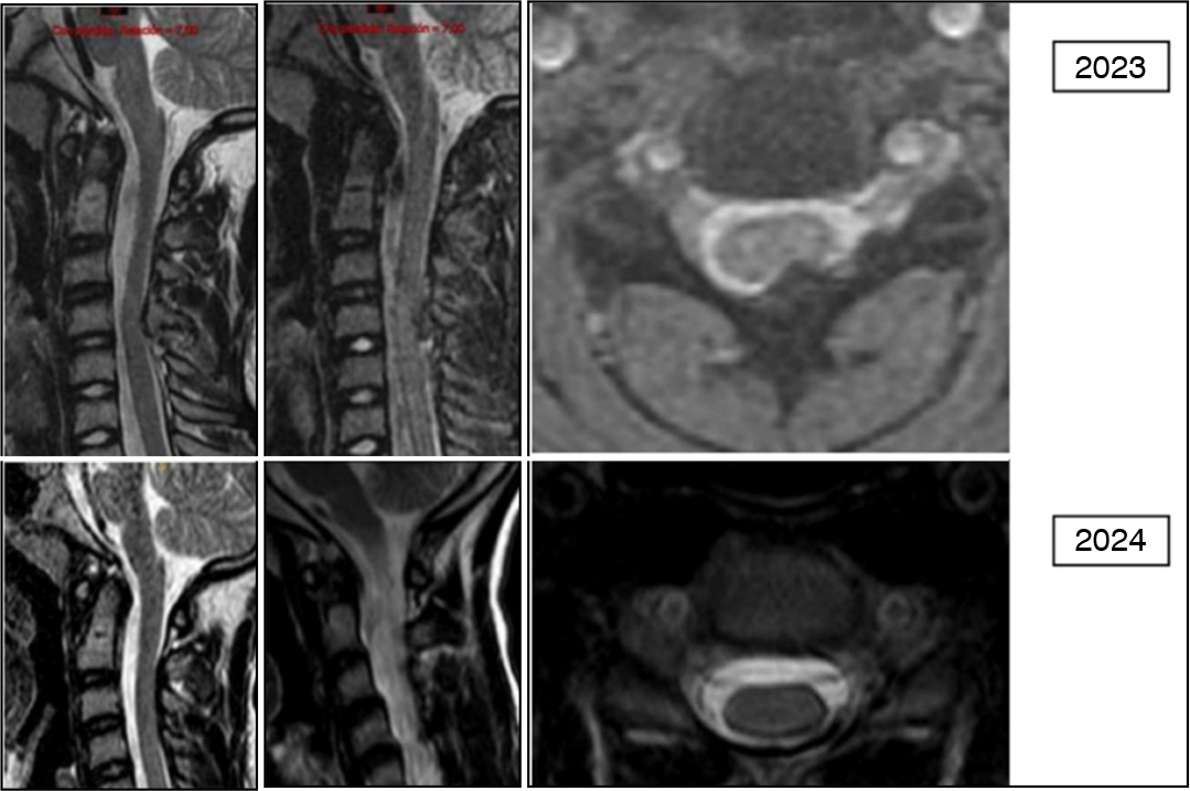
Eleven (11) of the 13 patients underwent surgery, predominantly with marginal resections (Figure 5); one patient underwent intralesional surgery (curettage). Two asymptomatic patients (with three tumors in total) were managed conservatively; both had Multiple Osteochondromatosis (MO), and one of them (Case 13, Table 1) showed spontaneous regression of an intracanalar OC during follow-up. (Figure 6)


Magnetic resonance images at two different points during the follow-up of case 13 (see Table 1) showing spontaneous regression of the intracanal OC.
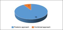
The surgical approach was almost always posterior (Figure 7). Posterior spinal instrumentation was performed in five patients, four received autologous bone grafts, and one had an additional interbody cage placed (Table 1). Three patients required postoperative immobilization (one with a cast, and two with orthoses).
Histopathological analysis confirmed the diagnosis of sessile osteochondroma in all cases.
There were two minor immediate postoperative wound complications (dehiscence). No patient required additional treatment.
The mean follow-up duration was 10 years and 6 months (range: 6 months-26 years). The mean age at last follow-up was 20 years and 4 months (range: 8-41 years); eight patients had reached skeletal maturity, and two had surpassed the peak of pubertal growth velocity.
Two patients with neurological deficits recovered neurologically (one completely and one partially; see Table 1). The patient with neurogenic bladder showed improvement but continued to experience urodynamic dysfunction.
Two spinal deformities developed later during follow-up - one required surgical intervention - and one case of neurologic hip subluxation also required surgical treatment.
There were no recurrences among the surgically treated patients. However, the patient who underwent intralesional resection showed residual tumor, which has been managed with observation only.
DISCUSSION
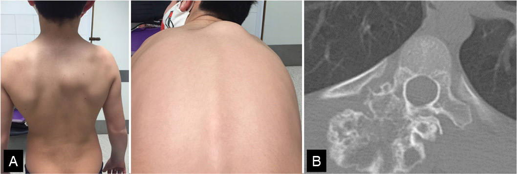
The indications for surgical resection of osteochondromas (OCs) in skeletally immature patients are well established.3 Approximately 1% to 7% of osteochondromas occur in the spine, predominantly in the cervical region,4 usually presenting as exophytic masses arising from the posterior elements.5-6 These tumors are often asymptomatic and clearly palpable, which usually prompts clinical consultation. (Figure 8)
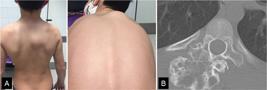
Case 2 in the series (Table 1). Large extracanalicular osteochondroma (103 cm3) of the thoracic spine. A: Clinical photos showing the size of the mass. B. Preoperative CT image.
In the case of spinal OCs, biopsy is not always necessary, as imaging studies are often sufficient for accurate diagnosis; it is only warranted when malignancy is suspected.7
Voluminous OCs of the limbs often produce symptoms that vary according to their location. Conversely, in the spine, large extracanalar OCs - such as the 7 cases in our series (Table 1) - typically result only in localized or axial pain8 or evolve as large, painless, palpable masses.9 Occasionally, they may induce secondary spinal deformities.10
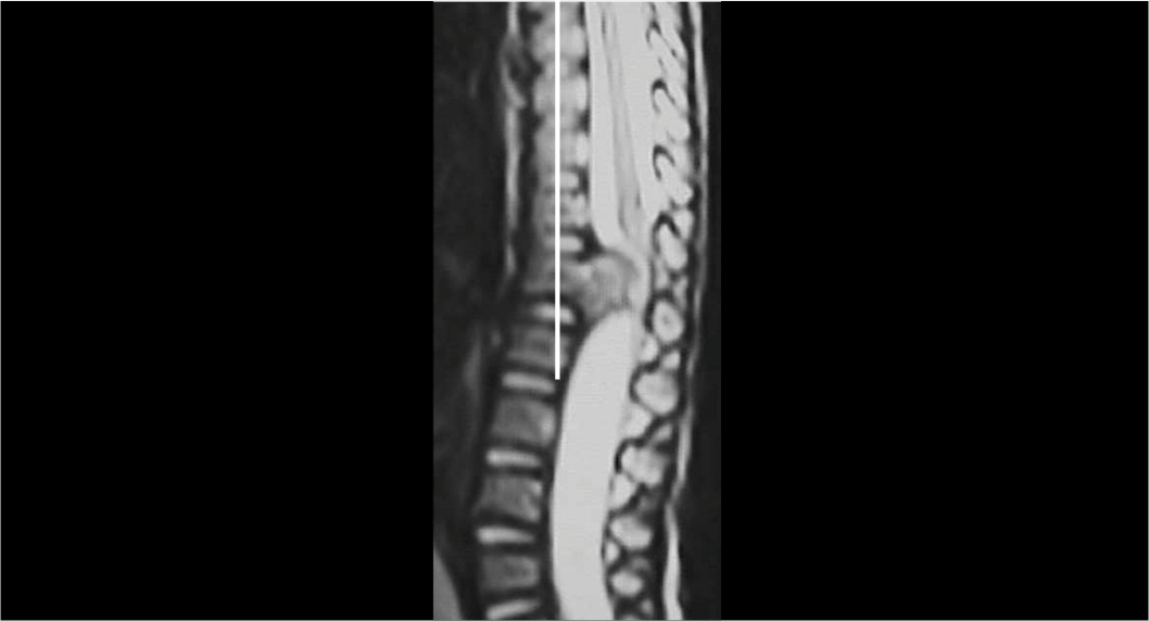
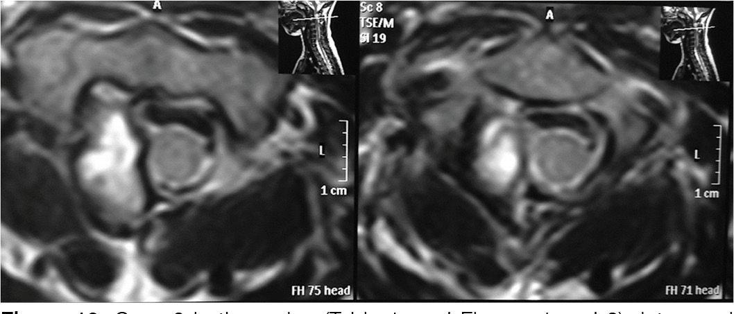
However, growth toward the spinal canal may lead to severe neurological deficits, especially in the cervical or thoracic spine (Figures 9 and 10). Two of our six patients with intracanalar OCs presented with significant neurological deficits, and one had a minor deficit (neurogenic bladder) (Table 1).11,12
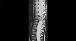
T2 sagittal MRI scan of the thoracic spine in Case 1 (Table 1), who presented with severe neurological deficit.

In large tumors or those with evident growth, there is - though rare - suspicion of malignant transformation, most commonly into chondrosarcoma, though osteosarcoma and other malignancies have also been described.13,14,15 The estimated risk of malignant transformation is <1% in solitary forms and between 2% and 5% in multiple osteochondromatosis (MO).16,17 Notably, three patients in this series had MO. (Table 1) Although chondrosarcomas in children and adolescents account for less than 5% of all cases, secondary chondrosarcomas represent more than half of these cases.17
In addition to tumor volume and multiplicity, the literature clearly indicates that spinal location and tumor recurrence increase the risk of malignant transformation.17
Differentiating between an OC and low-grade chondrosarcoma is based on clinical presentation - pain and mass growth are suspicious - and imaging findings: a size >5 cm, irregular margins, cortical disruption, soft tissue invasion, and cartilaginous cap thickness >2-3 cm should raise concern for malignant transformation.18
These considerations suggest that surgical resection of large, exophytic, spinal extracanalar OCs should be carefully evaluated. Moreover, in cases of doubt or suspicion of malignancy, wide excision is warranted, as this is the treatment of choice for secondary chondrosarcoma.19
In contrast, OCs growing into the spinal canal may cause neurological deficits even when small (Figure 10). In our series, although the volume difference between intraand extracanalar tumors was not statistically significant, there was a trend toward larger volume in the extracanalar group (mean: 37.75 cm3 vs. 19 cm3 for intracanalar tumors) (Table 1). A notable finding in this series was the percentage of spinal canal occupancy: the average occupancy was 54%, but all patients who presented with neurological deficits exceeded 40%. One patient with 43.79% occupancy at L5 (Case 5, Table 1) did not have overt neurological deficit but experienced severe radicular pain leading to functional impairment. There was a statistically significant difference in canal occupancy between patients with and without neurological deficits (p = 0.029).
The presence of neurological deficit symptoms alone justifies urgent surgical resection, regardless of tumor volume. In general, over 80% of patients experience improvement in symptoms compared to preoperative status.6,20,21 However, this is not universally true, as neurological deficits vary in duration, severity, and mode of onset, which may be progressive or sudden due to trauma involving an unrecognized and asymptomatic intracanalar OC.22,23 This is particularly relevant in patients with MO,24 as 20-27% may have occult cervical lesions.25,26 In our series, statistical analysis showed >95% probability of intracanalar OCs in patients with MO. Therefore, we agree with others that serial spinal MRI screening is necessary in this patient group.27 Still, only intracanalar OCs that enlarge or become symptomatic require surgical treatment; others do not.28 This position is further supported by the literature, which documents spontaneous regression,29-31 with rates ranging from 7.6% to 35%.32,33 This phenomenon is especially relevant in MO.32,34 One of our cases (Case 13, Table 1) showed spontaneous regression within the context of MO (Figure 6), supporting a strategy of screening in MO, surgery for symptomatic cases, and observation for small, asymptomatic lesions.28
Radiculopathies in the lumbosacral region are more common in adults than in children,35 though the prognosis for recovery is better in children, as seen in our Case No. 5. (Table 1)
Excision with tumor-free margins is the treatment of choice. Indeed, when surgery is indicated, marginal or wide excision without fusion appears sufficient,36 but in some cases, wide excision with internal fixation may be necessary.4
Recurrence
Local recurrence following complete surgical ablation of an osteochondroma is below 2%;37 however, in the adult spine, this rate may reach up to 8%.36 Notably, the risk of recurrence in the spine does not appear to be associated with the Enneking stage of the lesion, as recurrences have been reported even in Stage 1 (latent) lesions,36 and may occur many years after resection.38 In our series, one case of recurrence (Case No. 2, Table 1) actually involved an incomplete intralesional excision, resulting in residual tumor tissue. In this instance, the tumor volume (103 cm3) does not seem to have contributed to the recurrence; rather, the intralesional technique appears to be the main factor - suggesting that such approaches should be avoided whenever possible.
CONCLUSIONS
1. Surgical excision of bulky extracanal osteochondromas should be considered even in the absence of symptoms, due to the potential risk of malignant transformation.
2. Intracanal osteochondromas that exhibit growth and/or become symptomatic are clear candidates for surgical treatment, regardless of their volume.
3. Patients with Multiple Osteochondromatosis (MO) should be closely monitored due to the potential development of intracanal osteochondromas, although their mere presence does not always warrant surgical intervention.
4. Intralesional ablation should be avoided due to the associated risk of recurrence or residual tumor tissue. Marginal resection is generally the most appropriate approach; however, in selected cases, a wide resection with reconstruction may be necessary.
-
The study was conducted at Centro “Nicolás Andry”, Resistencia, Chaco, Argentina.
REFERENCES
- 1 Albrecht S, Crutchfield JS, SeGall GK. On spinal osteochondromas. J Neurosurg. 1992;77(2): 247-52.
- 2 Rajakulasingam R, Murphy J, Botchu R, James SL. Osteochondromas of the cervical spine-case series and review. J Clin Orthop Trauma. 2020;11(5): 905-9.
- 3 Unni KK, Inwards CY. Chapter 2: Osteochondroma (Osteocartilaginous Exostosis). In K. Krishnan Unni, Carrie Y editors: Dahlin’s bone tumors: general aspects and data on 10,165 cases; 6th ed., 2010 Mayo Foundation for Medical Education and Research, Lippincott Williams & Wilkins, Philadelphia. 2010;9-21.
- 4 Fowler J, Takayanagi A, Fiani B, Cathel A, Sarhadi KJ, Arshad M, et al. Diagnosis, management, and treatment options: a cervical spine osteochondroma meta-analysis. World Neurosurgery. 2021;149:215-225.
- 5 Tahasildar N, Sudesh P, Goni V, Tripathy SK. Giant osteochondroma of axis in a child with multiple hereditary exostoses: case report and review of literature. J Ped Orthop B. 2012;21(3):280-5.
- 6 Assan BR, Simon AL, Violas P, de Gauzy JS, Thepaut M, Ould-Slimane M, et al. Surgical outcomes of spinal osteochondroma in children: A multicentre observational study. Orthop Traumatol Surg Res. 2022,108(6):103239.
- 7 Weber MA, Bazzocchi A, Noebauer-Huhmann IM. Tumors of the spine: when can biopsy be avoided? Semin Musculoskelet Radiol. 2022;26(4): 453-68.
- 8 Villa A, Iacopino DG, Graziano F, Gulì C, Giugno A, Basile L, et al. A rare case of a giant cervical osteochondroma. Curr Pediatr Res. 2016;20(1-2):199-202.
- 9 Sharma C, Acharya M, Kumawat BL, Parekh J. Giant spinal exostosis. BMJ Case Rep. 2014;2014:2014203819.
- 10 Fiechtl JF, Masonis JL, Frick SL. Spinal osteochondroma presenting as atypical spinal curvature: a case report. Spine. 2003;28(13):E252-E255.
- 11 Elgamal EA. Complete recovery of severe tetraparesis after excision of large C1-osteochondroma. World J Neurol. 2013;3(3):79-82.
-
12 Mamdouhi T, Unadkat P, Edelman MC, Johnson AA, Levy CF, Mittler MA, et al. Solitary pediatric osteochondroma of the spine with cord compression. Cureus. 2022; 14(3): e23342. DOI 10.7759/cureus.23342.
» https://doi.org/10.7759/cureus.23342. - 13 Park YK. Chapter 20: Multiple Osteochondromatosis. In: E. Santini-Araujo et al. (eds.), Tumors and Tumor-Like Lesions of Bone: For Surgical Pathologists, Orthopedic Surgeons and Radiologists. Springer-Verlag London. 2015;273-6.
- 14 Schmale GA, Hawkins DS, Rutledge J, Conrad III EU. Malignant progression in two children with multiple osteochondromas. Sarcoma. 2010; 417105.
- 15 Bukara E, Buteera AM, Karakire R, Manirakiza F, Muhumuza S, Rudakemwa E, et al. Osteoblastic osteosarcoma arising beneath an osteochondroma in an 11-year-old male with multiple hereditary exostoses. Case Reports in Orthopedics. 2018;2018:8280415.
- 16 Sonne-Holm E, Wong C, Sonne-Holm S. Multiple cartilaginous exostoses and development of chondrosarcomas--a systematic review. Dan Med J. 2014;61(9):A4895.
-
17 Puri A, Gulia A, Kurisunkal VJ, Sukumar V, Rekhi B. Chondrosarcomas in adolescents: are they different? J Ped Orthop B. 2020;29(5):505-9. DOI: 10.1097/BPB.0000000000000641.
» https://doi.org/10.1097/BPB.0000000000000641. - 18 Choi JH, Ro JY. The 2020 WHO classification of tumors of bone: an updated review. Adv Anat Pathol. 2021;28(3):119-138.
-
19 Tsuda Y, Gregory JJ, Fujiwara T, Abudu S. Secondary chondrosarcoma arising from osteochondroma. Bone Joint J. 2019;101-B(10):1313-20. doi:10.1302/0301-620X.101B9.BJJ-2019-0190.R1
» https://doi.org/10.1302/0301-620X.101B9.BJJ-2019-0190.R1 - 20 Gille O, Pointillart V, Vital JM. Course of spinal solitary osteochondromas. Spine.2005;30(1): E13-E19.
- 21 Yakkanti R, Onyekwelu I, Carreon LY, Dimar JR. Solitary Osteochondroma of the spine-a case series: review of solitary Osteochondroma with Myelopathic symptoms. Global Spine J. 2017;8(4):323-39.
- 22 Okuyama K, Chiba M, Okada K, Sato K, Hoshi N. Huge solitary osteochondroma at T11 level causing myelopathy: case report. Spinal Cord. 1997;35(11):773-6.
- 23 Giudicissi-Filho M, de Holanda CVM, Borba LAB, Rassi-Neto A, Ribeiro CAA, de Oliveira JG. Cervical spinal cord compression due to an osteochondroma in hereditary multiple exostosis: case report and review of the literature. Surg Neurol. 2006;66 Suppl 3: S7-S11.
- 24 Kamiya Y, Horii E, Sakai Y, Inoue H. Cervical cord compression in pediatrics with hereditary multiple exostoses: a report of two cases and review of the literature. J Pediat Orthop B. 2016;25(3):267-70.
- 25 Roach JW, Klatt JW, Faulkner ND. Involvement of the spine in patients with multiple hereditary exostoses. J Bone Joint Am. 2009;91(8):1942-48.
- 26 Ashraf A, Larson AN, Ferski G, Mielke CH, Wetjen NM, Guidera, KJ. Spinal stenosis frequent in children with multiple hereditary exostoses. J Child Orthop. 2013;7(3):183-94.
- 27 Vu CL, Lindberg AW, Bompadre V, White KK, Bauer JM. Prospective spine at risk program for prevalence of intracanal spine lesions in pediatric hereditary multiple osteochondromas. Spine Deform. 2020;8(5):1069-74.
- 28 Aguer S, Dal Lago J, Cullari ML, Aguirre F, Levy E, Lloyd R. Osteocondroma cervical y mielopatía en niños: reporte de un caso y revisión de la bibliografía. Rev Asoc Argent Ortop Traumatol. 2024;89(6):651-56.
- 29 Valdivielso-Ortiz A, Barber I, Soldado F, Aguirre-Canyadell M, Enriquez G. Solitary osteochondroma: spontaneous regression. Pediatr Radiol. 2010;40(10):1699-701.
- 30 Passanise AM, Mehlman CT, Wall EJ, Dieterle JP. Radiographic evidence of regression of a solitary osteochondroma: a report of 4 cases and a literature review. J Pediatr Orthop. 2011;31(3):312-16.
- 31 Ábalo E, Wittwer C, Mallea A, Plater P. Regresión espontánea de un osteocondroma solitario en la rodilla: Presentación de un caso. Rev Asoc Argent Ortop Traumatol. 2015; 80(4):287-291.
- 32 Mordenti M, Shih F, Boarini M, Pedrini E, Gnoli M, Antonioli D, et al. The natural history of multiple osteochondromas in a large Italian cohort of pediatric patients. Bone. 2020;139:115499.
- 33 Adachi R, Nakamura T, Asanuma K, Hagi T, Uchiyama T, Sudo A. Thin cartilage cap may be related to the spontaneous regression in pediatric patients with osteochondroma. Curr Oncol. 2022;29(12):9884-9890.
- 34 Komura S, Matsumoto K, Hirakawa A, Akiyama H. Natural history and characteristics of hand exostoses in multiple hereditary exostoses. J Hand Surg Am. 2021; 46(9):815.e1-e12.
- 35 Agrawal A, Dwivedi SP, Joshi R, Gangane N. Osteochondroma of the sacrum with a correlative radiographic and histological evaluation. Pediatr Neurosurg. 2005;41(1):46-48.
- 36 Sciubba DM, Macki M, Bydon M, Germscheid NM, Wolinsky JP, Boriani S, et al. Long-term outcomes in primary spinal osteochondroma: a multicenter study of 27 patients. J Neurosurg Spine. 2015;22(6):582-588.
- 37 Tepelenis K, Papathanakos G, Kitsouli A, Troupis T, Barbouti A, Vlachos K, et al. Osteochondromas: An updated review of epidemiology, pathogenesis, clinical presentation, radiological features and treatment options. In vivo. 2021;35(2):681-91.
- 38 Hari A, Kavar B. Rare case of malignant transformation of a solitary spinal osteochondroma into recurrent metastatic chondrosarcoma. J Clin Neurosci. 2019;67: 280-88.
Edited by
-
Reviewed by:
Marcelo Risso


 CCA: upper cervical spine. CCSax: subaxial cervical spine. CT: thoracic spine. CTL: thoracolumbar spine. L: lumbar spine. LSS: lumbosacral spine.