Mutagenic biomarkers in erythrocytes of free-ranging and captive Kinosternon scorpioides from the Brazilian Amazon
Abstract
This study aimed to investigate the occurrence of micronuclei (MN) and erythrocyte nuclear abnormalities (ENAs) in natural and captive Kinosternon scorpioides populations and compare their frequencies with the environment type, sex, and seasonal period in an environmental protection area in the state of Maranhão, Amazon. Blood was collected from 112 specimens from regions with direct anthropogenic influence and indirect and control areas to produce blood smears for subsequent cytological analysis under an optical microscope. We found MN (N=91) and ENAs such as binucleated nuclei (N=14), notched (N=81), lobulated (N=35), segmented (N=16), and displaced (N=253) in animals in situ. No abnormalities were recorded in the erythrocytes of the captive specimens. The mean (±standard deviation) MN (1.13±0.47) differed from those of the other chelonians from the natural environment, and the displaced and binucleated nuclei manifested themselves at higher (51.6%) and lower (2.8%) frequencies, respectively. Lobulated nuclei frequency was significantly different between the areas (p<0.05); segmented nuclei (p<0.05) and displaced (p<0.05) had higher frequencies during the dry and rainy periods. Sex did not affect these abnormalities. No significant MN were found; however, erythrocytes with lobulated, segmented, and displaced nuclei were documented in animals from different environments and seasonal periods. Our study provides pioneering data on MN and nuclear anomalies in the erythrocytes of K. scorpioides and confirms the significant differences and relationships between them. The occurrence of abnormalities in wild animals demonstrates the need to maintain protected areas and the continuity of conservation actions aimed at Amazonian turtles. This contributes to reducing the impact on conservation units that employ the sustainable use of biodiversity in Brazil.
Key-words:
biomarkers; environmental stressors; micronuclei; turtle
Resumo
Este estudo teve como objetivo investigar a ocorrência de micronúcleos (MN) e anormalidades nucleares eritrocitárias (ANEs) em populações naturais e de cativeiro de Kinosternon scorpioides e comparar suas frequências com o tipo de ambiente, sexo e período sazonal em uma área de proteção ambiental no estado do Maranhão, Amazônia. O sangue foi coletado de 112 espécimes de regiões com influência antrópica direta e áreas indireta e de controle para produzir esfregaços de sangue para posterior análise citológica em microscópio óptico. Encontramos MN (N=91) e ANEs como núcleos binucleados (N=14), entalhados (N=81), lobulados (N=35), segmentados (N=16) e deslocados (N=253) em animais in situ. Nenhuma anormalidade foi registrada nos eritrócitos dos espécimes em cativeiro. A média (± desvio padrão) de MN (1,13±0,47) diferiu daquelas dos outros quelônios do ambiente natural, e os núcleos deslocados e binucleados se manifestaram em frequências maiores (51,6%) e menores (2,8%), respectivamente. A frequência de núcleos lobulados foi significativamente diferente entre as áreas (p<0,05); núcleos segmentados (p<0,05) e deslocados (p<0,05) apresentaram frequências maiores durante os períodos seco e chuvoso. O sexo não afetou essas anormalidades. Nenhum MN significativo foi encontrado; no entanto, eritrócitos com núcleos lobulados, segmentados e deslocados foram documentados em animais de diferentes ambientes e períodos sazonais. Nosso estudo fornece dados pioneiros sobre MN e anomalias nucleares nos eritrócitos de K. scorpioides e demonstra as diferenças e relações significativas entre eles. A ocorrência de anormalidades em animais silvestres confirma a necessidade de manutenção de áreas protegidas e a continuidade das ações de conservação voltadas para os quelônios amazônicos. Isso contribui para a redução do impacto em Unidades de Conservação que utilizam a biodiversidade de forma sustentável no Brasil.
Palavras-chave:
biomarcadores; estressores ambientais; micronúcleos; quelônios
1. Introduction
Kinosternon scorpioides (Linnaeus, 1766) is a widely distributed semiaquatic species that can be affected by seasonal variations and food availability. The species is known as “jurará” or “muçuã” in Brazil, has a reproductive range, and can be found in lakes, ponds, rivers and on the ground (1,2). Studies on the morphophysiological, reproductive, behavioral, ecological, environmental, and physiological aspects of this and other turtle species have already presented clear and encouraging results. Such information is essential for understanding the biology of this taxon and can act as a guideline for environmental policies focused on ecosystem conservation and recovery and natural Amazonian turtle populations.
Maintaining viable turtle populations in ecosystems requires protection, management, and monitoring efforts, which involve individuals, practical measures, effective conservation management techniques, and population biomonitoring, which must be applied with community support (3). Anthropogenic pressure, such as excess hunting, burning, and deforestation in protected areas, impacts K. scorpioides and other turtles (4). In the Amazon region, several K. scorpioides specimens have been subjected to degradation. In this scenario, understanding the impact of environmental stressors is relevant and allows for a qualitative understanding of their health and the environment. Despite the subtle changes in their DNA, they must be further elucidated because exposure to mutagenic agents can cause irreversible damage to genetic material, putting their health at risk (5).
Thus, biological biomarkers represent crucial instruments that signal molecular, biochemical, physiological, cellular, or other changes in the tissues and/or organs of an organism, indicating direct or indirect exposure to the harmful effects of environmental pollutants (6,7,8). The micronucleus (MN) test, an inexpensive, simple, and easy-to-apply technique, is crucial for evaluating mutagenic effects in blood cells, which has been frequently used in environmental studies to analyze structural changes and DNA stability in aquatic fauna (9).
MNs are cytoplasmic elements containing chromatin that arise during cell division when acentric chromosome fragments are left behind, complicating their connection with the main nucleus of daughter cells. Their formation results from genetic changes that manifest as chromosomal or mitotic spindle abnormalities. This irreversible process is passed on to future generations, reducing the genetic diversity of populations and harming animals residing in the affected regions (10). In this context, MNs evaluation is crucial in biomonitoring research because it provides reliable data on the environmental quality.
In this sense, studies in this area are important because turtles are excellent indicators of environmental quality and potential dangers to human health and biodiversity owing to their wide distribution and sensitivity to pollutants. In many cases, their physiology, behavior, and ecology are influenced by global environmental factors (11).
Therefore, this study aimed to investigate the presence of MN and erythrocyte nuclear abnormalities (ENAs) in free-ranging and captive K. scorpioides populations and compare their frequencies with the type of environment, sex, and seasonality in an Environmental Protection Area (APA) located in the state of Maranhão, which is a part of the Amazon.
2. Material and methods
2.1 Study area
The environmental protection area of Baixada Maranhense is in the State of Maranhão, which is a part of the Amazon and was placed on the Ramsar list on February 29, 2000 (12). It is among the most important Brazilian Ramsar sites and was included in the National Strategic Plan of Protected Areas - PNAP (13).
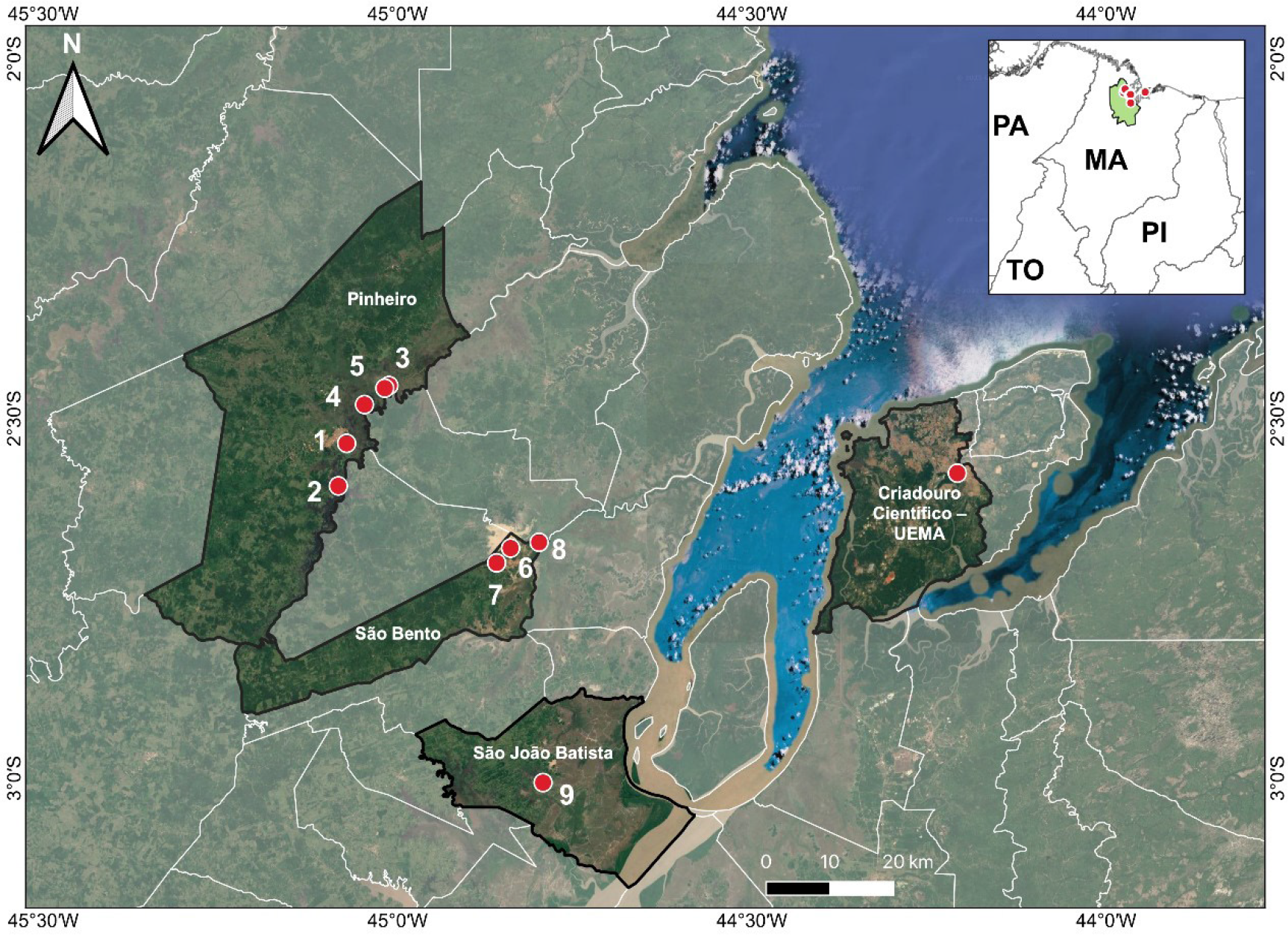
Based on the level of human interference and direct and indirect environmental effects, as evidenced by the presence or absence of deforestation, burning, mangrove and field devastation, predatory hunting, and urban waste, as well as the distribution of specimens by region and various environments, we grouped the sampling points into three categories: A1 (urbanized area, direct anthropogenic influence), A2 (area with vegetation, indirect influence), and A3 (UEMA scientific breeding ground, control group).
The A2 category is marked by the abundant presence of trees, shrubs, and other native plant species that occupy the site, which protect the soil against erosion and help preserve the ecological balance. The area is full of biodiversity, presenting diverse plant and animal species, which is characteristic of the Brazilian Amazon region. The local flora directly impacts the climate, hydrological cycle, air quality, and habitat of animals and other living organisms. This region exhibits minimal or no human interference.
In contrast, human activity is intense in the A1 category, considerably impacting the natural resources. There is a developed infrastructure with changes in land use, pollution, and noticeable environmental effects from industrial, commercial, and residential activities that have developed in its surroundings. Population density is high, thus changing the natural landscape, where native vegetation is replaced by urban areas and other constructions that directly affect the original natural environment. Economic and social activities in this region are accentuated and constant. Generally, it is characterized by strong interference in the natural environment, human actions, and changes in the landscape.
The animals in the control group were kept at the UEMA Scientific Breeding Center under the authorization of the Brazilian Institute of Environment and Renewable Natural Resources (IBAMA-MA), nº. 1899339/2008 (Figure 1). The aviary has an area of 159.92 m2 and consists of five stalls with galvanized iron screen walls, including the roof. Each stall occupies an area of 13.94 m2, forming a part of the total constructed area of approximately 91 m2. In addition, it has a free area of 68 m2, a ceramic-lined masonry tank, a ramp for animal access, available water, and a drainage system. The aviary contains trees, gravel, sand, and a large open space with natural ventilation, providing an environment similar to the natural habitat of the species. On alternate days, adult animals were handled to renew the water in the tanks, received food, and monitoring their health. They were identified individually and fed daily, preferably in the morning. While cleaning the area, the use of cleaning products was avoided and all animals were removed from their stalls for this purpose.

The locations of sampling points grouped as urbanized areas (1, 3, and 7) and with vegetation (2, 4, 5, 6, 8, and 9) in Pinheiro (MA), São Bento (MA) and São João Batista (MA) in the Baixada Maranhense, in addition to the UEMA Scientific Breeding Center, São Luís, Maranhão, Brazil. (Pará - PA; Maranhão - MA; Piauí - PI and Tocantins - TO).
The nine collection points are in strategic regions covered by the EPA. This was intended for better data amplitude, as well as mapping of anthropized or non-anthropized regions, such as the cities of Pinheiro, Maranhão (P1 - Maria Santa 2°32'5"S 45°4′19′W; P2 - upper Pericumã River 2°35'38"S 45°5'1"W; P3 - Sluice 2°27'16"S 45°0'44"W; P4 - lower Pericumã River 2°28'51"S 45°2'48"W; P5 - middle Pericumã River 2°27'29"S 45°1'5"W); São Bento, Maranhão (P6 - Campo do Chagas 2°40'49"S 44°50'26"W; P7 - Campo do Alegre 2°42'3"S 44°51'37"W; P8 - Defunto 2°40'20"S 44°48'1"W); São João Batista, Maranhão (P9 - Pução do Meio 3°0'22"S 44°47'41"W).
2.2 Environmental parameters of the water and soil
The abiotic variables of the lake water and field near the animal capture sites were measured between 8 and 10 am. The pH, temperature (T), and dissolved oxygen (DO) were measured using a multiparameter meter (Akso, AK88®), which was immersed in 4 m deep water. Lake water was divided into three water columns (three sample replicates), and the mean values were calculated. These parameters are important from the ecotoxicological point of view since they offer a safe assessment of the quality of water bodies, in addition to being commonly used in analyses integrated with other biomarkers of environmental interest.
Soil samples were collected for complete chemical analysis considering the elements K, Ca, Mg, Al, H+Al complex, base saturation (BS), cation exchange capacity (CEC), and micronutrients such as Fe, Mn, Cu, S, Zn, and organic matter (OM). Five simple samples were collected in a zigzag pattern at each point for a single composite sample composition. For this purpose, an auger was placed at 20 cm depth (14). Single portions were mixed to obtain a homogeneous compound, and 600 g was removed to obtain the final representative sample of the collection points. Subsequently, the samples were stored in closed, labeled plastic bags and sent to the laboratory for analysis.
2.3 Testing for MN and ENAs in K. scorpioides
The research protocol for biological analyses was submitted to the Ethics and Animal Experimentation Committee (EAEC) of the State University of Maranhão (UEMA), approved under opinion n.º 10/2021, and followed all recommended animal welfare principles. For capturing the animals, the study was licensed via the Biodiversity Information and Authorization System (SISBIO), registration n.º 85805-1. Individual animals were captured using artisanal circular funnel traps in August and October 2021; April, June, October, and December 2022; and March 2023. We captured 112 animals, including 89 free-ranging and 23 captive, of which 50 were males and 62 females.
The MN test was conducted according to the protocol proposed by Grisolia et al. (15) with some modifications. Several anomalies are associated with the erythrocyte nucleus of K. scorpioides, including lobulated, notched, segmented, binucleated, and displaced nuclei, in addition to MN. Before blood collection, all necessary precautions were taken to prevent stress in animals and sample contamination. Blood was then collected by puncturing the occipital venous sinus using a syringe with a hypodermic needle (0.7×25 mm2) after relaxing the animal muscles. Anesthetic use was avoided because it could interfere with the results of the biomarkers.
Two slides were produced per animal for 224 blood smears. In each of them, an aliquot of approximately 30 μL blood was used, which was subsequently dried at room temperature (between 25 °C and 32 °C) for 24 h, followed by fixation in ice-cold 10% methanol for 20 min and staining with 10% Giemsa solution diluted in 0.2 M phosphate buffer (KH2PO4). The slides were washed under running water and dried at room temperature, with readings performed under an optical microscope using the zigzag visualization technique at 100× magnification. A sampling effort of 30 min was used for each slide to examining 1.000 erythrocytes per slide, totaling 2.000 per turtle (16).
2.4 Data analysis
The data were subjected to Shapiro-Wilk and Levene tests to verify the normality and homoscedasticity of variance, respectively. Analysis of variance (one-way ANOVA) was performed to evaluate the differences between the environments, seasonal period, and sex, as well as summary statistics of the means and standard deviation; when a significant difference was found, they were subjected to the Tukey test using the Past 4.08 statistical program (Paleontological Statistics Software) considering a significance level of 5% (p<0.05).
3. Results
3.1 Environmental parameters of the water and soil of the Baixada Maranhense
The DO concentrations in the waters in A1 and A2 (table1), when related to the two sampling periods (dry and rainy), were low and did not comply with the quantities allowed by the Conselho Nacional do Meio Ambiente (CONAMA) (17,18).
Summary statistics of the physicochemical variables of the water of the different capture environments (A1 and A2) of Kinosternon scorpioides in the Baixada Maranhense in the months of August and October 2021; April, June, October and December 2022; and March 2023 in the dry and rainy periods in the Baixada Maranhense. SD = Standard deviation; pH = Hydrogenionic potential; DO = Dissolved oxygen.
In the soil samples, the mean K and Ca contents were high in A1 in the dry season, while Mg, BS, Al, H+Al, and CEC were high in A2 in the same period (Table 2). The mean Fe, Mn, Cu, and Zn contents were high in A2 in the dry season. The mean S content was also high in A1 during this period (Table 2).
Concentrations of chemical elements and micronutrients present in the soil corresponding to the different capture environments (A1 and A2) of Kinosternon scorpioides in the Baixada Maranhense in August and October 2021; April, June, October, and December 2022; and March 2023 in the dry and rainy seasons.
Granulometric studies revealed that A1 had the highest mean sand content (453.67±206.00 g/kg) in the rainy season, and A2 high silt/clay contents in both climatic periods (Table 3).
Granulometry of the soil of the habitats (A1 and A2) of the sampled specimens of Kinosternon scorpioides, obtained during the collections between the months of August and October 2021; April, June, October, and December 2022; and March 2023 in the dry and rainy periods in the Baixada Maranhense.
The mean OM level in A2 (29.16 g/kg) was higher than in A1 (15.95 g/kg). The numbers in A2 increased the CEC (21.94 to 47.93 cmolc/dm3). In A1, they ranged from 7.82 to 21.38 cmolc/dm3.
3.2 MN and ANE in K. scorpioides
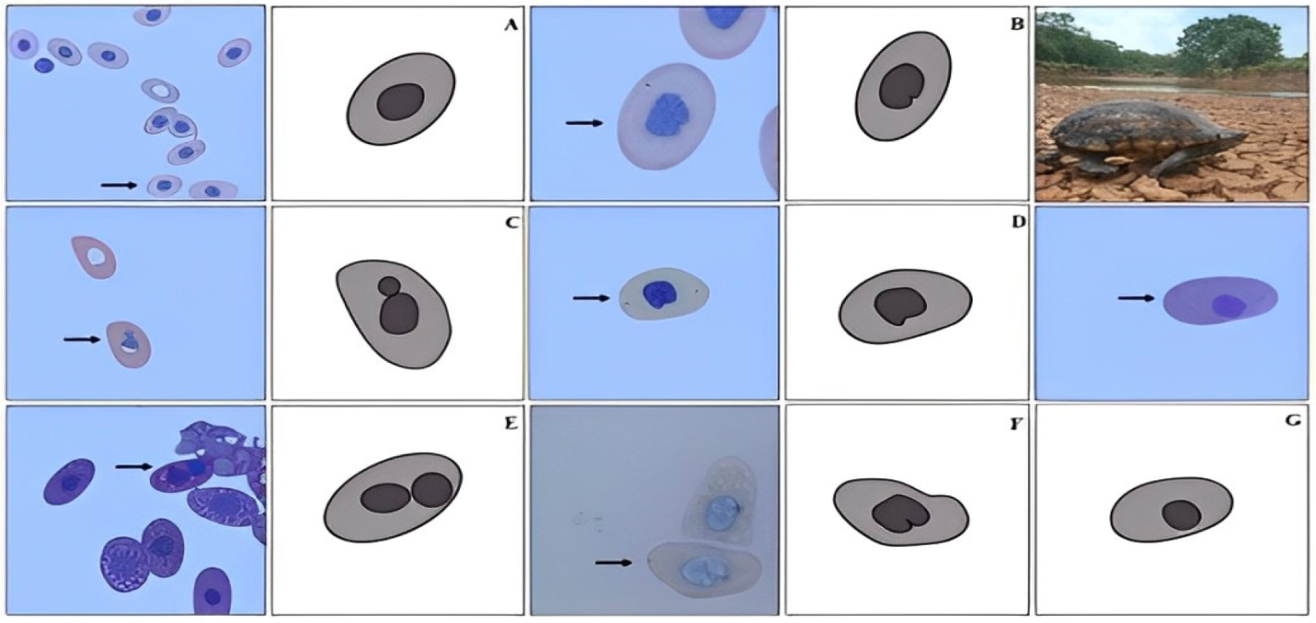
MN test results revealed that 26.7% (60/224) blood smears showed some alteration in erythrocyte nuclear morphology in addition to MN. Out of these samples, 33.3% (20/60) and 95% (57/60) showed MN and some form of ENA, respectively. More erythrocytes with MN were found in the animals from A2 (N=71) than in those from A1 (N=20). The ENAs identified were nuclei binucleated erythrocytes (BI), notched (NN), lobulated (LN), segmented (SN), and displaced (DN; Figure 2). We did not identify MN or other ENAs in the samples from the 46 captive animals. The values in Table 4 refer to those in the animals from areas A1 and A2.
Quantitative analysis of the types of erythrocyte nuclear anomalies (ENAs) recorded in samples of Kinosternon scorpioides captured from A1 and A2 in the Baixada Maranhense, Maranhão, Brazil.

Erythrocytes of Kinosternon scorpioides. Presentation of micronucleus (MN) and erythrocyte nuclear abnormalities (ENAs) of specimens captured in areas A1 and A2 in the Baixada Maranhense, with the use of 10% Giemsa stain and visualization under 100× magnification. Black arrows indicate: A - Normal erythrocytes; B - Erythrocyte with micronucleus; C - Binucleated erythrocyte; D - Notched nucleus; E - Lobulated nucleus; F - Segmented nucleus and G - Displaced nucleus.
Among the 490 erythrocytes counted, DN occurrence (N=253; 51.63%) was more frequent than BI (N=14, 2.85%; Table 4). Erythrocytes with SN, LN, and DN abnormalities were the most recurrent in most samples.
Comparing the means of the variables corresponding to MN and ENA according to environment, sex, and seasonal period indicated that the number of DN was higher in the animals from A1 (4.66±0.81). However, the number of LN were significantly different between the animals from A1 and A2 (p<0.05; Table 5). When the means of the variables were checked according to the seasonal period, the numbers of SN and DN in K. scorpioides erythrocytes were significantly higher in the dry season (5.27±0.90, p<0.05; Table 5). Therefore, environmental and climatic conditions directly affect the occurrence of erythrocyte anomalies. Sex did not interfere in the formation of these abnormalities, and the highest means detected showed similar values between males (4.30±0.76) and females (4.36±0.79) with DN in erythrocytes. The mean and standard deviation of MN detected in adult specimens of K. scorpioides (1.13±0.47) differed from that in other chelonians (Table 6).
Summary statistics of the frequencies of micronuclei and erythrocyte nuclear abnormalities (ENAs) in males and females of Kinosternon scorpioides (N=60) from the Baixada Maranhense (A1 and A2), in different periods (dry and rainy), in the months of August and October 2021; April, June, October and December 2022 and March 2023.
Comparison between means and standard deviation (SD) of MN found in erythrocytes of Kinosternon scorpioides with those in other chelonian species in marine and freshwater environments of Brazil.
4. Discussion
The estimated MN frequency in K. scorpioides coincided with that in other turtles, such as Trachemys callirostris, which presented 79 MN in erythrocytes. In addition to MN, our findings revealed other anomalies in the erythrocytes of K. scorpioides, contradicting the results of most studies that focused only on MN analysis, particularly in aquatic mammals (26,27). Previous studies have shown that the MN test effectively detects changes in reptile and bird erythrocytes, thus providing pertinent biological information in addition to acting as a signal of harmful effects in aquatic animals. In this scenario, the results of this study are particularly important, as no evidence of MN or other erythrocyte anomalies in the species under study is available.
Our results did not reveal MN of captive K. scorpioides. One factor linked to the occurrence of these abnormalities is the reduced number of animals in the area (28). Furthermore, stress factors that change the cell nucleus are uncommon in captive animals. Regarding the proportion of captive animals relative to free-ranging animals, Zapatta et al. (20) and Latorre et al. (21) reported proportions similar to that of this study (20 and 32 animals, respectively). Previous studies have reported reduced numbers and absence of MN in both captive and free-ranging Macroclemys temminckii and Kinosternon subrubum (29).
The average number of MN in animals from A2 was higher than from A1, although the difference was not statistically significant. These findings confirm the results reported by Castaño et al. (33) for specimens collected from regions with vegetation (3.33±0.62) and are similar to those reported by Zapata et al. (20), who identified a higher mean (8.04±7.08) in T. callirostris captured in rivers and subjected to stress factors. The scarcity of MN in free-ranging individuals could be attributed to its ability to adapt to regional environmental conditions and changes (30).
Although the animals in this study were captured from urban and vegetated areas in an APA in Maranhão, which presented varying levels of human interference, our reports indicated a low incidence of MN, suggesting that direct environmental impacts have not yet been manifested in these regions. These data are similar to those presented by Moron et al. (31) on Podocnemis expansa from an APA in Tocantins, Brazil. In previous studies conducted with P. hilarii and Caretta caretta (28), Casini et al. (25) demonstrated that MN showed significant responses from an environmental perspective and indicated possible effects on these species. The change in the origin of this anomaly indicates the quality of the environment, as it is common in animals from regions affected by environmental issues and imbalances, which compromise their survival (32).
Matson et al. (6) reported that industrial effluents act as environmental tensors in the impacted ecosystems. They identified the absolute values of MN in Emys orbicularis and C. mydas (N=23 and 1,254, respectively) when subjected to the negative impacts of agricultural activities in urban areas. These values exceeded those reported in our study for A1 (N=20). Furthermore, Castaño et al. (33) suggested that the high rates of nuclear anomalies in animals from urban and vegetated regions might be linked to human and agricultural activities that have considerable effects on these animals.
The results of the MN test did not reveal a significant difference between the sexes, confirming that it is an element that does not influence nuclear changes. Similar studies confirmed this same trend, such as that carried out by Borges et al. (34), who analyzed the health of Podocnemis unifilis kept in a sustainable development reserve in the state of Amazonas, Brazil, and identified erythrocytes with SN and LN. When examining different environments and times of the year, we observed notable differences in the nuclear abnormalities of erythrocytes with LN in K. scorpioides. This differentiates the data reported by Borges et al. (34) from that reported in this study.
The description of nuclear anomalies in our study revealed a high number of erythrocytes with DN, which appears to be frequent in free-ranging K. scorpioides captured from urban areas. This suggests that, in contrast to turtles from regions with large vegetation, those inhabiting urban areas are more likely to present this anomaly. Furthermore, the high frequency observed in K. scorpioides may be related to environmental conditions, particularly during the dry season in Maranhão.
Although previous studies suggest that this anomaly is rare, evidence in birds indicates frequent DN occurrence when exposed to pollutants. These erythrocytes exhibit different rotations and displacements of the nucleus in the cytoplasm (35,36), which has also been confirmed in K. scorpioides. Araújo et al. (37) reported notable anomalies in P. expansa cells affected by stress factors and neonates exposed to zinc oxide nanoparticles. This information highlights the importance of additional studies to detect pollutants in the blood of turtles to better characterize their effects on Chelonian populations, particularly K. scorpioides.
NN were the third most frequent anomalies in this species. According to Carrasco et al. (38), this was the second most frequent anomaly rate in fish from polluted areas. It was also reported by Quero et al. (39) whiles studying birds, where 74% specimens from a protected area in Argentina were represented by the NN, which is seen as a specific biomarker of genetic damage in passerines, since they are common in impacted environments.
Our study revealed a reduced number of binucleated erythrocytes in K. scorpioides. This type of erythrocyte has two nuclei in the cell, which can be separate or associated, with similar dimensions and colors (40). Several abiotic elements can affect the existence or absence of nuclear abnormalities in reptile erythrocytes. In our study, small changes in water temperature were noted at the points where the animals were captured. Çavas and Ergene-Gözükara (41) found that water heating caused DNA damage in Mugil cephalus, which was confirmed by the presence of MN and binucleate nuclei.
A joint evaluation of several methodologies used in environmental biomonitoring, combined with mutagenic biomarkers, provides solid and reliable conclusions regarding animal health and environment. This can help in selecting measures to be taken in areas affected by human activities. Due to the influence of these parameters on metabolism, growth, nutrient availability, and toxicity, environmental biomonitoring research should use aquatic animals as biomonitors (42).
In this context, regions affected by uncontrolled population growth have environmental resources compromised by human sources, such as domestic and industrial waste, which harm soil, fauna, flora, and water resources (43). Furthermore, the same author recorded the influence of these factors in an aquatic environment located in an urbanized region, which had a significant impact on water quality and animal life. In this study, the DO levels detected in the natural environment did not comply with environmental standards, unlike what we observed for pH. From an environmental perspective, these parameters are crucial indicators because when present in low concentrations in water, the survival of several species may become impossible (44).
In addition to water bodies, soil is also effective in detecting pollutants (44,45). They can retain trace elements, such as xenobiotics, which impact aquatic life. To monitor impacted areas, their chemical elements must be analyzed. Despite the low presence of Al, Pb, Fe, Cu, Mn, and Zn in nature, they are toxic because they are reactive and mutagenic, causing oxidative stress and irreparable DNA damage to DNA when in low concentrations (46,47).
We recorded high Ca and Mg concentrations in urban areas as well as H+Al in regions with vegetation. The absence of these components may indicate contaminated soil because when combined, they prevent these substances from causing toxic damage to plants, which serve as food for various animals. The surface transport of chemical elements in the soil facilitated by rain allows access to water bodies and, consequently, pollution, in addition to causing ecosystem imbalances.
High levels in green regions indicate that the soil has not yet been affected by pollutants and that it has been well preserved. According to Silva et al. (23), the high CEC of soil allows retains nutrients and ionic pollutants, thus reducing water pollution and negatively affecting animal health.
In this study, the OM levels in the soil were high. Santos and Zanello (48) also reported high OM levels in soils from Maranhão, confirming that its possible accumulation results in high CEC, as found in this study. Cu is commonly found in clayey soils owing to its ease of nutrient leaching. They are vital components of turtle reproduction, particularly egg production, oxygen transport, energy generation, and enzymatic activity (49).
In contrast, Zn deficiencies were more frequent in clayey soils in urban areas. Prezotti and Garçoni (19) argued that high doses of fertilizers, particularly phosphates, could considerably reduce the presence of Zn in Acrisols. High Cu and Zn levels are essential for the enzymatic functions of living organisms. However, at low concentrations, they can harm animal health.
The high levels of Fe and Na documented in urban areas may indicate the presence of pollutants. Although they are naturally found in soil, they can still be introduced directly through human and industrial activities. Lima et al. (44) observed high Fe concentrations in soils of a vegetated region in the state of Minas Gerais, Brazil. However, the lack of legal parameters for detecting and analyzing these elements in ecosystems makes it difficult to implement actions that preserve and mitigate their direct effects on these resources.
5. Conclusion
Our study provides pioneering data on MN and nuclear alterations in the erythrocytes of K. scorpioides, demonstrating significant differences and relationships between specimens captured from different environments and climatic seasons. We documented the absence of these abnormalities in captive individuals, unlike those living in the free-ranging, with no differences in mutagenic responses between sexes. It is important to emphasize that an appropriate sample size, clear geographic delimitation, and the use of reliable cytological biomarkers, such as those used in this study, are essential to obtain accurate, representative, and robust data, aiding the conservation and understanding of the health and biology of Amazonian turtles in different environments and seasons. Although nuclear anomalies were detected in the free-ranging specimens, they did not necessarily signal severe environmental impacts. However, they indicated the need for continuous biomonitoring of protected areas and maintenance of conservation measures aimed at Amazonian turtles to reduce the effects in protected areas that practice sustainable use of biodiversity in Brazil.
Data availability statement
Further information on the data and methodologies will be made available by the corresponding author upon request.
References
-
1 Cabrera MR, Colantonio SE. 1997. Taxonomic revision of the South American subspecies of the turtle Kinosternon scorpioides Journal Herpetology. 1997; 31:507-513. Available from: https://doi.org/10.2307/1565602
» https://doi.org/10.2307/1565602 -
2 Medeiros AM, Rodrigues CAL, Pereira EP, Cunha FAG, Ferreira DIS, Chaves EP, Chaves LPFA, Lima BC, Pereira LA, Sousa AL. Reproductive biology and habitat characterization of the Scorpion Mud Turtle Kinosternon scorpioides (Linnaeus, 1776) in the Amazon-Cerrado ecotone region, northeastern Brasil. Brazilian Journal of Biology. 2024; 84(e285306):1-13. Available from: https://doi.org/10.1590/1519-6984.285306
» https://doi.org/10.1590/1519-6984.285306 -
3 Júnior GS, Balestra RAM, Luz VLF. Breve histórico da conservação dos quelônios amazônicos no Brasil. In: Balestra RAM. Manejo conservacionista e monitoramento populacional de quelônios amazônicos. Brasília: Ibama, p. 11-14, 2016.Available from: https://www.ibama.gov.br/sophia/cnia/livros/PANqueloniosamazonicos.pdf
» https://www.ibama.gov.br/sophia/cnia/livros/PANqueloniosamazonicos.pdf -
4 Pereira LA, Sousa AL, Cutrim MVJ, Moreira EG. Características ecológicas do habitat de Kinosternon Scorpioides scorpioides Linnaeus, 1766 (Reptila, Chelonia, Kinosternidae) no município de São Bento - Baixada Maranhense (Maranhão, Brasil). Boletim do Laboratório de Hidrobiologia. 2007; 20:9-14. Available from: https://cajapio.ufma.br/index.php/blabohidro/article/view/2026
» https://cajapio.ufma.br/index.php/blabohidro/article/view/2026 -
5 Valente D, Costa AIC, Carvalho LVB, Dos Santos MVC, Castro VS, Rodrigues DRF, De Falco A. Utilização de biomarcadores de genotoxidade e expressão gênica na avaliação de trabalhadores de postos de combustíveis expostos a vapores de gasolina. Revista Brasileira de Saúde Ocupacional. 2017; 42(suplemento 1):1-21. Available from: https://doi.org/10.1590/23176369000124415
» https://doi.org/10.1590/23176369000124415 -
6 Matson CW, Palatnikov G, Izlamzadeh A, Mc Donald TJ, Autenrieth RL, Donnelly KC, Bickham JW. 2005. Chromosomal damage in two Species of aquatic turtles (Emys orbicularis and Mauremys caspica) inhabiting contaminated sites in Azerbaijan. Ecotoxicology. 2005; 14, 1-13. Available from: https://doi.org/10.1007/s10646-005-0001-0
» https://doi.org/10.1007/s10646-005-0001-0 -
7 Ryan PB, Burke TA, Hubal EAC, Cura JJ, McKone TE. Using Biomarkers to Inform Cumulative Risk Assessment. Environmental Health Perspectives. 2007; 115, 833-840. Available from: https://pmc.ncbi.nlm.nih.gov/articles/PMC1867975/
» https://pmc.ncbi.nlm.nih.gov/articles/PMC1867975/ -
8 Ramsdorf WA, Vicari T, Almeida MIM, Artoni RF, Cestari MM. 2012. Handlingof Astyanax sp, for biomonitoring in Cangüiri Farm within a fountainhead (Iraí River Environment Preservation Area) through the use of genetic biomarkers. Environmental Monitoring and Assessment. 2012; 184, 5841-5849. Available from: https://doi.org/10.1007/s10661-012-2752-4
» https://doi.org/10.1007/s10661-012-2752-4 -
9 Flores M, Yamaguchi MU. Teste do micronúcleo: uma triagem para avaliação genotóxica. Revista Saúde e Pesquisa. 2008; 1(3):337-340. Available from: https://periodicos.unicesumar.edu.br/index.php/saudpesq/article/view/907
» https://periodicos.unicesumar.edu.br/index.php/saudpesq/article/view/907 -
10 Obiakor M, Okonkwo JC, Nnabude P, Ezeonyejiaku CD. Eco-ecotoxicology: micronucleus assay in fish erythrocytes as in situ aquatic pollution. Journal of Animal Science Advances. 2012; 2:123-133. Available from: https://api.semanticscholar.org/CorpusID:33690814
» https://api.semanticscholar.org/CorpusID:33690814 -
11 Moll D, Moll EO. The ecology, exploitation and conservation of river turtles. New York: Oxford University Press, 2004; 393p. Available from: https://doi.org/10.1093/oso/9780195102291.001.0001
» https://doi.org/10.1093/oso/9780195102291.001.0001 -
12 Ministério do Meio Ambiente (MMA), Lista Ramsar - Sítios Ramsar no Brasil. 2000. Available from: https://rsis.ramsar.org/ris/1020
» https://rsis.ramsar.org/ris/1020 -
13 Brasil. Decreto nº. 5.758, de 13 de abril de 2006, que institui o Plano Nacional Estratégico de Áreas Protegidas, Brasília: Diário Oficial. Available from: https://www.planalto.gov.br/ccivil_03/_Ato2004-2006/2006/Decreto/D5758.htm
» https://www.planalto.gov.br/ccivil_03/_Ato2004-2006/2006/Decreto/D5758.htm -
14 Martinhão DGS, Lobato E. 2004. Cerrado - Correção do Solo e Adubação. Brasília, DF: Embrapa Informação Tecnológica, 2004, 416p. Available from: https://www.embrapa.br/busca-de-publicacoes/-/publicacao/555355/cerrado-correcao-do-soloe-adubacao
» https://www.embrapa.br/busca-de-publicacoes/-/publicacao/555355/cerrado-correcao-do-soloe-adubacao -
15 Grisolia CK, Rivero CLG, Starling FLRM, Silva ICR, Barbosa AC, Dorea JG. Profile of micronucleus frequencies and DNA damage in different species of fish in a eutrophic tropical lake. Genetics and Molecular Biology. 2009; 32:138- 143. Available from: https://doi.org/10.1590/S1415-47572009005000009
» https://doi.org/10.1590/S1415-47572009005000009 -
16 Fonseca LA. Biomarcadores de estresse e carcinogênese: um estudo em Chelonia mydas Vitoria: Universidade Federal do Espírito Santo, 2014. Tese de Doutorado em Ciências Biológicas. Available from: https://repositorio.ufes.br/server/api/core/bitstreams/d92f0833-e498-4015-8182-207d119b1be2/content
» https://repositorio.ufes.br/server/api/core/bitstreams/d92f0833-e498-4015-8182-207d119b1be2/content -
17 Brasil. Resolução CONAMA nº. 357, de 17 de março de 2005. Publicada no Diário Oficial nº. 53 em 18 de março de 2005. Available from: https://www.siam.mg.gov.br/sla/download.pdf?idNorma=2747
» https://www.siam.mg.gov.br/sla/download.pdf?idNorma=2747 -
18 Brasil. Resolução CONAMA nº. 430, de 13 de maio de 2011. Publicada no Diário Oficial nº. 92 em 16 de maio de 2011. Available from: https://conama.mma.gov.br/?option=com_sisconama&view=processo&id=1835
» https://conama.mma.gov.br/?option=com_sisconama&view=processo&id=1835 -
19 Prezotti LC, Guarçoni AM. 2013. Guia de interpretação de análise de solo e foliar. Vitória: Incaper, 2013, 104 p. Available from: https://www.infoteca.cnptia.embrapa.br/infoteca/bitstream/doc/1042994/1/Doc206.pdf
» https://www.infoteca.cnptia.embrapa.br/infoteca/bitstream/doc/1042994/1/Doc206.pdf -
20 Zapata LM, Bock BC, Orozco LY, Palacio JA. Application of the micronucleus test and comet assay in Trachemys callirostris erythrocytes as a model for in situ genotoxic monitoring. Ecotoxicology Environmental Safety. 2016; 127:108-116. Available from: https://doi.org/10.1016/j.ecoenv.2016.01.016
» https://doi.org/10.1016/j.ecoenv.2016.01.016 -
21 Latorre MA, López González EC, Siroski PA, Poletta GL. Basal frequency of micronuclei and hematological parameters in the Side-necked Turtle, Phrynops hilarii (Duméril & Bibron, 1835). Acta Herpetologica. 2015; 10-1:31-37. Available from: https://doi.org/10.13128/Acta_Herpetol-14988
» https://doi.org/10.13128/Acta_Herpetol-14988 -
22 Frossard A, Vieira LV, Carneiro MTWD, Gomes LC, Chippari-Gomes AR. 2020. Accumulation of trace metals in eggs and hatchlings of Chelonia mydas Journal of Trace Elements in Medicine and Biology. 2020; 62:126654. Available from: https://doi.org/10.1016/j.jtemb.2020.126654
» https://doi.org/10.1016/j.jtemb.2020.126654 -
23 Silva CC, Klein RD, Barcarolli IF, Niachini A. Metal contamination as a possible etiology of fibro papillomatosis in juvenile female green sea turtles Chelonia mydas from the Southern Atlantic Ocean. Aquatic Toxicology. 2016; 170:42-51. Available from: https://doi.org/10.1016/j.aquatox.2015.11.007
» https://doi.org/10.1016/j.aquatox.2015.11.007 -
24 Herrera, V. H. Q., Baena, J. P. 2017. Genotoxic Biomarkers in Erythrocytes of Lepidochelys olivácea (Cheloniidae) from Colombia. Acta Biológica Colombiana 22(3), 322-330. Available from: https://doi.org/10.15446/abc.v22n3.58688
» https://doi.org/10.15446/abc.v22n3.58688 -
25 Casini S, Caliani I, Giannetti M, Marsili L, Maltese S, Coppola D, Biachi N, Campani T, Ancora S, Caruso C, Furii G, Parga M, D’Agostino A, Fossi MC. First ecotoxicological assessment of Caretta caretta (Linnaeus, 1758) in the Mediterranean Sea using an integrated nondestructive protocol. Science of The Total Environment. 2018; 631-632, 1221-1233. Available from: https://doi.org/10.1016/j.scitotenv.2018.03.111
» https://doi.org/10.1016/j.scitotenv.2018.03.111 -
26 Udroiu I. The micronucleus test in piscine erythrocytes. Aquatic Toxicology. 2006; 79:201-20. Available from: https://doi.org/10.1016/j.aquatox.2006.06.013
» https://doi.org/10.1016/j.aquatox.2006.06.013 -
27 Carvalho-Neta RNF, Pinheiro-Sousa DB, De Macedo Sobrinho IC, Yarbrough Horton E, Almeida ZS, Tchaicka L, Sousa AL. Genotoxic and hematological parameters in Colossoma macropomum (Pisces, Serrasalmidae) as biomarkers for envinromental impact assessment in a protected área in northeastern Brasil. Environmental Science and Pollution Research International. 2015; 4(1):1. Available from: https://doi.org/10.1007/s11356-015-4748-4
» https://doi.org/10.1007/s11356-015-4748-4 -
28 Poletta GL, Larriera A, Kleinsorge E, Mudry MD. Caiman latirostris (broad-snoutedcaiman) as asentin el organism for genotoxic monitoring: Basal values determination of micronucleus and comet assay. Mutation Research. 2008; 650:202-209. Available from: https://doi.org/10.1016/j.mrgentox.2007.12.001
» https://doi.org/10.1016/j.mrgentox.2007.12.001 -
29 Zúñiga-González G, Torres-Bugarıín O, Zamora-Perez A, Gómez-Meda BC, Ramos Ibarra ML, Martínez-González S, GonzálezRodrıguez A, Luna Aguirre J, Ramos-Mora A, Ontiveros-Lira D, Gallegos-Arreola MP. Differences in the number of micronucle ated erythrocytes among young and adult animals including humans: Spontaneous micronuclei in 43 species. Mutation Research. 2001; 494:161-167. Available from: https://doi.org/10.1016/s1383-5718(01)00180-2
» https://doi.org/10.1016/s1383-5718(01)00180-2 -
30 Cafofo-Silva EG, Delariva RL, Affonso IP. Distribuição espaço temporal de Scinax fuscovarius (Lutz, 1925) (Anura, Hylidae) em Maringá - PR, Brasil. Revista em Agronegócios e Meio Ambiente. 2009; 2, 431-445. Available from: https://periodicos.unicesumar.edu.br/index.php/rama/article/view/1142
» https://periodicos.unicesumar.edu.br/index.php/rama/article/view/1142 - 31 Moron SE, Lima LBD, Vieira LPL, Morais PB, Andrade RLT. Monitoramento e caracterização da qualidade da água do rio Javaés. In: Pinheiro RT. 2019. Biodiversidade na região da ilha do Bananal, Tocantins: EDUFT, 2019, 150p.
-
32 Gabiatti S. Avaliação da genotoxidade do rio Caçador, Seara - SC através da formação de micronúcleos em Astyanax sp, para análise da presença de contaminantes. In: Oliveira RJ. Águas e florestas: desafios para conservação e utilização. Guarujá: Cientifica Digital, 2021, 400p. Available from: https://doi.org/10.37885/210504527
» https://doi.org/10.37885/210504527 -
33 Castanõ GV, Cabagna MZ, Prieto Y, Manzano AS. Analysis of genotoxicity in erithrocytes of turtles (Phrynops hilarii) from anthropized and natural sites of entre ríos, Argentina. Journal of Basic & Appllied Genetics. 2020; 31:15-22. Available from: http://dx.doi.org/10.35407/bag.2020.31.01.02
» http://dx.doi.org/10.35407/bag.2020.31.01.02 -
34 Borges ÂO, Erickson J, Silva LA, Fantin C, Domingos-Moreira FXV. Mercury bioaccumulation, genotoxic and biochemical biomarkers revel the health status of yellow-spotted Amazon River turtles (Podocnemis unifilis) in na environmental protection área in the Amazon. Acta Amazonica. 2022; 52:254-263. Available from: https://doi.org/10.1590/1809-4392202201230
» https://doi.org/10.1590/1809-4392202201230 -
35 Gonçalves VF. Anomalias nucleares eritrocitárias como ferramentas de biomonitoramento por aves no Cerrado. Uberlândia: Universidade Federal de Uberlândia, 2022. Tese de Doutorado em Ecologia e Conservação de Recursos Naturais, Universidade Federal de Uberlândia, Brasil, 84p. Available from: https://repositorio.ufu.br/handle/123456789/35157
» https://repositorio.ufu.br/handle/123456789/35157 -
36 Souza JM, Montalvão AR, Silva AR, Rodrigues ASL, Malafaia CA. Pioneering study on cytotoxicity in Australian parakeets (Melopsittacus undulates) exposed to tannery efluente. Chemosphere. 2017; 175:521- 533. Available from: https://doi.org/10.1016/j.chemosphere.2017.02.087
» https://doi.org/10.1016/j.chemosphere.2017.02.087 -
37 Araújo APC, Lima VS, Vieira JEAV, Mesak C, Malafaia G. First report on the mutagenicity and cytotoxicity of Zno nanoparticles in reptiles. Chemosphere. 2019; 235:556-564. Available from: https://doi.org/10.1016/j.chemosphere.2019.06.164
» https://doi.org/10.1016/j.chemosphere.2019.06.164 -
38 Carrasco KR, Tilbury KL, Myers MS. Assessment of the Piscine Micronucleus Test as an in situ biological indicator of Chemical Contaminant Effects. Canadian Journal of Fisheries and Aquatic Sciences. 1990; 47:2123-2136. Available from: https://doi.org/10.1139/f90-237
» https://doi.org/10.1139/f90-237 -
39 Quero AAM, Ferré D, Zarco A, Cuervo PF, Gorla NBM. Erythrocyte micronucleus cytome assay of 17 wild bird species from the central Monte desert, Argentina. Environmental Science and Pollution Research. 2016; 23:25224-25231. Available from: https://doi.org/10.1007/s11356-016-7638-5
» https://doi.org/10.1007/s11356-016-7638-5 -
40 Jindal R, Verma S. In vivo genotoxicity and cytotoxicity assessment of cadmium chloride in peripheral erythrocytes of Labeo rohita (Buchanan-Hamilton, 1822). Ecotoxicology and Environmental Safety. 2015; 118:1-10. Available from: https://doi.org/10.1016/j.ecoenv.2015.04.005
» https://doi.org/10.1016/j.ecoenv.2015.04.005 -
41 Çavas T, Ergene-Gözükara S. Teste de micronúcleo em células de peixes: um bioensaio para monitoramento in situ da poluição genotóxica no ambiente marinho. Environmental and Molecular Mutagenesis. 2005; 46:64-70. Available from: https://doi.org/10.1002/em.20130
» https://doi.org/10.1002/em.20130 -
42 Moro GV, Torati LS, Luiz DB, Matos FT. 2013. Monitoramento e manejo da qualidade da água em pisciculturas. [recurso eletrônico]. In: Rodrigues APO, Lima AF, Alvez AL, Rosa DK, Torati LS, Santos VRV (Editores técnicos). Piscicultura de água doce: multiplicando conhecimentos. Brasília: Embrapa, 2013, 440p. Available from: https://www.infoteca.cnptia.embrapa.br/infoteca/bitstream/doc/1083545/1/cap.5.pdf
» https://www.infoteca.cnptia.embrapa.br/infoteca/bitstream/doc/1083545/1/cap.5.pdf -
43 Gomes JNS. Qualidade da água do rio Pericumã e sua relação com ocupação urbana em Pinheiro - Ma. Belém: Universidade Federal do Pará, 2017. Dissertação de Mestrado em Recursos Hídricos, Universidade Federal do Pará, Brasil, 108p. Available from: https://repositorio.ufpa.br/jspui/handle/2011/10757
» https://repositorio.ufpa.br/jspui/handle/2011/10757 -
44 Lima MC, Giacomelli MB, Stupp V, Roberge FD, Barrera PB. Especiação de cobre e chumbo em sedimento do rio Tubarão (SC) pelo método Tessier. Química Nova. 2001; 24(6):734 - 742. Available from: https://doi.org/10.1590/S0100-40422001000600005
» https://doi.org/10.1590/S0100-40422001000600005 -
45 Jesus HC, Costa EA, Mendonça ASF, Zandonade E. Distribuição de metais pesados em sedimentos do sistema estuarino da Ilha de Vitória - ES. Química Nova. 2004; 27(3):378 - 386. Available from: https://doi.org/10.1590/S0100-40422004000300004
» https://doi.org/10.1590/S0100-40422004000300004 -
46 Essington ME. 2004. Soil and Water Chemistry: An integrative approach. Soil and Water Chemistry: An integrative approach. 2004; CRC Press:534. Available from: https://slunik.slu.se/kursfiler/MV0218/20103.1920/Essington_1st_edition.pdf
» https://slunik.slu.se/kursfiler/MV0218/20103.1920/Essington_1st_edition.pdf -
47 Lushchak VI. Environmentally induced oxidative stress in aquatic animals. Aquatic Toxicology. 2011: 101(1:13 - 30. Available from: https://doi.org/10.1016/j.aquatox.2010.10.006
» https://doi.org/10.1016/j.aquatox.2010.10.006 -
48 Santos ES, Zanello S. 2016. Análises físico-químicas das águas e de solos do rio canguri. Available from: http://www.diaadiaeducacao.pr.gov.br/portals/pde/arquivos/1701-8.pdf
» http://www.diaadiaeducacao.pr.gov.br/portals/pde/arquivos/1701-8.pdf -
49 Souza NLN, Carneiro MTWD, Pimentel EF, Frossard A, Freire JB, Endringer DC, Ferreira Júnior PD. 2018. Trace elements influence the hatching success and emergence of Caretta caretta and Chelonia mydas Journal of Trace Elements in Medicine and Biology. 2018; 5. Available from: https://doi.org/10.1016/j.jtemb.2018.06.007
» https://doi.org/10.1016/j.jtemb.2018.06.007
Edited by
-
Editor:
Luiz Auguto B. Brito