Antimicrobial resistance in ovine production: maternal-descendant similarities in Coliforms and Enterococcus spp.
Abstract
Reports are scarce on the increase of antimicrobial resistance and the transfer of resistance from progenitor to progeny in production animals. Given this scenario, it is essential to understand how bacterial resistance mechanisms spread in ovine culture. The aim of this study is to identify the occurrence of antimicrobial resistance in Coliforms and Presumptive (P.) Enterococcus spp. present in progenitors and their descendants during the birth and weaning periods. Twenty-six pregnant ewes (Ovis aries) with an average body weight of 40 ± 2.0 kg and a body condition score of 3.5 ± 0.3 were used. Blood and fecal samples were collected from both dams and offspring for complete blood count analysis and antimicrobial susceptibility testing (AST). The AST was performed against the antibiotics penicillin, tetracycline, enrofloxacin, ampicillin, streptomycin, erythromycin and ceftiofur, targeting the bacterial genera Coliforms and Enterococcus spp. The results have shown that in the birth and weaning period, the progenitor and progeny had similarity to the susceptibility tests. Antibiotics such as erythromycin and penicillin presented high resistance levels. Furthermore, ceftiofur, an antibiotic not used in sheep farming, showed isolated cases of resistance. Although these occurrences were low and not widespread, the findings highlight the potential impact of pre-existing bacterial resistance mechanisms on the effectiveness of newer antimicrobials. It is concluded that there is a maternal-descendant similarity between both bacteria. The antibiotics ceftiofur, tetracycline, streptomycin, ampicillin and enrofloxacin showed lower resistance; erythromycin and penicillin showed higher resistance. Specific studies are recommended for each ovine production to control the resistance offered by erythromycin and penicillin.
Key-words:
Antibiotic; ewe; lamb; sensitivity; susceptibility.
Resumo
Há uma escassez de relatos sobre o aumento da resistência antimicrobiana e a transferência dessa resistência de progenitores para descendentes em animais de produção. Diante desse cenário, é essencial compreender como os mecanismos de resistência bacteriana se disseminam na criação de ovinos. O objetivo deste estudo foi identificar a ocorrência de resistência antimicrobiana em Coliformes e Enterococcus spp. presuntivos presentes em progenitores e seus descendentes durante os períodos de nascimento e desmame. Foram utilizadas vinte e seis ovelhas prenhes (Ovis aries), com peso corporal médio de 40 ± 2,0 kg e escore de condição corporal de 3,5 ± 0,3. Amostras de sangue e fezes foram coletadas tanto das matrizes quanto dos filhotes para análises de hemograma completo e testes de sensibilidade antimicrobiana (TSA). O TSA foi realizado contra os antibióticos penicilina, tetraciclina, enrofloxacina, ampicilina, estreptomicina, eritromicina e ceftiofur, tendo como alvo os gêneros bacterianos Coliformes e presuntivo de (P.) Enterococcus spp. Os resultados mostraram que, nos períodos de nascimento e desmame, os progenitores e seus descendentes apresentaram semelhança nos testes de susceptibilidade. Antibióticos como eritromicina e penicilina apresentaram altos níveis de resistência. Além disso, o ceftiofur, um antibiótico não utilizado na ovinocultura, demonstrou casos isolados de resistência, embora tenham sido baixos e não generalizados, esses achados destacam o potencial impacto de mecanismos bacterianos de resistência pré-existentes sobre a eficácia de antimicrobianos mais recentes. Conclui-se que há uma similaridade materno-descendente entre as bactérias analisadas. Os antibióticos ceftiofur, tetraciclina, estreptomicina, ampicilina e enrofloxacina apresentaram menor resistência, enquanto eritromicina e penicilina mostraram níveis mais elevados. Recomenda-se a realização de estudos específicos em cada sistema de produção ovina para o controle da presença de resistência à eritromicina e penicilina.
Palavras-chave:
Antibiótico; cordeiro; ovelha; sensibilidade; susceptibilidade.
1. Introduction
Antimicrobial resistance (AMR) is a critical global issue, a complex phenomenon driven by various factors that favor its proliferation. This is one of the main concerns in public health, as the use of antimicrobials at the population level, particularly in animals, raises alarms about the imminent resistance that impacts both animal and human health. Therefore, collective action among producers, researchers and distributors is necessary to contain the growing threat of ARM worldwide (1, 2).
Among the most commonly used antibiotics in sheep farming are Penicillin, ampicillin, as well as others less frequently used, such as Ceftiofur, which are β-lactam antibiotics that inhibit bacterial cell wall synthesis by binding to penicillin-binding proteins (PBPs) (3, 4). Streptomycin (an aminoglycoside) and tetracycline act on the 30S ribosomal subunit, disrupting protein synthesis. Streptomycin causes misreading of mRNA, whereas tetracycline blocks aminoacyl-tRNA binding, thereby inhibiting translation (5, 6). Enrofloxacin, a fluoroquinolone, interferes with bacterial DNA replication by inhibiting DNA gyrase, leading to cell death (7, 8). Erythromycin, a macrolide antibiotic, impairs peptidyl-tRNA translocation, affecting protein synthesis and bacterial replication (8).
Enterococcus spp. and coliform bacteria, such as Escherichia coli, are widely recognized for their relevance in public health and animal production. Enterococcus spp., Gram-positive bacteria, exhibit high resistance and the ability to survive under adverse environmental conditions. They are found in soil, water and plants, and are part of the intestinal microbiota of healthy humans and animals (6, 9). E. coli, a Gram-negative bacterium, is one of the main pathogens involved in foodborne outbreaks and predominantly inhabits the intestinal tract (10). The increasing occurrence of resistant strains in both genera has raised significant concerns in animal and human health research, as well as in institutions dedicated to monitoring and controlling antimicrobial resistance.
While studies related to fetal development and growth until weaning have primarily focused on the nutritional matrix and its effects on offspring performance (11), there remains the need for more comprehensive research on AMR to truly benefit ruminant production. Recent findings challenge the long-standing belief that the uterus is a sterile environment, suggesting that bacterial colonization may begin in utero (11, 12). Despite this, some experts agree that vertical transmission of bacteria occurs mainly during parturition and the postpartum period, a topic that remains controversial (13, 14). Furthermore, the vertical transmission of antibiotic-resistant bacteria between ewes and lambs represents a significant gap in the literature. Addressing this issue is essential not only for ensuring animal health and welfare but also for advancing sustainable sheep meat production, safeguarding food safety and protecting public health. Thus, the objective is to identify the incidence of antimicrobial resistance of Coliform and P. Enterococcus spp bacteria in both progenitors and their descendants during the periods of birth and weaning.
2. Material and methods
2.1. Animals and handling
This study was approved by the Ethical Commission on Animals Use CENA / USP (Protocol nº 002/2019) and performed at the Bioterium of Animal Nutrition Laboratory (LANA) within the Center for Nuclear Energy in Agriculture, University of São Paulo (CENA / USP).
Twenty-six clinically healthy Santa Inês ewes, with an average body weight of 40 ± 2.0 kg and a body condition score of 3.5 ± 0.3, were used in the study. A total of twenty-six (Singleton pregnancy) were evaluated, with an average birth weight of 3.7 ± 0.5 kg and an average weaning weight of 12.63 ± 0.8 kg. The ewes were fed on a standard diet composed of ground corn and soybean meal in a 70:30 ratio, respectively. They were maintained in Panicum maximum cv. Aruana pastures until two weeks before lambing and supplemented with coast-cross (Cynodon dactylon (L.) Pers) hay, mineral salt and water ad libitum. The lambs were kept with their progenitors for sixty days. 15 days after birth, creep feeding was initiated using a structure accessible only to the lambs, providing hay, mineral salt and the standard diet.
The chemical analyses for Organic matter (MO; ID no. 934.01), dry matter content (DM; ID no. 934.01), crude protein (CP; ID no. 2001.11), ether extract (EE; ID no. 2003.5) and ash fraction (A; ID no. 942.05) were performed according to methods approved by the Official Association of Analytical Chemists (15). The Lignin (sa)-Lignin was determined by the solubilization of cellulose with sulfuric acid. Neutral detergent fiber (assayed with a heat stable amylase and expressed exclusive of residual ash - aNDFom-NDF) and acid detergent fiber (also expressed exclusive of residual ash-ADFom-ADF) were determined using a fiber analyzer (Tecnal TE-149, Piracicaba, Brazil) (16) (Table 1).
2.2. Sample collection (meconium, feces, and blood)
In lambs (progeny), meconium samples collected up to 2 h after birth and feces were collected directly from the rectum. Simultaneously, fecal samples from the ewe (progenitor) were collected during both periods. The collection was conducted using sterile swabs, which were stored in tubes containing Amies transport medium for the subsequent AST of Coliforms and P. Enterococcus spp.
Blood samples were collected after birth and weaning periods, through a jugular venous puncture, using needles coupled to Vacutainer tubes containing ethylenediaminetetraacetic acid 0.05 mL (EDTA) for the blood count performance and without EDTA for the biochemical parameter performance. Blood samples were centrifuged at 2.000 rpm for 10 minutes; Plasma and serum were separated and stored at -20 °C for further analysis.
2.3. Microbiological analysis
Each fecal sample was homogenized in a sterile tube containing 5 mL sterile Nutrient Broth (K25610037, Kasvi, Brazil). Then, 100 µL of this suspension was aseptically streaked with MacConkey agar plate (K25-610028, Kasvi, Brazil) to isolate Coliforms and a bile esculin azide agar plate (K25-610210, Kasvi, Brazil) to isolate P. Enterococcus spp. Plates were then incubated for 16-20 hours at 37°C.
After incubation, pink colonies on MacConkey agar were presumptively identified as Coliforms, while white colonies with black halos on bile esculin azide agar were identified as Enterococcus spp. From each sample, one coliform colony and two Enterococcus colonies were isolated, selected among the largest and most prominent colonies on the plates as representatives of the predominant bacterial populations. The isolates were suspended in sterile saline solution (8.5 g/L) until the turbidity matched the 0.5 McFarland standard. Using a sterile swab, the suspension was inoculated onto Mueller-Hinton agar plates, where six antibiotic discs were evenly distributed (Table 2). The plates were incubated at 35°C for 16 to 20 hours.
Antibiotics and their cut points to the sensitive, intermediate and resistant determination of Coliforms and Presumptive Enterococcus spp. bacteria.
The diameter of the zone of inhibition (mm) was calculated and compared with the values in Table 2 to determine whether the Coliforms or P. Enterococcus spp. were susceptible to specific antibiotics. The disc content for these antibiotics and the interpretive zone of inhibition for resistance were based on data from EUCAST (2018), CLSI (2018), document M100-S30 and Cefar Diagnóstica Ltda (17, 18).
2.4. Blood analysis
Blood samples with EDTA were used for complete blood count analysis in a hematology analyzer (Davol poch-100 iV, São Paulo, Brasil). The following parameters were determined: red blood cells (RBC), hemoglobin concentration (HGB), hematocrit (HCT), medium corpuscular volume (MCV), medium corpuscular hemoglobin (MCH), platelets count (PLT) and white blood cells (WBC). Differential leucocyte counts were performed using light microscopic by counting 100 cells on stained blood smears with Fast Panotic stain (Fast Panotic - LABORCLIN® LTDA, Pinhais, Paraná, Brasil) to determinate hematologic parameters and lymphocytes, neutrophils, eosinophils and monocytes. The biochemical determinations on serum were performed using comercial kits (LABTEST® Lagoa Santa, MG, Brasil) by spectrophotometry in automated equipment (spectrophotometer Thermo Fisher Scientific, Waltham, Massachusetts, USA). The biochemical exams included glucose (cinetic method); urea (enzymatic colorimetric method); total protein (colorimeter-biuret method) and albumin (bromocresol colorimeter-green method).
2.5. Statistical analysis
Antimicrobial susceptibility test data (sensitive, intermediate and resistant) between the progenitor and the progenies, and periods (birth and weaning) for the Coliforms and P. Enterococcus spp. bacteria were compared with the contingency table and analyzed by the Chi-squared test on a probability to 5% (df 2 = 5.99; P = 0.05) (19). The biochemical data were analyzed using a randomized design with repetitive measures over time (birth and weaning) on the Mixed procedure in the software SAS v. 9.1® (SAS Institute Inc., Cary, North Carolina, USA).
3. Results
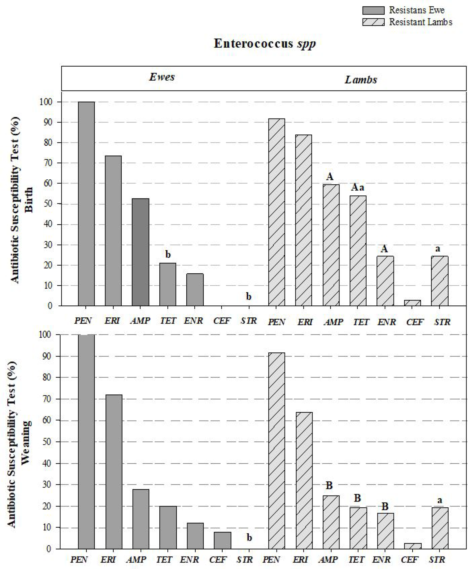
The results show that ewes and lambs presented P. Enterococcus spp. and Coliforms bacteria and, on the antimicrobial susceptibility test, most of the antibiotics were characterized on the three evaluated levels (sensible, intermediate and resistant). During the birth and weaning periods, ewes and lambs exhibited P. Enterococcus spp. bacteria with high resistance to erythromycin and penicillin. Lambs showed greater resistance to tetracycline and streptomycin compared to their progenitors at birth, and to streptomycin at weaning (P<0.05). When comparing the two periods (birth and weaning), a reduction in bacterial resistance to tetracycline, ampicillin and enrofloxacin was observed in the lambs (P<0.05, Figure 1).

Antimicrobial susceptibility testing (AST) in Santa Inês ewes and lambs at birth and weaning period. Graphics describe the percentage (%) in antibiotics resistant level, on Presumptive Enterococcus spp. bacteria at birth and weaning period. ab shows the antibiotic difference between ewes and lambs from the same category (birth or weaning; P<0.05). AB shows the differences between the antibiotics in lambs within birth and weaning period or in ewe within birth and weaning period (P<0.05). PEN: Penicillin, ERI: Erythromycin, AMP: Ampicillin, TET: Tetracycline, ENR: Enrofloxacin, CEF: Ceftiofur, STR: Streptomycin.
On the other hand, regarding susceptibility levels, ewes showed higher sensitivity of P. Enterococcus spp. to streptomycin compared to lambs at both birth and weaning, as well as greater sensitivity to tetracycline at birth (P<0.05). Lambs, in contrast, exhibited increased bacterial sensitivity to ampicillin and tetracycline by the weaning period (P<0.05).
In the susceptible category, ewes demonstrated higher percentages of sensitivity to ampicillin, tetracycline, enrofloxacin and streptomycin (P>0.05), with differences observed for tetracycline (76.32% in ewes versus 43.24% in lambs; P<0.05) and streptomycin (94.74% in ewes versus 59.46% in lambs; P<0.05; Table 3). No significant differences were observed in the intermediate category (P>0.05; Table 3). Lambs presented higher percentages of intermediate levels to erythromycin, tetracycline, enrofloxacin and streptomycin, especially at weaning.
Antibiotic susceptibility test (AST) percentages of ewes versus lambs and the birth and weaning periods.
In the resistant category, both groups showed high resistance to penicillin (≥ 91.67%), although lambs presented higher resistance rates to tetracycline and enrofloxacin; differences were only observed in streptomycin at weaning (19.44% in lambs versus 0.00% in ewes; P<0.05; Table 3).
The Coliform bacteria showed high resistance to penicillin, with resistance levels of 87.80% (ewes) and 92.68% (lambs) at birth, and 100% during the weaning period in both categories. No significant differences were observed between ewes and lambs during the same period (birth or weaning), highlighting the sharing of bacterial resistance between progenitors and their offspring. Coliform bacteria also exhibited an increase in resistance to ceftiofur from birth to weaning (P<0.05). In contrast, resistance to ampicillin in ewes and to both ampicillin and enrofloxacin in lambs significantly decreased from birth to weaning (P<0.05; Figure 2).

Antimicrobial susceptibility testing (AST) in Santa Inês ewes and lambs at birth and weaning period. Graphics describe the percentage (%) in antibiotics resistance level, on Coliforms bacteria at birth and weaning period. ab shows the antibiotic difference between ewes and lambs from the same category (birth or weaning; P<0.05). AB shows the differences between the antibiotics in lambs within birth and weaning period or in ewe within birth and weaning period (P<0.05).
At other susceptibility levels, it was observed that Coliform bacteria in ewes exhibited increased sensitivity to ampicillin and decreased sensitivity to ceftiofur from birth to weaning (P<0.05). Similarly, lambs also exhibited a decrease in sensitivity to ceftiofur, mirroring the response seen in their dams (P<0.05; Table 3).
In the susceptible category, Coliforms spp. isolated from ewes showed a higher percentage of sensitivity to ampicillin and tetracycline at birth when compared to isolates from lambs. However, during the weaning period, ewes and lambs presented similar sensitivity rates across the antibiotics tested, with no expressive variations (P>0.05).
In the intermediate category, no statistically significant differences were observed between the groups (P> 0.05). Nevertheless, lambs exhibited slightly higher percentages for ampicillin, enrofloxacin (both 12.90 %) and streptomycin (29.03 %) at weaning compared to ewes. For the other antimicrobials, intermediate levels remained low and comparable between the groups.
In the resistant category, both ewes and lambs exhibited high resistance rates to penicillin (≥ 87.80 %) and moderate resistance levels to erythromycin during both periods (≥ 46.43 %) evaluated. Lambs showed greater resistance to ampicillin at birth (65.85 %), while resistance percentages for the remaining antibiotics were similar within each susceptibility category (P> 0.05). These findings suggest subtle differences (not statistically) in the resistance profiles between adult ewes and young lambs, possibly related to age, microbiota composition and prior antimicrobial exposure.
Hematological and biochemical analyses were performed to assess sanitary status, and the results are presented in Table 4 of this study. No clinical signs of disease were observed in either ewes or lambs during the evaluation period. Their hematological and biochemical parameters remained within species-specific reference ranges. Lambs exhibited naturally low leukocyte and glucose levels at birth, while ewes showed elevated urea levels during the peripartum period. These fluctuations reflect normal physiological adaptations to birth and weaning rather than pathological conditions. This highlights the importance of interpreting laboratory values within the context of the animal’s physiological state and production stage, which is essential for effective health monitoring and early detection of potential disorders in ovine production systems.
Period effects (Birth and weaning) on hemogram measurements and blood biochemistry on Santa Inês ewes and lambs.
4. Discussion
The intestinal microbiota colonization was traditionally thought to begin only after direct contact with the feces and the progenitor microbiota (13). However, studies performed in humans and animals have shown the presence of microorganisms in fetal membranes, amniotic fluid, womb (20, 21, 22) and meconium (23), which indicates bacterial colonization prior to birth. Research on animals and human samples suggest that the microbial colonization process is particularly meaningful at the beginning of life, once this period consists of a critical window to the immunological and physiological system development (24).
Our results have shown the bacteria P. Enterococcus spp. and Coliforms in antimicrobial susceptibility tests during the birth and weaning periods; highlighting the maternal-descendant resemblance of bacterial resistance, displayed at birth and manteined until the weaning period. P. Enterococcus spp. is naturally resistant to semisynthetic penicillin, added to an acquired resistance to chloramphenicol, erythromycin and other drug classes such as aminoglycosides, tetracycline and fluoroquinolones, as evaluated in our study (25, 26).
The existence of a placental microbiota remains controversial; however, recent studies suggest that metabolite transport derived from commensal bacteria can occur across the fetal membrane barrier. The progenitor intestinal microbiota plays a fundamental role in maternal-fetal transfer, showing the efficiency in modulating fetal development (27). The baby bacterial microbiome is 63% compatible with his/her mother, indicating a bacterial colonization transfer (28).
Based on our results, the Coliforms resistance on antibiotics with large-scale use, such as penicillin, was higher than 90%. Similar results were observed in European Union Countries, with antimicrobial susceptibility of Coliforms, Enterococcus spp and other commensals tested in poultry, swines and bovines. That same study found a low or inexistent resistance in Coliforms isolated from newer antimicrobials used solely in humans, while old antimicrobials widely applied on veterinarian medicine, such as penicillin, (29) ampicillin and tetracycline, showed strong resistance (30).
Studies investigating antimicrobial resistance in swine demonstrated that coliform isolates from piglets were more resistant to ampicillin and azithromycin when their respective dams also carried resistant strains (31, 32). These findings suggested the occurrence of vertical transmission of antimicrobial resistance traits from sow to offspring. However, despite these indications, the mechanisms underlying perinatal transfer of resistant bacteria or resistance genes remained poorly understood, emphasizing a significant gap in the current scientific knowledge (33). The initial colonization of the neonatal microbiota is influenced by the progenitor and the environmental microbiota during the birth period (34). While the perinatal transference in animals and humans may be questioned, on the other hand, resistant bacteria can transmit their resistance genes through vertical or horizontal transmission to other bacterial species (35).
Although the animals used in this study had no prior history of treatment with penicillin or erythromycin, a high level of bacterial resistance to these antimicrobials was observed among the isolates of Coliforms spp. and P. Enterococcus spp. This finding suggests that the resistance detected is not necessarily associated with the direct use of these drugs but may result from mechanisms such as intrinsic resistance, co-selection or horizontal gene transfer. Briefly explained, intrinsic resistance refers to the natural ability of certain bacteria to withstand specific antimicrobials. Co-selection occurs when the use of one antibiotic indirectly promotes resistance to other structurally related drugs. Horizontal gene transfer, in turn, allows resistance genes to be exchanged between bacteria, facilitating the spread of resistance (36, 37).
Penicillin, a β-lactam antibiotic, has limited activity against many Gram-negative bacteria, such as Coliforms spp, due to the presence of an outer membrane that restricts drug penetration, along with the frequent production of β-lactamases enzymes that hydrolyze the β-lactam ring, thereby inactivating the antibiotic (3). In Enterococcus spp., resistance to penicillin is often considered as intrinsic, due to the low affinity of PBPs. These mechanisms may persist within the microbiota even in the absence of direct selective pressure (38).
Regarding erythromycin, a macrolide that inhibits bacterial protein synthesis by binding to the 50S ribosomal subunit, resistance observed in Enterococcus spp. and Coliforms spp. may be related to changes in membrane permeability, ribosomal target modification or activation of efflux pumps (8).
Thus, mobile genetic elements such as plasmids, transposons and integrons, which mediate horizontal gene transfer, circulate among commensal and pathogenic bacteria present in the environment, food, water, or even through indirect contact with other animals previously exposed to antimicrobials (39). Co-selection can further support the persistence and dissemination of these resistance genes, even in the absence of direct use of penicillin or erythromycin (40).
Therefore, the resistance observed in this study may reflect not only intrinsic characteristics of the bacterial species but also the complex dynamics of resistance gene dissemination in agricultural environments, regardless of direct exposure to the specific antimicrobials in question.
Based on the data herein obtained, we observed the presence of bacteria resistant to ceftiofur, an antibiotic not used in the evaluated herd. In this case, we considerate the existence of bacteria with the same capacity to recognize newer antibiotics, which presents similar or same actions to those against which they already demonstrate resistance. For instance, ceftiofur, which causes cellular lysis and interrupts cross-linking with the peptidoglycan and cell wall formation, may be a negative answer from bacteria already accustomed to similar antimicrobial attacks (3, 41) such as penicillin and ampicillin, which showed high percentages of resistance in this study.
Finally, it is important to highlight that an intermediate level does not guarantee a complete resistance or a future susceptibility; rather, it represents an average point in the susceptibility test. Oscillation was evident in our results, indicating that environmental characteristics, animal specific traits and other less understood factors influence this level of higher sensibility or stronger resistance. The evolution of bacterial resistance is frequently related to the incorrect use of many medicines. This issue concerns scientists worldwide, as resistance is hindering the treatment of several infectious diseases. Therefore, continuous monitoring, responsible antimicrobial use and further research into resistance mechanisms are essential to preserve the efficacy of current therapies and ensure the sustainability of animal and public health interventions (42).
5. Conclusion
This study identified similar antimicrobial resistance patterns in Coliforms and P. Enterococcus spp. isolated from sheep and their offspring during birth and weaning. Isolates showed greater susceptibility to ceftiofur, tetracycline, streptomycin, ampicillin and enrofloxacin, as well as higher resistance to erythromycin and penicillin. These findings highlight the need for targeted investigations in different production systems to support effective AMR control and responsible antimicrobial use in sheep farming.
Acknowledgment
The authors are grateful for the financial support provided by the Brazilian National Council for Scientific and Technological Development (CNPq) and the São Paulo Research Foundation (FAPESP).
Data availability statement
The data generated and analyzed during this study will be made available upon request.
References
-
1 World Health Organization (WHO). Antimicrobial Resistance: Global Report on Surveillance. Geneva: World Health Organization; 2014. Available from: https://apps.who.int/iris/handle/10665/112642 [Accessed 10 July 2024]
» https://apps.who.int/iris/handle/10665/112642 -
2 Office International des Epizooties (OIE). Resistência antimicrobiana. Disponível em: https://www.woah.org/en/what-we-do/global-initiatives/antimicrobial-resistance/ [Accessed 01 July 2024].
» https://www.woah.org/en/what-we-do/global-initiatives/antimicrobial-resistance -
3 Alcock BP, Raphenya AR., Lau TTY, et al. CARD: antibiotic resistome surveillance with the comprehensive antibiotic resistance database. Nucleic Acids Res. 2020;48 (D1): D517-D525. https://doi.org/10.1093/nar/gkz935
» https://doi.org/10.1093/nar/gkz935 -
4 Zhang T, Niu G, Boonyayatra S, Pichpol D. Antimicrobial resistance profiles and genes in Streptococcus uberis associated with bovine mTSAitis in Thailand. Front Vet Sci. 2021;8:705338. https://doi.org/10.3389/fvets.2021.705338
» https://doi.org/10.3389/fvets.2021.705338 -
5 Melo MR, Almeida E, Hofer E, et al. Antibiotic resistance of Vibrio parahaemolyticus isolated from pond-reared Litopenaeus vannamei marketed in Natal, Brazil. Braz J Microbiol. 2011;42(4):1463-1469. https://doi.org/10.1590/S1517-83822011000400032
» https://doi.org/10.1590/S1517-83822011000400032 -
6 Grabowski Ł, Gaffke L, Pierzynowska K, et al. Enrofloxacin: the ruthless killer of eukaryotic cells or the lTSA hope in the fight against bacterial infections? Int J Mol Sci. 2022;23(7):3648. https://doi.org/10.3390/ijms23073648
» https://doi.org/10.3390/ijms23073648 -
7 Riboldi GP, Frazzon J, d'Azevedo PA, Frazzon AP. Antimicrobial resistance profile of Enterococcus spp. isolated from food in Southern Brazil. Braz J Microbiol. 2009;40(1):125-128. https://doi.org/10.1590/S1517-83822009000100021
» https://doi.org/10.1590/S1517-83822009000100021 -
8 Kaszanyitzky ÉJ, Tenk M, Ghidán Á, Fehérvári GY, Papp M. Antimicrobial susceptibility of enterococci strains isolated from slaughter animals on the data of Hungarian resistance monitoring system from 2001 to 2004. Int J Food Microbiol. 2007;115:119- 123. https://doi.org/10.1016/j.ijfoodmicro.2006.10.004
» https://doi.org/10.1016/j.ijfoodmicro.2006.10.004 -
9 López M, Sáenz Y, Rojo-Bezares B, et al. Detection of vanA and vanB2-containing enterococci from food samples in Spain, including Enterococcus faecium strains of CC17 and the new singleton ST425. Int J Food Microbiol. 2009;133:172-178. https://doi.org/10.1016/j.ijfoodmicro.2009.05.020
» https://doi.org/10.1016/j.ijfoodmicro.2009.05.020 -
10 Acha PN, Szyfres B. Zoonosis y enfermedades transmisibles comunes al hombre y a los animales: Volumen 1: bacteriosis and micosis. 3rd ed. Washington: Organização Panamericana de la Salud; 2001. 398 p. (Publicación Científica y Técnica, 580). https://doi.org/10.1590/S0102-311X2005000300038
» https://doi.org/10.1590/S0102-311X2005000300038 -
11 Guo W, Bi SS, Wang WW, Zhou M, Neves ALA, Degen AA, Guan LL, Long RJ. Maternal rumen and milk microbiota shape the establishment of early-life rumen microbiota in grazing yak calves. J Dairy Sci. 2023;106(3):2054-2070. https://doi.org/10.3168/jds.2022-22655
» https://doi.org/10.3168/jds.2022-22655 -
12 Messman RD, Lemley CO. Bovine neonatal microbiome origins: A review of proposed microbial community presence from conception to colostrum. Transl Anim Sci. 2023;7:txad057. https://doi.org/10.1093/tas/txad057
» https://doi.org/10.1093/tas/txad057 -
13 Perez-Muñoz ME, Arrieta MC, Ramer-Tait AE, Walter J. A critical assessment of the "sterile womb" and "in utero colonization" hypotheses: implications for research. Microbiome. 2017;5:48. https://doi.org/10.1186/s40168-017-0268-4
» https://doi.org/10.1186/s40168-017-0268-4 -
14 Rackaityte E, Halkias J, Fukui EM, Mendoza VF, Hayzelden C, Crawford ED, et al. Viable bacterial colonization is highly limited in the human intestine in utero. Nat Med. 2020;26(4):599-607. https://doi.org/10.1038/s41591-020-0761-3
» https://doi.org/10.1038/s41591-020-0761-3 -
15 AOAC, Association of Official Analytical Chemists - International. Official Methods of Analysis. 23st ed. Gaithersburg, MD, USA; 2023. https://www.aoac.org/official-methods-of-analysis/
» https://www.aoac.org/official-methods-of-analysis/ -
16 Mertens DR. Measuring fiber and its effectiveness in ruminant diets. US Dairy Forage Research Center, USDA-ARS, Madison, WI; 2002. www.nutritionmodels.com/papers/MertensPNC2002.pdf
» www.nutritionmodels.com/papers/MertensPNC2002.pdf -
17 EUCTSA, The European Committee on Antimicrobial Susceptibility Testing, 2018. Available from: https://www.eucTSA.org Accessed on: 01 july 2024
» https://www.eucTSA.org -
18 CLSI, Clinical and Laboratory Standards Institute, Inc. [Internet]. 2018 [cited 2024 Jul 01]. Available from: https://clsi.org/
» https://clsi.org/ - 19 Sampaio, IBM. Estatística aplicada à experimentação animal. Fundação de Ensino e Pesquisa em Medicina Veterinária e Zootecnia. 3.ed. Belo Horizonte - MG, 2002, 265p.
-
20 Stinson LF, Boyce MC, Payne MS, Keelan JA. The not so sterile womb: evidence that the human fetus is exposed to bacteria before birth. Front Microbiol. 2019;10:1124. https://doi.org/10.3389/fmicb.2019.01124
» https://doi.org/10.3389/fmicb.2019.01124 -
21 Salter SJ, Cox MJ, Turek EM, et al. Reagent and laboratory contamination can critically impact sequence-based microbiome analyses. BMC Biol. 2014;12:87. https://doi.org/10.1186/s12915-014-0087-x
» https://doi.org/10.1186/s12915-014-0087-x -
22 Winters AD, Romero R, Greenberg JM, et al. Does the amniotic fluid of mice contain a viable microbiota? Front Immunol. 2022;13:820366. https://doi.org/10.3389/fimmu.2022.820366
» https://doi.org/10.3389/fimmu.2022.820366 -
23 Al-Balawi M, Morsy FM. Prenatal versus postnatal initial colonization of healthy neonates' colon ecosystem by the Enterobacterium Escherichia coli. Microbiol Spectr. 2021;9(2):e0050621. https://doi.org/10.1128/Spectrum.00379-21
» https://doi.org/10.1128/Spectrum.00379-21 -
24 Martínez I, Maldonado-Gomez MX, et al. Experimental assessment of the importance of colonization history in shaping the gut microbiota in early life. eLife. 2018;7:e36521. https://doi.org/10.7554/eLife.36521
» https://doi.org/10.7554/eLife.36521 -
25 Mancuso G, Midiri A, Gerace E, Biondo C. Bacterial Antibiotic Resistance: The Most Critical Pathogens. Pathogens. 2021 Oct 12;10(10):1310. https://doi.org/10.3390/pathogens10101310
» https://doi.org/10.3390/pathogens10101310 -
26 Kristich CJ, Rice LB, Arias CA. Enterococcal Infection-Treatment and Antibiotic Resistance. 2014 Feb 6. In: Gilmore MS, Clewell DB, Ike Y, et al., editors. Enterococci: From Commensals to Leading Causes of Drug Resistant Infection [Internet]. Boston: Massachusetts Eye and Ear Infirmary; 2014-. Available from: https://www.ncbi.nlm.nih.gov/books/NBK190420/
» https://www.ncbi.nlm.nih.gov/books/NBK190420/ -
27 Macpherson AJ, de Agüero MG, Ganal-Vonarburg SC. How nutrition and the maternal microbiota shape the neonatal immune system. Nat Rev Immunol. 2017;17(8):508-517. https://doi.org/10.1038/nri.2017.58
» https://doi.org/10.1038/nri.2017.58 -
28 Maqsood R, Rodgers R, Rodriguez C. Discordant transmission of bacteria and viruses from mothers to babies at birth. Microbiome. 2019;7:156. https://doi.org/0.1186/s40168-019-0766-7
» https://doi.org/0.1186/s40168-019-0766-7 -
29 Haulisah NA, Hassan L, Bejo SK, et al. High levels of antibiotic resistance in isolates from diseased livestock. Front Vet Sci. 2021;8:652351. https://doi.org/10.3389/fvets.2021.652351
» https://doi.org/10.3389/fvets.2021.652351 -
30 De Jong A, Bywater R, Butty P, et al. A pan-European survey of antimicrobial susceptibility towards human-use antimicrobial drugs among zoonotic and commensal enteric bacteria isolated from healthy food-producing animals. J Antimicrob Chemother. 2009;63(4):733-744. https://doi.org/10.1093/jac/dkp012
» https://doi.org/10.1093/jac/dkp012 -
31 O’Neill L, García Manzanilla E, Ekhlas D, et al. Antimicrobial resistance in commensal Escherichia coli of the porcine gTSArointestinal tract. Antibiotics (Basel). 2023;12:1616. https://doi.org/10.3390/antibiotics12111616
» https://doi.org/10.3390/antibiotics12111616 -
32 Burow E, Rostalski A, et al. Antibiotic resistance in Coliforms from pigs from birth to slaughter and its association with antibiotic treatment. Prev Vet Med. 2019;165:52-62. https://doi.org/10.1016/j.prevetmed.2019.02.008
» https://doi.org/10.1016/j.prevetmed.2019.02.008 -
33 Chen F, Qingqing W, Wei-Dong W, Yan-Dong. Microbiota intestinal: um moderador integral em saúde e doença. Front Microbiol. 2018;9:151. https://doi.org/10.3389/fmicb.2018.00151
» https://doi.org/10.3389/fmicb.2018.00151 -
34 Seale, J., Millar, M., 2014. Perinatal vertical transmission of antibiotic-resistant bacteria: a systematic review and proposed research strategy. BJOG: An International Journal of Obstetrics and Gynaecology, 121, 923-928. https://doi.org/10.1111/14710528.12746
» https://doi.org/10.1111/14710528.12746 -
35 Holmes AH, Moore LS, Sundsfjord A, et al. Understanding the mechanisms and drivers of antimicrobial resistance. Lancet. 2016;387:176-187. https://doi.org/10.1016/S0140-6736(15)00473-0
» https://doi.org/10.1016/S0140-6736(15)00473-0 -
36 Murray LM, Hayes A, Snape J, et al. Co-selection for antibiotic resistance by environmental contaminants. npj Antimicrob Resist. 2024;2:9. https://doi.org/10.1038/s44259-024-00026-7
» https://doi.org/10.1038/s44259-024-00026-7 -
37 Goh YX, Anupoju SMB, Nguyen A, Zhang H, Ponder M, Krometis LA, Pruden A, Liao J. Evidence of horizontal gene transfer and environmental selection impacting antibiotic resistance evolution in soil-dwelling Listeria. Nat Commun. 2024;15:54459. https://doi.org/10.1038/s41467-024-54459-9
» https://doi.org/10.1038/s41467-024-54459-9 -
38 Hollenbeck BL, Rice LB. Intrinsic and acquired resistance mechanisms in Enterococcus Virulence. 2012;3(5):421-433. https://doi.org/10.4161/viru.21282
» https://doi.org/10.4161/viru.21282 -
39 Verraes C, Van Boxstael S, Van Meervenne E, et al. Antimicrobial resistance in the food chain: A review. Int J Environ Res Public Health. 2013;10(7):2643-2669. https://doi.org/10.3390/ijerph10072643
» https://doi.org/10.3390/ijerph10072643 -
40 Seiler C, Berendonk TU. Heavy metal driven co-selection of antibiotic resistance in soil and water bodies impacted by agriculture and aquaculture. Front Microbiol. 2012;3:399. https://doi.org/10.3389/fmicb.2012.00399
» https://doi.org/10.3389/fmicb.2012.00399 -
41 Gordo I. Evolutionary change in the human gut microbiome: from a static to a dynamic view. PLoS Biol. 2019;17:2. https://doi.org/10.1371/journal.pbio.3000126
» https://doi.org/10.1371/journal.pbio.3000126 -
42 Centers for Disease Control and Prevention (CDC). Antibiotic Resistance Threats in the United States, 2019 [Internet]. Atlanta (GA): Centers for Disease Control and Prevention (US); 2019 [cited 2024 Jul 29]. Available from: https://www.cdc.gov/drugresistance/pdf/threats-report/2019-ar-threats-report-508.pdf
» https://www.cdc.gov/drugresistance/pdf/threats-report/2019-ar-threats-report-508.pdf
Edited by
-
Editor:
Rondineli P. Barbero