Radiographic patterns and positioning recommendations for skull radiology in the brown brocket, Subulo gouazoubira (Fischer,1814)
Abstract
Incidents related to human activities, such as roadkill and illegal hunting, result in a high number of small deer being referred to triage and rehabilitation centers in Brazil, with multiple fractures being the most common injuries. Rapid and standardized radiographic diagnosis is essential for the emergency management of these animals. This study evaluated the most efficient radiographic projections for examining the cranial structures of the brown brocket deer (Subulo gouazoubira), using heads of eight individuals that died during the rehabilitation process at Wildlife Rehabilitation Center (Wildlife Rehabilitation Center) and which, after radiographic analysis procedures, were osteologically prepared for comparison. The most suitable projections proved to be the lateral, lateral with natural obliquity, dorsoventral, and rostrocaudal views, with the ventrodorsal view at 20° with 45° ventroflexion indicated as complementary for evaluating the tympanic bulla. The results highlight the importance of specific positioning protocols for accurate visualization of the nasal cavity, paranasal sinuses, and tympanic bullae. This study establishes, for the first time, a standardized emergency radiographic protocol for cervids with suspected cranial trauma, contributing to faster diagnoses and more assertive clinical decisions in rehabilitation centers.
Keywords:
cervid; diagnostic imaging; head
Resumo
Incidentes relacionados a atividades humanas, como atropelamentos e caça ilegal, resultam em um elevado número de cervídeos de pequeno porte encaminhados a centros de triagem e reabilitação no Brasil, sendo as multifraturas as lesões mais comuns. O diagnóstico radiográfico rápido e padronizado é essencial para o manejo emergencial desses animais. Este estudo avaliou as projeções radiográficas mais eficientes para examinar as estruturas cranianas do veado-catingueiro (Subulo gouazoubira), utilizando cabeças de oito indivíduos que foram a óbito ao longo do processo de reabilitação no Núcleo de Reabilitação da Fauna Silvestre/Centro de Triagem de Animais Silvestres da Universidade Federal de Pelotas (Núcleo de Reabilitação da Fauna Silvestre/Centro de Triagem de Animais Silvestres da Universidade Federal de Pelotas) e que, após procedimentos de análise radiográfica, foram preparados osteologicamente para comparação. As projeções mais adequadas se mostraram ser a látero-lateral, látero-lateral com obliquidade natural, dorsoventral e rostrocaudal, sendo a ventrodorsal a 20° com ventroflexão de 45° indicada como complementar para avaliação da bula timpânica. Os resultados reforçam a importância de protocolos específicos de posicionamento para a visualização precisa da cavidade nasal, seios paranasais e bulas timpânicas. Este trabalho estabelece, pela primeira vez, um protocolo radiográfico emergencial padronizado para cervídeos com suspeita de traumatismo craniano, contribuindo para diagnósticos mais ágeis e decisões clínicas mais assertivas em centros de reabilitação.
Palavras-chave:
cabeça; cervídeo; diagnóstico por imagem.
1. Introduction
Subulo gouazoubira Fischer, 1814, commonly known as the brown brocket deer, is one of the eight cervid species found in Brazil (1), with documented presence across all brazilian biomes (2, 3). Although the species is globally classified as “Least Concern” (LC) (4) it holds varying conservation statuses within Brazil due to its broad geographic distribution and the condition of local populations. In the state of Rio Grande do Sul, it is listed as “Vulnerable” (VU); in Paraná, it is categorized as “Data Deficient” (DD); and in Rio de Janeiro, it is considered “Endangered” (EN) (5-7). Among the main threats to its populations are habitat loss, poaching, predation by domestic dogs in rural areas, and roadkill incidents (8). Through the Brazilian Institute of the Environment and Renewable Natural Resources (IBAMA), wildlife screening and rehabilitation centers located in various Brazilian states provide care for animals found injured or seized from illegal capture and breeding. The caseload of cervids in these centers is high, primarily due to vehicle collisions and other anthropogenic interactions (9). Among these patients, multiple fractures are among the most frequent injuries (10), often leading to death. Mortality among rescued individuals may be linked to the complexity and inherent risks of managing cervids in captivity, given their biological characteristics of low stress tolerance and poor adaptability to handling, which hinders essential diagnostic, treatment, and rehabilitation procedures (10). Therefore, a rapid and accurate diagnosis is a key factor in achieving successful case resolution and initiating timely treatment, ultimately maximizing the chances of survival and full rehabilitation.
In wildlife veterinary medicine, clinicians must be knowledgeable about and apply appropriate handling techniques and clinical examination techniques to assess the patients efficiently, while minimizing animal stress and ensuring team safety (11). In domestic species such as dogs and cats, handling and restraint are facilitated by domestication, and the species' anatomy is well understood. Additionally, there are standardized protocols for radiographic examinations that enable the rapid acquisition of high-quality diagnostic images (12). These protocols define appropriate restraint methods, body positioning, and beam incidence angles to optimize the projection of anatomical structures of interest.
For wild animals, although there are several documented protocols for radiographic and ultrasonographic anatomy (13,14), these resources are often limited in scope when considering the extensive biodiversity of fauna in need of conservation. Consequently, significant knowledge gaps remain, even for widely distributed species. This is the case for the brown brocket deer (S. gouazoubira), for which existing radiographic and osteological studies have primarily focused on age estimation (15) and descriptions of the thoracic limb bones (16). More recently (17), conducted a comprehensive study on S. gouazoubira and Mazama nana, highlighting critical aspects of cranial bone morphology using computed tomography. While CT provides high-definition anatomical detail, conventional radiography remains the most commonly used imaging modality in wildlife rehabilitation centers in Brazil. However, radiographic interpretation is more challenging due to the superimposition of anatomical structures, which is highly influenced by head positioning during image acquisition. Furthermore, anatomical references based on other cervid species often lack species-specific traits, limiting their applicability and accuracy in diagnostic settings (18).
The present study aims to determine the ideal positioning of the animal for radiographic examination of the head, as well as to demonstrate and identify the normal anatomical patterns of the cranial bony structures visualized in radiographic images.
2. Material and methods
A total of eight deceased specimens of S. gouazoubira (5 males and 3 females) originating from the Pampa Biome, in the vicinity of the municipality of Pelotas, RS, Brazil, were used in this study. S. gouazoubira. The specimens were legally received by the Wildlife Rehabilitation Center (NURFS/CETAS/UFPel) under IBAMA authorization, and their use for scientific purposes is institutionally exempt from CEUA evaluation. Therefore, the project fits the criteria for ethical review exemption, involving only material from animals that were not subjected to any procedures while alive. Only animals that had died naturally during treatment attempts without evident head injuries were selected. The carcasses were frozen at -20°C immediately after death and thawed at room temperature prior to processing. Once thawed, the animals were externally inspected, measured, and identified by species and sex. At that time, it was not possible to determine their precise stage of physical maturity, as all individuals had similar body sizes within the range described for adult animals (8).
The radiographic system used consisted of an Orange 1060HF X-ray generator and a direct digital acquisition plate (AGFA DX-D 40G). The focal distance was set at 67 cm, with an exposure of 65 kV and 1.6 mAs. An established radiographic protocol for dogs and cats (19) was initially tested, encompassing ten radiographic views targeting specific projections and focal areas: right lateral, right oblique lateral (mandible in natural obliquity), right oblique lateral (maxilla), lateral oblique with 45° rotation (tympanic bulla), ventrodorsal, dorsoventral, ventrodorsal with 20° rotation and 45° ventroflexion (tympanic bulla), rostrocaudal (nasal cavity and paranasal sinuses), rostrocaudal with 20° rotation (foramen magnum), and open-mouth rostrocaudal. Positioning for each projection considered the natural angulations and movements permitted by each joint and feasible for use in live animals under sedation.
To interpret the anatomical structures projected in the images, the previously radiographed heads were disarticulated at the atlanto-axial joint and partially dissected to remove soft tissues. They were then macerated in water until all remaining tissues were removed. The preserved bones were disinfected with 20% diluted sodium hypochlorite, followed by bleaching using 40% hydrogen peroxide. Anatomical identification was based on the Nomina Anatomica Veterinaria (20) for the terminology used.
3. Results and discussion
3.1 Laterolateral projection
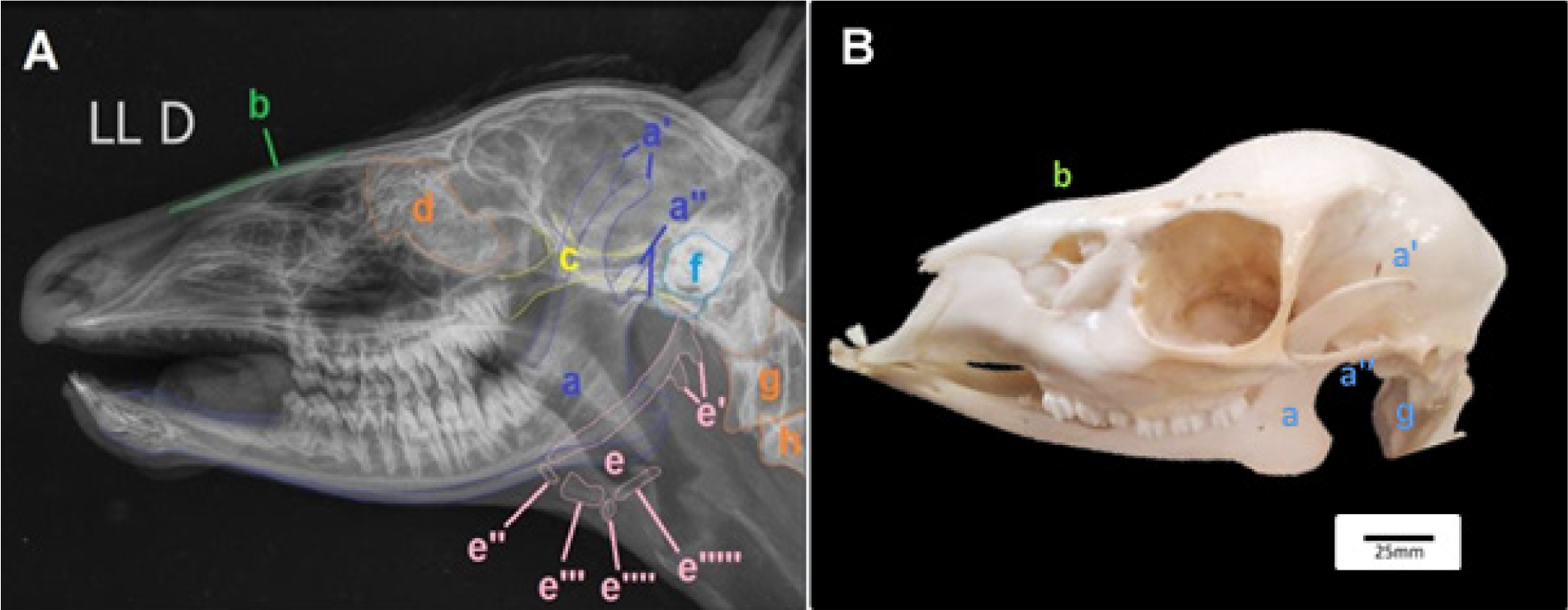
The laterolateral projection proved useful for the evaluation of the nasal cavity and paranasal sinuses, similarly to what is observed in small animals. Clearly visible structures are indicated in Figure 1.
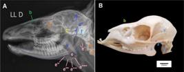
(A) Right laterolateral radiograph (LL) of the S. gouazoubira skull. 2: (B) Left lateral view of the S. gouazoubira skull. a. Mandible; a’. Coronoid process; a’’. Mandibular condyle; b. Nasal bone; c. Zygomatic arch; d. Ethmoidal concha; e. Hyoid apparatus: e’. Stylohyoid, e’’. Epihyoid, e’’’. Ceratohyoid, e’’’’. Basihyoid, e’’’’’. Thyrohyoid; f. Tympanic bullae; g. Atlas; h. Axis. D - right.
3.2 Laterolateral oblique (mandible in natural obliquity) and laterolateral oblique (maxilla)
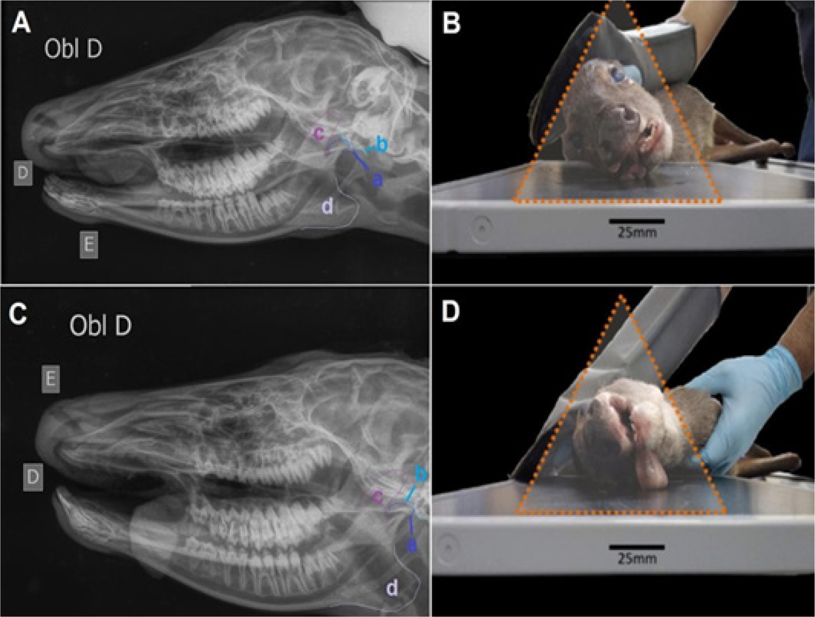
The laterolateral oblique projections proved useful for assessing the teeth, alveolar bone, mandibular ramus and body, and temporomandibular joint (Figure 2), similarly to small animal radiography. However, evaluation of the tympanic bullae was challenging due to anatomical superimposition. Greater obliquity was therefore required to achieve individual visualization of these structures.

A. Right laterolateral oblique radiograph (Obl) of S. gouazoubira with the right mandible in contact with the image receptor (D - right side, E - left side). B. Illustration of the positioning used in image A. C. Right laterolateral oblique radiograph with the maxilla in contact with the image receptor (D - right side, E - left side). D. Illustration of the positioning used in image C. Structures clearly visualized: a. Mandibular condyle; b. Temporal articular surface; c. Coronoid process; d. Angular process.
No significant advantages were observed in the oblique projection with the maxilla in contact with the plate when compared to the mandibular projection. Since the incidence that places the mandibular portion closest to the image receptor can be more easily achieved due to the natural obliquity of the head in a resting position, this view may be considered more efficient due to easier handling and lower risk of technical errors.
3.3 Laterolateral oblique (45° rotation) (tympanic bullae)
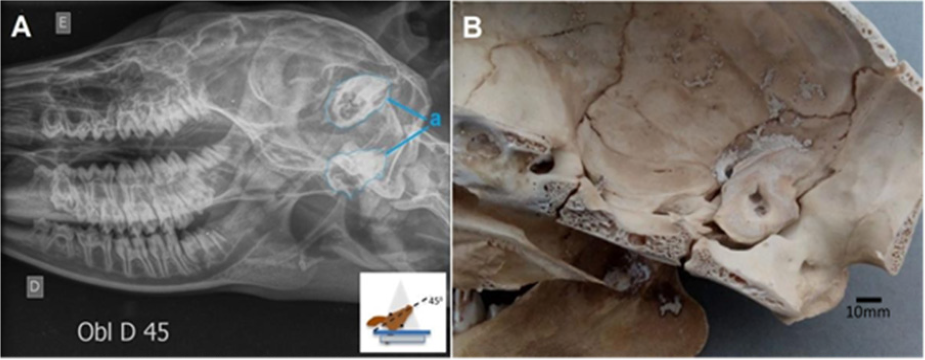
The laterolateral oblique projection highlights the tympanic bulla (Figure 3) in a more ventral topography when performed at a 45-degree angle.
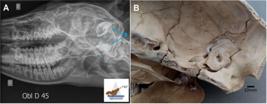
A. Right laterolateral radiograph with 45-degree angle of obliquity (Obl D45) of S. gouazoubira.B. Internal surface of the braincase of S. gouazoubira.a. Tympanic bullae. E (left side) D (right side).
3.4 Ventrodorsal and dorsoventral projections
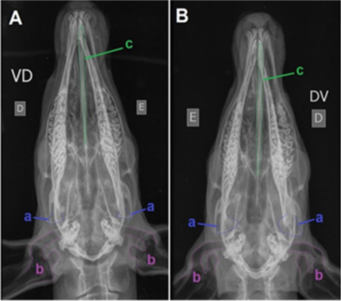
Both dorsoventral and ventrodorsal projections were useful for evaluating the nasal cavity, paranasal sinuses, auditory canals, and other structures commonly assessed in small animal radiography (Figure 4). However, they were less effective for evaluating the tympanic bullae, and these projections can be used complementarily. The positioning for the dorsoventral incidence proved easier to apply compared to the ventrodorsal, as it did not require the use of ropes for positioning and the animal could be placed in a more comfortable decubitus position. The shape of the species' head prevents magnification of structures.
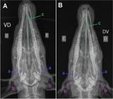
A. Ventrodorsal radiograph (VD) of S. gouazoubira. B. Dorsoventral radiograph (DV) of S. gouazoubira. a. Mandibular condyle; b. Auditory canal; c. mandibular ramus. (E) left side (D) right side.
3.5 Ventrodorsal 20° with (45° ventroflexion) (tympanic bullae)
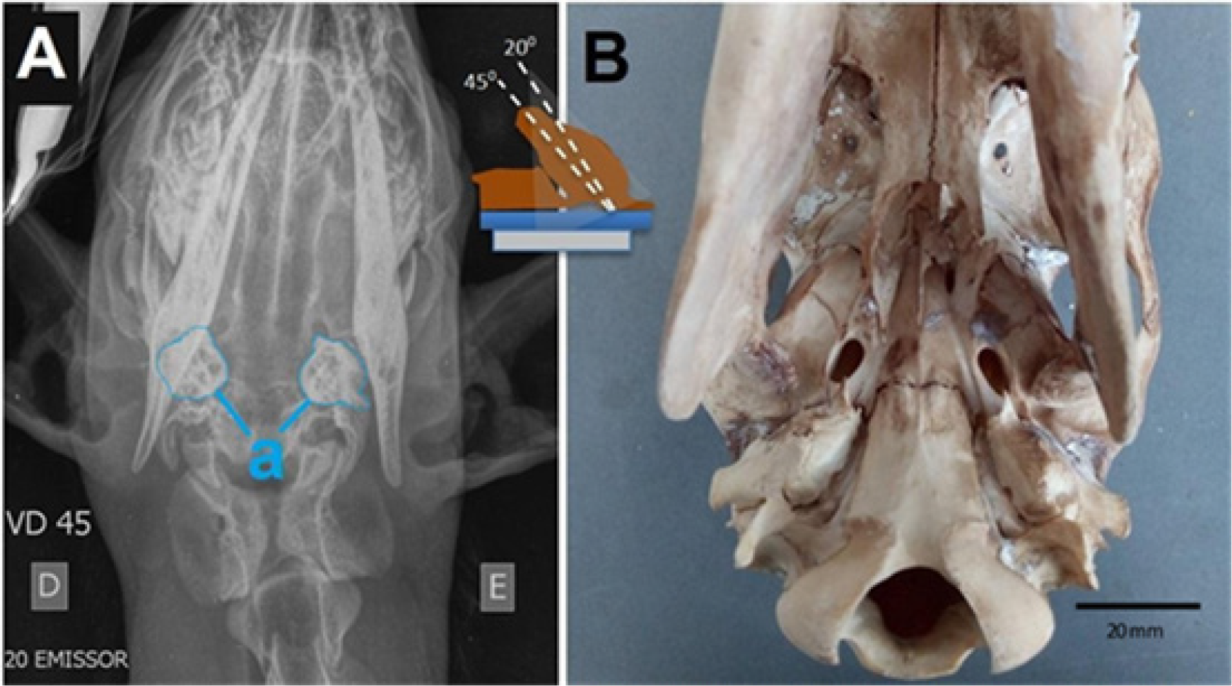
To better evaluate the tympanic bullae, some adjustments were made to the dorsoventral incidence. In this case, cervical flexion was performed at 45 degrees, and the X-ray emitter was positioned at a 20-degree angle, with the beam directed in a caudoventral-craniodorsal incidence (Figure 5).
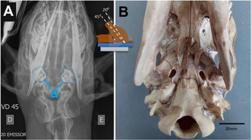
A. Ventrodorsal radiograph with 45° (VD 45) cervical flexion and 20° inclination of the X-ray beam of S. gouazoubira. B. Ventral view of S. gouazoubira skull. a. Tympanic bullae. (E) left side (D) right side. Schematic representation of the head position and beam incidence as indicated in the upper central portion of the figure.
3.6 Rostrocaudal (nasal cavity and paranasal sinuses)
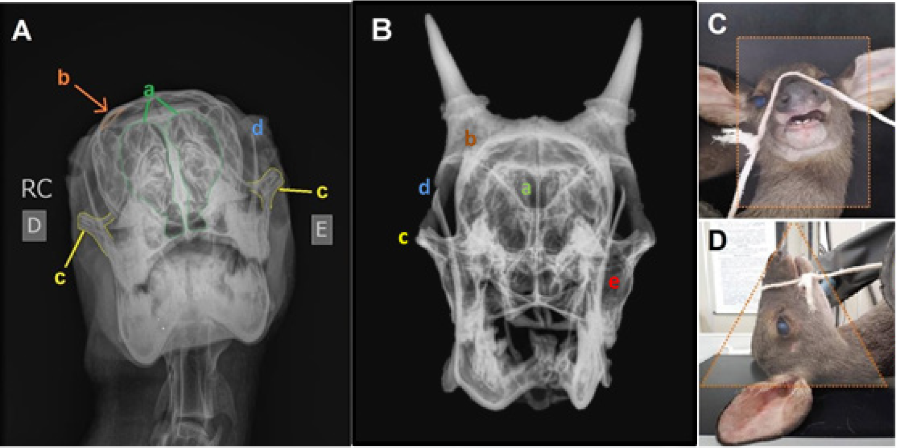
The rostrocaudal projection proved useful for evaluating the nasal cavity and for individualizing the nasal conchae (Figure 6), similarly to small animals.

A. Rostrocaudal radiograph (skull and mandible) of S. gouazoubira B. Rostral radiograph view (bone preparation - male) of S. gouazoubira. C. Illustration of the positioning used in image A. D. Illustration of the positioning used in image A. a. Nasal cavity; b. Frontal sinus; c. Zygomatic process; d. Coronoid process; e. Maxillary sinus (E) left side (D) right side.
3.7 Rostrocaudal 20° (Foramen magnum)
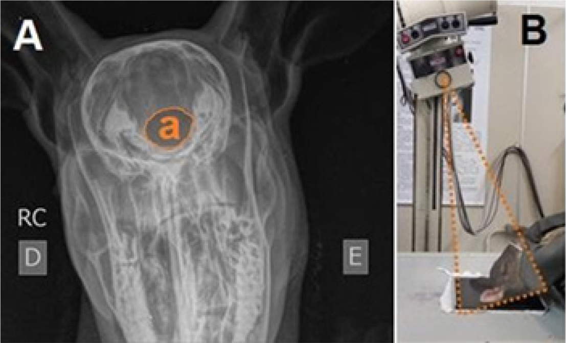
To obtain the Foramen magnum image, the head must be in ventroflexion and the emitter inclined at 20 degrees (Figure 7). This projection showed good individualization of the foramen, although it has limited significance in clinical routine, as no reports of disorders such as Occipital Displasia seen in dogs (21) have been found in cervids.
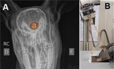
A. Rostrocaudal radiograph (skull and mandible) of S. gouazoubira. B. Illustration of the positioning used in image A. a. Foramen magnum. (E) side (D) right side.
3.8 Rostrocaudal open mouth
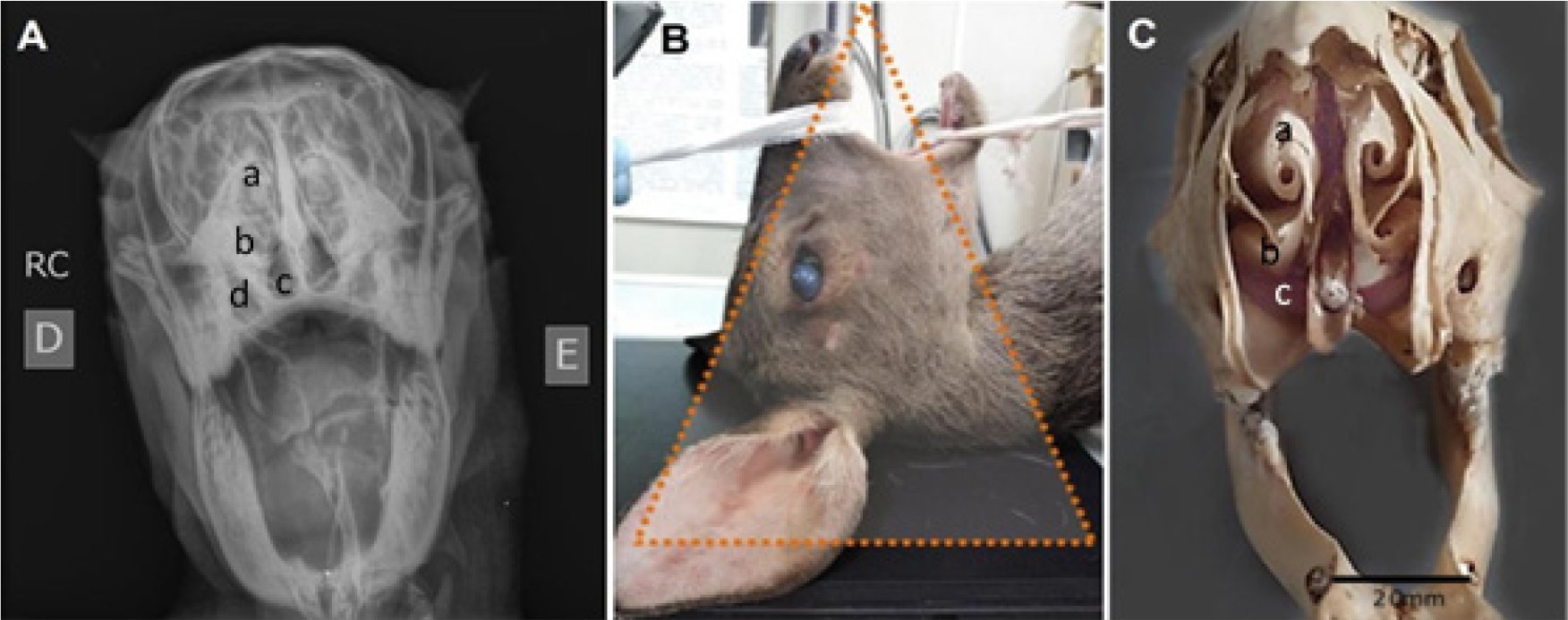
The rostrocaudal open-mouth projection did not provide structural differentiation when compared to the closed-mouth view, and visualization of the tympanic bullae was not achieved as it commonly is in dogs (Figure 8). This limitation is primarily attributed to the restricted degree of mouth opening in this species when compared to carnivores, as well as to the less prominent morphology of the bullae relative to that observed in domestic dogs.
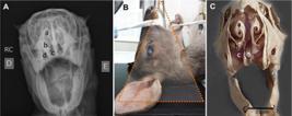
A. Rostrocaudal radiograph (skull and mandible) with an open mouth of S. gouazoubira. a. dorsal nasal conchae; b. ventral nasal conchae; c. Ventral nasal meatus; d. Maxillary sinus. B. Illustration of the positioning used in image A. C. Rostral view of skull osteological preparation (premaxillary bonés absent) of S. gouazoubira. (E) left side (D) right side.
3.9 General discussion
Due to the wide diversity of species within the Cervidae family, several studies addressing skull morphology through osteological preparations, radiographic, and/or computed tomography images have been conducted worldwide (15,17,22,23). Most of these studies (17,22,23) describe skull bone morphology and present radiographic images; however, they do not explore clinical applicability, as they rely on osteologically prepared skulls without soft tissues. This methodological difference limits direct comparison with live-animal imaging, which is the focus of the present study. For example, previous authors (23) indicated the ventrodorsal projection as the most appropriate for visualizing the vomer, nasal septum, nasal cavity, and premolar and molar teeth, a conclusion that does not align with our findings. Such discrepancies are likely related to the use of radiographs obtained exclusively from osteological specimens, lacking the mandible and hyoid apparatus, which in live animals create natural superimpositions that significantly affect image interpretation.
In Brazil, a previous study (15) conducted radiographic and histological assessments of the gonads of M. bororo and S. gouazoubira to estimate ages between 1 and 17 months old and to evaluate gonadal development. The author also presented excellent radiographic images for assessing dental eruption. Aside from the technique of placing the radiographic plate inside the oral cavity up to the limits of the labial commissures, the positioning and radiographic adjustments used in live animals were similar to those employed in the present study, with minor differences in focal distance (0.75 m; 45-60 kV versus 0.67 m; 65 kV in the present work). Given the small size of S. gouazoubira, specimen handling, positioning, and image acquisition protocols were logistically comparable to those used for a lupoid-type dog.
The use of fixed exposure parameters (65 kV; 1.6 mAs) was selected because the skull exhibits minimal variation in tissue thickness among its standard projections. Within this anatomical region, expected differences in attenuation between views are subtle, and the digital radiographic system compensates automatically for such minor fluctuations. Thus, a standardized protocol ensures consistent and comparable image quality throughout the study. The focal distance of 0.67 m likewise followed the technical recommendations of the radiographic equipment manufacturer. Although variations in source-image distance can influence magnification in skull radiography, the effect in the present work was minimal because specimens were positioned uniformly and the object-detector distance remained short and consistent. As a result, magnification remained within acceptable and reproducible limits across all projections. Overall, the obtained images demonstrated excellent quality for visualizing bony structures due to their natural contrast with air-filled cavities of the head.
Importantly, the present study also provides a practical reference standard for rapid radiographic assessment during emergency protocols in cervids presenting with suspected cranial trauma in rehabilitation centers. The detailed projection guidelines, combined with information on superimpositions caused by soft tissues, mandible, and hyoid apparatus, allow clinicians to select the most informative incidences while minimizing repositioning and handling time-critical factors for stressed or debilitated wild animals. Therefore, this radiographic pattern serves as a valuable tool for improving diagnostic efficiency, supporting clinical decision-making, and enhancing patient stabilization during emergency evaluations.
4. Conclusion
Based on the radiographic findings from testing various positioning techniques to obtain the best images of specific cranial and facial structures, it can be concluded that the S. gouazoubira, despite its high vulnerability to stress from handling, can be easily positioned when sedated, provided that positioning priorities are followed based on clinical suspicion. The recommended basic positioning techniques are: laterolateral, laterolateral oblique, dorsoventral, and rostrocaudal, with the additional option of performing the Ventrodorsal 20° (with cervical flexion of 45°) if there is suspicion regarding the tympanic bullae region. By comparing our findings with previous works based on osteological preparations, we highlight important differences in image interpretation associated with soft tissue superimposition in live specimens. Beyond contributing to anatomical and radiographic knowledge of neotropical cervids, the present study establishes a practical and reproducible reference for rapid screening in emergency protocols for wildlife rehabilitation centers. These standardized projections can support early detection of cranial trauma, guide clinical decision-making, and improve the overall care of cervids admitted for evaluation following suspected head injuries.
Data availability statement
The data supporting the findings of this study are available from the corresponding author upon reasonable request.
Generative AI use statement
The authors declare that Artificial Intelligence tools available through the OpenAI GPT platform were used exclusively to assist with orthographic corrections, textual and grammatical agreement adjustments, and reference formatting. The use of these tools did not influence the scientific content, data interpretation, or conclusions of the study. Full responsibility for the conception of the work, data analysis, and final scientific writing rests entirely with the authors.
References
- 1 Duarte JMB, Reis ML. Plano de ação nacional para conservação dos cervídeos ameaçados de extinção. Brasília: Instituto Chico Mendes de Conservação da Biodversidade. 2012; 126.
- 2 Rossi RV, Bodmer R, Duarte JMB. et al. Amazonian brown brocket deer Mazama nemorivaga (Cuvier,1817). Neotropical Cervidology, Biology and Medicine of Latin American Deer. Funep/IUCN. 2010; 393.
- 3 Black-Décima PA. Brown Brocket Deer Mazama gouazoubira (Fisher 1814) Neotropical cervidology: biology and medicine of Latin America deer. 2010; 190-201.
-
4 Black-Decima PA, Vogliotti A. Mazama gouazoubira . Lista vermelha de espécies ameaçadas da IUCN. 2016; Disponível em: https://bibliotecadigital.ufrgs.br/bitstream/handle/10183/283242/001194883.pdf?sequence=1&isAllowed=y
» https://bibliotecadigital.ufrgs.br/bitstream/handle/10183/283242/001194883.pdf?sequence=1&isAllowed=y - 5 Bergallo HG, Rocha CFD, Alves MAS. et al. A fauna Ameaçada de extinção no Estado do Rio de Janeiro. EDURJ. 2000; 66.
- 6 Fontana CS, Bencke GAR. Livro vermelho da fauna ameaçada de extinção no Rio Grande do Sul. EDIPUCRS. 2003; 632.
- 7 Mikich SB, Bérnil SRS. Livro Vermelho da fauna ameaçada no Estado do Paraná. Instituto Ambiental do Paraná. 2004; 763.
-
8 Duarte JMB. et al. Avaliação do risco de Extinção do Veado-catingueiro Mazama gouazoubira (Fischer 1814), no Brasil. Biodiversidade Brasileira, 2012; 2 (3): 50-58. https://doi.org/10.37002/biodiversidadebrasileira.v2i1.238
» https://doi.org/10.37002/biodiversidadebrasileira.v2i1.238 -
9 Nisbet HO. et al. Treatment results of traumatic injuries in 20 Roe Deer (Capreolus capreolus): a retrospective study. Kafkas Universitesi Veteriner Fakultesi Dergisi, 2010; 16 (4):17-622. https://vetdergikafkas.org/uploads/pdf/pdf_KVFD_700.pdf
» https://vetdergikafkas.org/uploads/pdf/pdf_KVFD_700.pdf -
10 Kaneps JA. Orthopedic conditions of small ruminants: llama, sheep, goat, and deer. Veterinary Clinics of North America: Food Animal Practice, Advances in Ruminant Orthopedics, Philadelphia, 1996; 12 (1): 211-232. https://doi.org/10.1016/S0749-0720(15)30444-8
» https://doi.org/10.1016/S0749-0720(15)30444-8 -
11 Reeder DM, Kramer KM. Stress in Free-Ranging Mammals: IntegratingPhysiology, Ecology, and Natural History. Journal of Mammalogy. 2005; 86 (2). Disponível em: https://doi.org/10.1644/BHE-003.1
» https://doi.org/10.1644/BHE-003.1 -
12 Mannheim J. et al. Standardization of Small Animal Imaging-Current Status and Future Prospects. Molecular Imaging and Biology. 2017; 20. 1-16. Disponível em: https://doi.org/10.1007/s11307-017-1126-2
» https://doi.org/10.1007/s11307-017-1126-2 -
13 Taube MJ. et al. Heart measurements of non-human primates by radiographic techniques. Ciência Animal Brasileira. 2019; 20. Disponível em: https://doi.org/10.1590/1089-6891v20e-52786
» https://doi.org/10.1590/1089-6891v20e-52786 -
14 Willians J. Orthopedic radiography in exotic animal practice. Vet. Clin. North Am. Exot. Anim. Pract. 2002; 1. 43-44. Disponível em: https://doi.org/10.1016/S1094-9194(03)00043-4
» https://doi.org/10.1016/S1094-9194(03)00043-4 -
15 Souza RAM. Determinação da idade e biologia reprodutiva de Mazama rafinesque, 1817 (Artiodactyla, Cervidae) [Tese] [Internet]. Paraná: - Universidade Federal do Paraná; 2019. [acesso em 2018 nov 12]. Disponível em: https://hdl.handle.net/1884/61888
» https://hdl.handle.net/1884/61888 -
16 Souza JP. et al. Osteología del Miembro Torácico de la Corzuela Parda Mazama gouazoubira (G. Fischer, 1814) (Cetartiodactyla: Cervidae). International Journal of Morphology. 2017; 35 (3): 22-89. http://dx.doi.org/10.4067/S071795022017000300031
» http://dx.doi.org/10.4067/S071795022017000300031 -
17 Silva GM, Stipp ACM, Oliveira, LR. et al. Descriptive skull morphology of Subulo gouazoubira and Mazama nana (Artiodactyla, Cervidae). Ciência Rural, Santa Maria. 2025; 55 (2): https://doi.org/10.1590/0103-8478cr20230692
» https://doi.org/10.1590/0103-8478cr20230692 -
18 Schimming BC. et al. Osteology and radiographic anatomy of the hind limbs in Marsh deer (Blastocerusdichotomus). PesquisaVeterinária Brasileira. 2015; 35 (12): 997-1001. https://doi.org/10.1590/S0100-736X2015001200009
» https://doi.org/10.1590/S0100-736X2015001200009 -
19 Thrall DE. Diagnóstico de Radiologia Veterinária. 2019; 7. 01-1000. https://app.minhabiblioteca.com.br/books/9788595150515
» https://app.minhabiblioteca.com.br/books/9788595150515 - 20 International committee on veterinary gross anatomical nomenclature (icvgan). Terminologia anatômica: Nomina anatômica veterinária. Rio de Janeiro: Interamericana; 2017.
-
21 Baroni CO. et al. Morphology and morphometry of the foramen magnum in Toy Poodle and Yorkshire terrier dogs. Ciência Rural. 2011; 41 (7): 1239-1244. https://doi.org/10.1590/S0103-84782011000700021
» https://doi.org/10.1590/S0103-84782011000700021 -
22 Keneisenuo OP. et al. A comparative study on the morphology, radiography and computed tomography of the skull bones of barking deer (Muntiacus muntjak) and sambar deer (Rusa unicolor). Folia Morphologica. 2021. Disponível em: https://doi.org/10.5603/FM.a2021.0015
» https://doi.org/10.5603/FM.a2021.0015 -
23 Choudhary OP, Singh I. Morphogical and radiographic studies on the skull of Indian blackbuck (Antilope cervicapra). Int. J. Morphol. 2016; 16(2): 775-783. Disponível em: http://dx.doi.org/10.4067/S0717-95022020000501473
» http://dx.doi.org/10.4067/S0717-95022020000501473