Hoof morphometry of Santa Inês and Dorper sheep raised in Northeastern Brazil
Abstract
This study aimed to determine the hoof measurements in Santa Inês and Dorper sheep breeds, establishing a morphometric standard for evaluation. The thoracic (TL) and pelvic (PL) limbs from the right antimere of twenty male sheep, slaughtered at 30–35 kg and 150–180 days of age, were used. Their hooves were deungulated and, with the aid of a digital caliper, measurements of the wall, heel, bulb, and sole (height, length, width, and thickness) were taken. The hoof angle was measured with a protractor. No difference was found in the hoof measurements of the same limbs among sheep of the same breeds. In contrast, a comparison between the limbs showed that the PL was larger than the TL, mainly in terms of the heel height, bulb thickness, and sole and bulb widths in the Santa Inês breed and the heel height and abaxial wall and sole lengths in the Dorper breed. The lateral hoof of the PL showed the greatest differences among the measured parameters, as well as heel height in both limbs. The ratio between the abaxial wall and heel heights varied between 1.35 and 1.67:1 in the breeds. A comparative analysis of the measurements between the breeds showed a difference in the conformation of the TL hooves, depicting a significant difference in the heel and abaxial wall heights, wall thickness, and angle. In the PL, this difference was found in all variables, except the sole width. Based on hoof metrics of the right antimere of sheep, it can be identified that, in general, Dorper sheep hooves are larger than those of Santa Inês.
Keywords:
biangulated; horn capsule; morphometry; phalanges; podiatry
Resumo
Este trabalho teve por objetivo determinar as medidas dos cascos de ovinos das raças Santa Inês e Dorper, estabelecendo um padrão morfométrico para avaliação. Utilizou-se membros torácicos (MT) e pélvicos (MP) do antímero direito de vinte ovinos machos, abatidos com 30 a 35 Kg e 150 a 180 dias de vida. Os cascos foram deungulados e, com o auxílio de um paquímetro eletrônico, foram mensuradas as medidas da parede, talão, bulbo e sola (altura, comprimento, largura e espessura). Além disso, com o uso de um transferidor, foi determinado o ângulo dos cascos. Entre ovinos das mesmas raças, não houve diferença nas medidas dos cascos dos mesmos membros, enquanto a comparação entre os membros demonstrou que o MP foi maior que o MT, principalmente quanto: à altura do talão, à espessura do bulbo, à largura da sola e do bulbo no Santa Inês e à altura do talão e da parede abaxial e ao comprimento da sola no Dorper. O casco lateral do MP foi o que apresentou mais diferenças entre os parâmetros mensurados, assim como a altura do talão nos dois membros. A relação entre as alturas da parede abaxial e do talão variou entre 1,35 e 1,67:1 entre as raças. Quanto à análise comparativa das medidas entre as raças, verificou-se uma diferença de conformação dos cascos dos MT, com diferença significativa na altura do talão e da parede abaxial, espessura da parede e ângulo. Enquanto no MP, observaram-se diferenças significativas em todas as variáveis, exceto largura de sola. Com base nas métricas dos cascos do antímero direito dos ovinos, pode-se identificar que, em geral, os cascos de ovinos da raça Dorper são maiores que os da Santa Inês.
Palavras-chave:
biungulado; estojo córneo; falanges; morfologia; podologia
1. Introduction
The ovine hoof comprises four main regions: heel, bulb, sole, and wall. The heel is a rounded, soft, and elastic structure located in the hoof’s posterior portion between the wall and the coronary band, extending plantarly to form the bulb. The sole is more complex than the bulb because it constitutes the hoof base and connects to the wall through the white line. The wall encapsulates the sensitive inner tissues and the bones within the hoof, is more rigid and less elastic than the sole, and grows up to 3 mm per month, on average (1,2,3).
A detailed understanding of the hoof dimensions and their functional aspects is of crowing importance, as the conformation of these structures can significantly influence animal longevity and productive performance, while also predisposing to lameness through its direct impact on weight distribution in locomotion biomechanics (4, 5, 6). As the main support of the animal on the ground, hooves act as shock absorbers and are subject to wear, growth abnormalities, and diseases that vary according to breed (2), age (1), body weight (7), diet, and environmental conditions, especially temperature, management system, slope, and abrasiveness of the ground or flooring (3, 8, 9).
Establishing breed-specific parameters makes it possible to define the objective criteria for identifying the most appropriate timing, method, and frequency of hoof trimming, particularly in intensive systems. This enables the definition of normality metrics, facilitating the early identification of morphological changes (1, 2, 8, 9). Breed may influence the hoof size and the growth pattern; hence, adopting a universal trimming method suitable for all sheep breeds is challenging (2).
Hoof measurements can be obtained both in live animals (1,4,5,9,10) and post-slaughter (2). A morphological characterization or growth evaluation can be performed using basic instruments, such as digital calipers, rulers, protractors, and/or plastic goniometers (4,11,12), which can detect the size variations among the hooves of different limbs (4). Alternatively, more advanced equipment, such as magnetic resonance imaging, may be used to identify more subtle changes like differences in size between the hooves of the same limb (5), which can indicate predisposition to hoof disorders.
The evaluation of ovine hoof characteristics was initially limited to a simple scoring system based on the presence or absence of normal or abnormal hoof conformation or damaged and deformed sole, bulb, or wall (13). To increase sensitivity in the early detection of foot problems, subjective assessments were proposed, utilizing visual inspection and a three-level scoring scale (0 to 2) that compiles information on the phalangeal length, heel shape, fetlock shape, and hoof width and opening (5). However, detailed studies involving the actual hoof measurements in sheep remain scarce. Available studies exhibit a significant variability in relation to breeds, age groups, management systems, limbs, claws, techniques, and measurement reference points, consequently hindering the comparison of results. What is clearly known is that oversized hooves predispose animals to podiatric disorders, making it important to define the ideal morphometric values for hoof health maintenance in different sheep breeds (1, 2, 4, 5,10, 12,14).
Foot lesions show a high incidence in Northeastern Brazil, where Dorper and Santa Inês breeds are widely present in flocks. They become more severe under conditions of heavy rainfall, grazing in wet areas, clay soils with poor drainage, rocky terrain, night-time pen confinement, and introduction of infected animals. Corrective hoof trimming is one of the main prophylactic strategies performed under these conditions (3,15,16).
Therefore, this study aims to determine the hoof measurements of healthy Santa Inês and Dorper sheep to establish a morphometric standard for evaluating hooves in these animal groups.
2. Material and methods
All right antimere limbs of 20 healthy male sheep randomly selected from flocks were used, weighing between 30 and 35 kg live weight and aged between 150 and 180 days. We used 10 Dorper and 10 Santa Inês sheep, totaling to 80 hooves. The animals belonged to two different flocks, with none receiving interventions or treatments in the hooves or in any other system that could have interfered with the natural hoof growth and wear, according to records from the farms’ zootechnical logs.
The Santa Inês sheep were raised in the municipality of Aquidabã (10° 16′ 58″S, 37° 1′ 12″ W) in the Agreste region of Sergipe, while the Dorper flock was raised in Nossa Senhora da Glória (10° 13′ 0″ S, 37° 25′ 27″ W) in the Sertão region of Sergipe. The municipalities are located 50 km apart in areas with a predominance of red and yellow clay soil (massapê type) characterized by flat to gently undulating relief. Both municipalities present a semiarid climate, yielding an annual average rainfall of 700 mm3 and a rainy season extending from March to August (Figure 1).

Map of the state of Sergipe, highlighting the municipalities of Nossa Senhora da Glória, in the Sertão (green), and Aquidabã, in the Agreste region (red), 50 km apart, where Dorper and Santa Inês sheep were raised, respectively.
The flocks were raised under a semiintensive system, kept on Panicum maximum cv. Massai pasture during the day and housed in earthen-floor pens at night. The soil was clayey with small gravel. The properties had similar relief and topography. The animals received 300 g/day of concentrate feed composed of corn (60 %) and soybean (40 %) and had ad libitum access to mineral salt and water.
The sheep were maintained on the farms for growth and finishing for 150–180 days between June and December, and slaughtered between October and November 2020 when they reached a 30–35 kg live weight. The slaughter was performed in a municipal slaughterhouse in Itabaiana City, Sergipe, under official municipal inspection according to the recommendations of the Brazilian Ministry of Agriculture, Livestock, and Food Supply.
After slaughter, the limbs were collected, refrigerated, and sent to the Animal Anatomy Laboratory of the Department of Veterinary Medicine at the Federal University of Sergipe, São Cristóvão campus, Sergipe, for the measurement of the right thoracic and pelvic hoof parameters. The hooves were deungulated after immersion in water at 60°C for 10 min. Using a 150 mm digital caliper (Zaas Precision®, Brazil), the heel and abaxial (Figure 2A) and axial (Figure 2B) wall heights and the sole and bulb length and width (Figure 2C) were measured. To obtain the hoof wall, sole, and bulb thickness, an incision was made along the coronary border of the hooves using a hand saw to remove the adjacent structures attached to the distal phalanges (17). With a 12 cm, 180° plastic protractor with a 100 mm scale (Waleu®, Brazil), the dorsal hoof angle of each limb was measured in relation to a flat surface, considering the average of the lateral (digit IV) and medial claws (digit III) of each limb (Figure 2D).
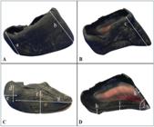
Measurement sites (mm) of the hoof parameters evaluated in Dorper and Santa Inês sheep. (A) Abaxial view of the hoof indicating the measurement sites for (a) heel height and (b) abaxial wall. (B) Axial wall view indicating the measurement site for (c) axial wall height. (C) Solar view of the hoof where the lines indicate the measurement sites for (d) bulb width, (e) bulb length, (f) sole width, and (g) sole length. (D) Internal view of the hoof showing the measurement sites for (h) bulb thickness, (i) sole thickness, (j) dorsal hoof angle, and (k) wall thickness.
The data obtained were subjected to a 2 × 2 factorial statistical analysis (2 breeds × 2 limbs). The variables were analyzed separately for both the medial (digit III) and lateral hoof (digit IV) parameters using the analysis of variance at a 5 % probability level and Tukey’s test at a 5 % probability level with RBio statistical software (18). The hoof measurements from each breed were statistically compared to assess the differences between the lateral and medial hooves of the same limb (digit III × IV), as well as between the thoracic and pelvic limbs (digit III × III and digit IV × IV), in addition to the comparison of the hoof measurements between the breeds.
3. Results
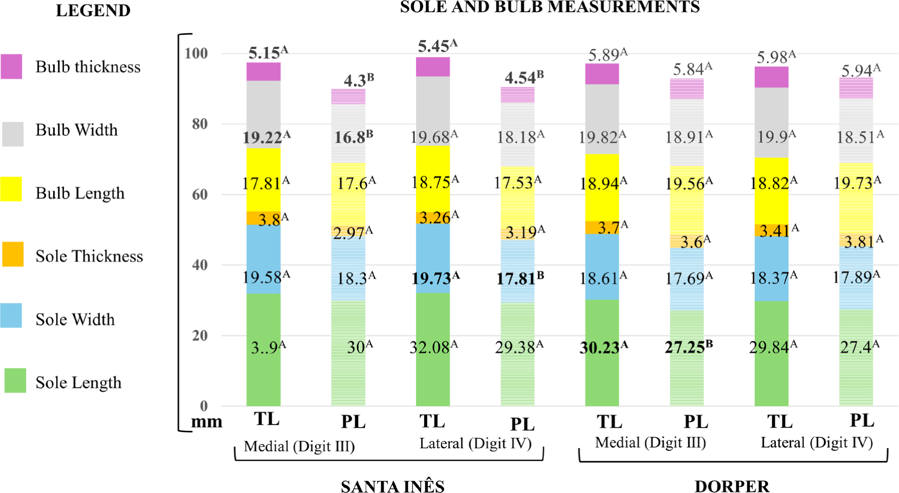
All of the measured parameters of the lateral (digit IV) and medial (digit III) hooves of the same limb and the hoof angle between the limbs showed no statistical difference (p>0.05) in either breed (Figures 3, 4, and 5).

Mean values (mm) of axial wall height, abaxial wall height, heel height, and wall thickness of the lateral (digit IV) and medial (digit III) hooves of the right thoracic (TL) and pelvic (PL) limbs of Santa Inês and Dorper sheep weighing between 30 and 35 kg and aged 150 to 180 days, raised in Sergipe, Northeastern Brazil. Values in bold followed by different letters, within bars of the same color, indicate differences according to Tukey’s test (p<0.05).

Mean values (mm) of bulb and sole thickness, width, and length of the lateral (digit IV) and medial (digit III) hooves of the right thoracic (TL) and pelvic (PL) limbs of Santa Inês and Dorper sheep weighing between 30 and 35 kg and aged 150 to 180 days, raised in Sergipe, Northeastern Brazil. Values in bold followed by different letters, within bars of the same color, indicate differences according to Tukey’s test (p<0.05).

Mean values of the dorsal wall angle of the hooves of the right thoracic (TL) and pelvic (PL) limbs of Santa Inês and Dorper sheep weighing between 30 and 35 kg and aged 150 to 180 days, raised in Northeastern Brazil. Blue lines represent the angle of Dorper sheep and green lines the angle of Santa Inês sheep. Means followed by different letters, within lines of the same color, indicate differences according to Tukey’s test (p<0.05).
The comparison of the medial (digit III) and lateral (digit IV) hoof measurements between the limbs showed that the values were greater in the thoracic limbs than in the pelvic limbs. The parameters showing the statistical differences (p<0.05) in the Santa Inês sheep were the heel height and the bulb thickness of both hooves, the sole width of the medial hoof (digit III), and the bulb width of the lateral hoof (digit IV). In the Dorper sheep, the differences were observed in the heel height of both hooves and in the abaxial wall height and sole length of the lateral hoof (digit IV). The heel height differed in all four hooves of both breeds (Figures 3 and 4). The ratio between the abaxial wall height and heel height ranged from 1.35:1 to 1.67:1 across breeds, showing lower ratios and less variation in the thoracic limbs (Table 1).
Mean values (mm) of the ratio between abaxial wall height (AW) and heel height (H) of the lateral hoof (digit IV) and medial hoof (digit III) of the right thoracic limbs of Santa Inês and Dorper sheep weighing between 30 and 35 kg and aged 150 to 180 days, raised in Sergipe, Brazil, 2020.
The comparative analysis of the hoof measurements between the breeds showed a difference in the conformation of the right thoracic limb hooves of the Dorper and Santa Inês sheep, especially in the heel height, wall thickness, abaxial wall height, and hoof angle in both claws (Table 2).
Means and standard deviations (mm) of height, length, wall thickness, sole and bulb dimensions, and hoof angle of the lateral hoof (digit IV) and medial hoof (digit III) of the right thoracic limbs of Santa Inês and Dorper sheep weighing between 30 and 35 kg and aged 150 to 180 days, raised in Sergipe, Brazil, 2020.
The data on the pelvic limb hoof morphometry demonstrated statistical differences (p<0.05) between the breeds for all variables, except for the sole width. The Dorper sheep presented significantly higher values in both hooves for the bulb length and thickness and the wall thickness. The abaxial and axial wall heights were higher in the lateral hoof (digit IV), whereas the heel height and the bulb and sole widths were greater in the medial hoof (digit III). The only parameter significantly higher in Santa Inês sheep was the sole length of the medial hoof (digit III) (Table 3).
Means and standard deviations (mm) of height, length, wall thickness, sole and bulb dimensions, and hoof angle of the lateral hoof (digit IV) and medial hoof (digit III) of the right pelvic limbs of Dorper and Santa Inês sheep weighing between 30 and 35 kg and aged 150 to 180 days, raised in Sergipe, Northeastern Brazil, 2020.
Considering the 10 parameters showing significant differences in the mean values (mm) of the hooves of both limbs in the two breeds, the lateral hoof (digit IV) of the pelvic limb exhibited the greatest number of parameters, with differences in the mean values (70 %, n = 7/10), followed by the medial hoof (digit III) of the same limb (50 %, n = 5/10). Of these parameters, 30 % (3/10) were the same for both hooves: bulb length, bulb thickness, and wall thickness. The two hooves of the thoracic limb showed more similar measurements, differing in only 30 % (3/10) and in the same parameters (i.e., heel height, abaxial wall height, and wall thickness). The only parameter in which the measurements differed in all four hooves was wall thickness. In 90 % (9/10) of the parameters, the mean values (mm) were greater in the Dorper hooves, while in 10 % (1/10), they were greater in the Santa Inês hooves (Tables 2 and 3).
4. Discussion
The study’s literature review showed that information on this subject in sheep is scarce. No morphometric data were found for the parameters analyzed here in the Santa Inês and Dorper breeds.The use of limbs from only one antimere (right) is justified by the low angular variability of the joints during movement and the execution of flexion/extension, abduction/adduction, and internal/external rotation between the right and left limbs (6). This choice does not interfere with the hoof growth (1) or wear, except under adverse environmental conditions (3, 9) that may cause hoof alterations. However, it is noteworthy that only healthy animals were used in this work. Therefore, these data are important to support the literature on sheep, even considering the specificity of the hoof measurements of the animals used here in terms of breed, age, sex, and management system, which differ from those addressed in the few studies available on sheep (1, 2, 4,14).
Reliable and easily reproducible methods should be sought to obtain these measurements using accessible and easy-to-handle equipment, such as digital calipers. However, despite its apparent practicality, its use in regions like the bulb may not be recommended because it is more suitable for the hoof wall and sole measurements. Measuring the hoof wall angle using a protractor may present great variation (4). The use of a goniometer is suggested to be more appropriate because it shows good results when applied in canine orthopedics (11).
In this study, the measurement difficulty was minimized by working with slaughterhouse specimens, which facilitated the instrument handling and positioning. The measurements in live animals may present difficulties in the restraint and reproducibility of data, as reported by Bhardwaj et al. (4), who restrained sheep in dorsal recumbency after unsuccessful attempts to measure them in a standing position. Even when performed on a flat surface, this position made it difficult to visualize the reference points for measurement.
Other techniques were used in live sheep, but with limited success. These include using a ruler to measure the distance between the coronary band and a horizontal groove made with a saw on the lateral wall of the hoof. This technique has limitations caused by the variation in the coronary band thickness and the fact that it is not a straight line, which makes it difficult to repeat the vertical measurement point toward the sole (12). The three markings used as the measurement reference points for assessing the hoof growth (i.e., an “X” on the skin above the coronary band after clipping and two marks, one horizontal and one vertical, of 0.5 mm, on the hoof wall made with a file) either disappeared or varied in size, consequently reducing the accuracy, as indicated by the occurrence of negative growth rates (1). Therefore, although working with slaughterhouse specimens provides a greater reliability in the obtained values, the measurements may differ in live animals depending on the restraint quality, reference points used, width, and portion of the coronary band chosen as a reference (1, 4,12).
The greater the measurement precision, the more studies and analyses can be performed on sheep hooves. For example, in a study on Merino rams during the first 6 months of life, Tehupuring et al. (14) suggested that the sole area of the left thoracic limb is closely related to the body weight during growth, while the area of the right thoracic limb will result from the interaction between age and body weight. Costa et al. (9) evaluated the abrasion levels on three types of flooring by measuring the hoof parameters in Santa Inês ewes confined for 120 days. They found that concrete flooring is more abrasive than sand or rubber flooring. In our study, this variation in the abrasion between flocks was minimized by the similarity of the terrain where both groups were raised, even though they were from different municipalities.
The values obtained for the Santa Inês and Dorper breeds can serve as the reference measurements for the hooves of males at 6 months of age, that is, in their prepubertal phase. These data are particularly relevant for animals with potential to become future breeders. Furthermore, these information may contribute to the development of specific measuring instruments for sheep hooves, such as angulators adapted to the species, serving as a reference during hoof trimming, similar to those available for cattle. Note that the angulator used in cattle, although widely applied in different breeds, was developed based on the parameters from the Holstein animals raised in Europe (19), differing, for example, from the angulation observed in the beef cattle raised in Brazil (20).
As suggested, hoof trimming in sheep must be performed using the reference of a 55° dorsal wall angle relative to the ground (21). This value is close to that found in the Santa Inês breed (52.9 ± 3.39 and 54.1 ± 3.71) but significantly higher than in the Dorper breed (44.9 ± 4.84 and 47.1 ± 4.08), which may lead to exposure of sensitive tissues. This difference in the hoof wall angle was also reported when comparing Merinoland (50.1° and 49.2°) and Rhoen (47.6° and 45.7°) sheep, demonstrating the different hoof angle conformations among breeds and highlighting the importance of understanding breed-specific characteristics prior to any evaluation and/or intervention (10).
No differences are generally observed between the hooves of the same limb in sheep, as also seen in the breeds studied here, allowing trimming to start on either claw and trimming both to approximately the same size. In situations where differences occur (4), care must be taken to trim the shorter hoof first to avoid excessive tissue removal (22).
In cattle, the ratio between the dorsal wall and heel heights is 2:1 (22). In sheep, this proportion is 1.25:1, with a range of 1.05:1–1.23:1 in different Iranian breeds (2, 23). These values are lower than those observed in the Santa Inês and Dorper sheep raised in Sergipe, which is probably due to age differences and less hoof wear because in the cited studies (2, 23), the animals were over 2 years old. In contrast, the sheep from Sergipe were up to 6 months of age.
Some thoracic limb parameters were greater than those of the pelvic limbs in the sheep of both breeds studied, as also reported in Iranian sheep breeds (2), Merinoland, Rhoen (10), and Merino (4), suggesting that this may be a species-wide pattern. In the Noduz sheep, the thoracic hooves showed more adequate conformation compared with the pelvic hooves, which presented excessive growth and slipping (5). In our results, the Santa Inês and Dorper sheep follow this pattern, in which the larger thoracic limb measurements may be caused by lower abrasion, consequently requiring more frequent trimming. In contrast, the pelvic limbs with smaller measurements may be predisposed to erosions, cracks, and lesions. The hoof angle and the periople height of the thoracic limbs, which were greater than those in the pelvic limbs, in addition to a higher abaxial wall: heel ratio in the pelvic limbs of the Santa Inês and Dorper sheep in this study, support the suggestion of greater hoof wear in the pelvic limbs, as also reported in Iranian sheep (23).
Studies on sheep show no significant differences in the growth rates between the thoracic and pelvic hooves (1, 12). Therefore, the differences found in the hoof size between the limbs in this work are the result of wear. Larger values resulting from less wear in the thoracic limbs suggest the following: their hooves supporting 60 % of their body weight (24) is not sufficient to cause greater wear, but rather stimulates a faster growth rate, as reported in cattle (25). Even though they support less weight (40 %), the pelvic limbs are responsible for generating propulsion during locomotion that overloads these hooves and may increase wear (8, 23). In both breeds, the hoof with the greatest size variation was the lateral pelvic hoof, probably because it suffers greater pressure and wear due to the type of terrain where the sheep were raised.
Different breeds raised under the same management system may present distinct foot health problems, as observed in the flocks in Paraíba, where the Dorper sheep had a significantly higher prevalence of foot rot (17.5 %) when compared with the Santa Inês sheep (6.79 %), in addition to a greater number of digits affected (15). Hypothetically, adopting a standardized trimming technique for both breeds in that system could lead to this outcome, especially if the Santa Inês hoof size was used as the reference, because even though the Dorper sheep presented a greater wall thickness, a standardized cut could result in a greater exposure of the white line region.
Considering that the Dorper sheep showed higher mean values in the hoof parameters (Figures 3 and 4), especially in the pelvic limb, they are presumed to have a faster hoof growth rate than the Santa Inês sheep. This may imply a greater predisposition of the Dorper sheep to the problems related to the excessive hoof growth when raised in the same environment. One example is the overgrowth of the abaxial wall over the sole, which leads to the accumulation of debris, soil, and moisture between them and may justify the need for more frequent trimming in this breed, depending on the degree of wear provided by the production environment. This idea is consistent with reports on the Iranian sheep breeds, which showed that the Chaleshtori ewes had higher values for most traits studied. In comparison, the Afshari and Makoui ewes had lower values, suggesting that they may require more conservative trimming compared to the Chaleshtori ones (2). Consequently, the hoof trimming methods may need to be adjusted to the breed-specific characteristics to avoid potential tissue damage.
In this work, the comparison between breeds showed that the Dorper sheep yield higher values (p<0.05) for the heel and abaxial wall heights in the pelvic limb, indicating that these animals may have developed this structure as a result of the selection process to which they were subjected such that they could adapt to native rangelands (26), where selection occurred, characterized by irregular, dry, and compacted soil. This favors the compensatory development of the heel height, as also reported in the Nelore, Curraleira, and Pantaneira cattle and buffaloes raised in Brazil (20). Another important factor is that the weight placed on the hoof wall tends to be proportionally distributed between the sole and heel region, which may be advantageous for this breed because this region contains digital cushions that absorb impact during locomotion. This reduces predisposition to slippering and/or foot lesions in this region, thereby highlighting the need to preserve it during trimming in this breed. In contrast, the hoof region in the Santa Inês sheep is lower, which may reduce impact absorption during locomotion, predisposing to lesions and conditions like heel erosion. Genetic improvement programs should consider hoof development, especially of regions absorbing the greatest impact during locomotion.
The hoof wall thickness in the Dorper sheep was greater compared to the Santa Inês sheep in both limbs, indicating a greater resistance of the Dorper hooves. This may be related to the hoof’s ultrastructural characteristics, including the number, length, and diameter of the horn tubules that influence the amount of keratin produced by the horn capsule and consequently increase the wall thickness (7). Furthermore, the environment in which the breed was developed may have contributed to the greater wall thickness. This characteristic may determine the resistance of these hooves to traumatic lesions and reduce predisposition to infectious wall lesions compared with the Santa Inês sheep because the insulating barrier is greater. However, in very humid environments, thick hooves may absorb more water, leading to tissue softening and predisposing animals to foot lesions, as evaluated in cattle (10, 27).
The sole length, width, and thickness, the bulb width, length, and thickness, and the axial wall height of the thoracic hooves showed no significant differences (p>0.05) between the breeds, possibly indicating a characteristic inherent to the ovine species or reflecting an adaptation process of the Dorper sheep to the Northeastern territory considering that the breed was developed in the 1930s in South Africa and introduced to Northeastern Brazil in 1997 after the evaluation of its adaptability to semiarid conditions (26).
In contrast, in pelvic hooves, only the sole width showed no significant difference. The bulb length, width, and thickness in the Dorper sheep were greater than those in the Santa Inês sheep, which may influence greater impact absorption during locomotion, consequently reducing the lesion occurrence in this breed. This is because the bulb region houses the digital cushions, which are specialized anatomical structures that are responsible for absorbing the locomotor impact (7). However, this is only a hypothesis because these data alone do not allow a clear conclusion regarding this correlation. Another hypothesis is that greater impact absorption may allow the Dorper sheep to forage more effectively in irregular terrains and consume a wider variety of plant species due to their lower selectivity (26).
The sole length of the medial hoof (digit III) of the Santa Inês sheep was the only parameter in which this breed exceeded the Dorper sheep. The heel height of the same digit also showed significant differences between the breeds. However, these results may be related to the hoof wear variations inherent to the species and the environment in which the animals were raised. Although the management conditions were the same, the locomotor pattern differences among individuals, such as greater or lesser movement, may have influenced the hoof wear. For example, in blue sheep, the most pressured hoof quadrant of the thoracic limb shifts from the caudomedial to the craniomedial region as the slope increases. This is not observed in the pelvic limb relative to the craniomedial quadrant. In all slopes, the vertical force shifted from the lateral to the medial quadrant and from the caudal to the cranial quadrant (8).
The axial and abaxial wall heights of the lateral hoof (digit IV) in the Santa Inês sheep were lower compared to the other measurements, which may be explained by the fact that this area has greater contact with the ground and therefore undergoes greater wear. As noted, this region is prone to increased wear during locomotion. Thus, during sheep trimming, care should be taken to remove only the excess horn tissue, avoiding excessive removal to prevent lesions arising from natural wear during locomotion.
The sole thickness showed a statistical difference only in the medial hoof (digit III) of the Santa Inês sheep. This difference may be related to the adaptation of these animals to the greater pressure exerted on this digit compared to the lateral hoof (digit IV). This difference was probably not found in the thoracic limb because the pelvic limbs are responsible for generating propulsion during locomotion, which may lead to greater contact with the ground and greater wear (8, 24).
5. Conclusion
This study determined the hoof capsule measurements of the Santa Inês and Dorper sheep raised under the same housing, management, and climatic conditions, an aspect still being scarcely described in the literature. Through the analysis of variance, we confirmed that morphometric differences exist between the hoof capsules of the two breeds. This result has practical relevance because it can provide reference values for the healthy hooves of prepubertal males (6 months old) and guide the development of breed-specific trimming techniques. However, further studies must be conducted to compare the hoof growth and wear patterns in these and other sheep breeds and evaluate the mineral composition and histology of the hooves, thereby advancing knowledge on their physiology and resistance.
Data availability statement
The complete dataset supporting the results of this study is available upon request from the corresponding author.
Acknowledgment
We thank CAPES for the scholarship granted.
References
-
1 Shelton J, Usherwood NM, Wapenaar W, Brennan ML, Green LE. Measurement and error of hoof horn growth rate in sheep. Journal of Agricultural Science, 2012;150(3):373-378. DOI: http://dx.doi.org/10.1017/S0021859611000748
» http://dx.doi.org/10.1017/S0021859611000748 -
2 Azarpajouh S, Marchewka J, Segura Correa JC, Calderón Díaz JA. Anatomical characterization of hoof growth pattern in six Iranian sheep breeds and its possible implication for trimming recommendations. Tropical Animal Health and Production, 2018;50(6):1343-1348. DOI: http://dx.doi.org/10.1007/s11250-018-1566-y
» http://dx.doi.org/10.1007/s11250-018-1566-y -
3 Best CM, Roden J, Phillips K, Pyatt AZ, Behnke MC. New insight into the prevalence and risk factors for three distinct hoof conformation traits in UK commercial sheep flocks. Veterinary Sciences, 2021;8:176. DOI: http://dx.doi.org/10.3390/vetsci8090176
» http://dx.doi.org/10.3390/vetsci8090176 -
4 Bhardwaj V, Dhungyel OP, Silva K, Dhand NK, Whittington RJ. An objective method for assessment of foot conformation in sheep. Small Ruminant Research, 2018;167:22-28. DOI: http://dx.doi.org/10.1016/j.smallrumres.2018.08.003
» http://dx.doi.org/10.1016/j.smallrumres.2018.08.003 -
5 Al-Barwari III, Karakus, F. An evaluation on hoof deformations in ewes. Journal of Research in Agriculture and Animal Science, 2023;10(12):34-39. Available at: https://www.questjournals.org/jraas/papers/v10-i12/10123439.pdf
» https://www.questjournals.org/jraas/papers/v10-i12/10123439.pdf -
6 Silva B, João F, Amado S, Alvites RD, Maurício AC, Esteves B, Sousa AC, Lopes B, Sousa P, Dias JR, Veloso A, Pascoal-Faria P, Alves N. Biomechanical gait analysis in sheep: kinematic parameters. Frontiers in Bioengineering and Biotechnology, 2024;12:1370101. DOI: http://dx.doi.org/10.3389/fbioe.2024.1370101
» http://dx.doi.org/10.3389/fbioe.2024.1370101 -
7 Lima EMM, Borges JRJ, Lima FB, Silva FOC, Leonardo AS, Barreto-Viana ARC. Morfometria do casco de bovinos nelorados em diferentes sistemas de criação. Bioscience Journal, 2013;29(2):412-8. Available at: https://seer.ufu.br/index.php/biosciencejournal/article/view/13568/12284
» https://seer.ufu.br/index.php/biosciencejournal/article/view/13568/12284 -
8 Liu X, Kui H, Qian Z, Ren L. Hoof pressure distribution pattern of Blue Sheep during walking on different slopes: a subject-specific analysis. Frontiers in Veterinary Science, 2021;8:633509. DOI: http://dx.doi.org/10.3389/fvets.2021.633509
» http://dx.doi.org/10.3389/fvets.2021.633509 -
9 Costa JHS, Lopes Neto JP, Furtado DA, Ribeiro NL, Santos LFD, Neves RS, Medeiros GR, Miranda JR. Behavior and wear of the hooves of confined sheep in different types of floors. Revista Brasileira de Ciências Agrárias, 2022;17(1):e1368. DOI: http://dx.doi.org/10.5039/agraria.v17i1a1368
» http://dx.doi.org/10.5039/agraria.v17i1a1368 -
10 Lambertz C, Friedrich C, Moors E, Brandt H, Erhardt G, Gauly M. A comparison of claw conformation and claw horn structure of two sheep breeds, and their relationship to footrot incidence. Small Ruminant Research 2014;117(1):103-107. DOI: http://dx.doi.org/10.1016/j.smallrumres.2013.11.017
» http://dx.doi.org/10.1016/j.smallrumres.2013.11.017 -
11 Jaegger G. Marcellin-Little DJ, Levine D. Reliability of goniometry in Labrador Retrievers. American Journal of Veterinary Research, 2002;63(7):979-986. DOI: http://dx.doi.org/10.2460/ajvr.2002.63.979
» http://dx.doi.org/10.2460/ajvr.2002.63.979 -
12 Dekker A, Moonen P, Pol J. Linear hoof defects in sheep infected with foot-and-mouth disease. The Veterinary Record, 2005;156(18):572-575. DOI: http://dx.doi.org/10.1136/vr.156.18.572
» http://dx.doi.org/10.1136/vr.156.18.572 -
13 Kaler J, Medley GF, Grogono-Thomas R, Wellington EM, Calvo-Bado LA, Wassink GJ, King EM, Moore LJ, Russell C, Green LE. Factors associated with changes of state of foot conformation and lameness in a flock of sheep. Preventive Veterinary Medicine, 2010;97(3-4): 237-244. DOI: http://dx.doi.org/10.1016/j.prevetmed.2010.09.019
» http://dx.doi.org/10.1016/j.prevetmed.2010.09.019 -
14 Tehupuring BC, Widjiati S, Hendrawan VF, Luqman EM. Relations of weight and age to the front feet sole area of merino ram (Ovis aries). Journal of Veterinary Anatomy, 2021;14(2):73-78. DOI: http://dx.doi.org/10.21608/jva.2021.197239
» http://dx.doi.org/10.21608/jva.2021.197239 -
15 Aguiar GMN, Simões, SVD, Silva TR, Assis ACO, Medeiros JMA, Garino Júnior, F, Riet-Correa, F. Foot rot and other foot diseases of goat and sheep in the semiarid region of northeastern Brazil. Pesquisa Veterinária Brasileira, 2011;31(10):879-884. DOI: https://doi.org/10.1590/S0100-736X2011001000008
» https://doi.org/10.1590/S0100-736X2011001000008 -
16 Carvalho VS, Santos MC, Guimarães JE, Oliveira Filho JC, Costa Neto AO, Macêdo JTSA, Pedroso, PMO. Doenças podais em ovinos no estado da Bahia. Pesquisa Veterinária Brasileira, 2018;38(7):1250-8. DOI: http://dx.doi.org/10.1590/1678-5150-PVB-5504
» http://dx.doi.org/10.1590/1678-5150-PVB-5504 -
17 Ollhoff RD, Ortolani EL. Comparação do crescimento e do desgaste do casco em bovinos taurinos e zebuínos. Ciência Rural. 2001;31(1):67-71. DOI: https://doi.org/10.1590/S0103-84782001000100011
» https://doi.org/10.1590/S0103-84782001000100011 - 18 Bhering LLR. A tool for biometric and statistical analysis using the r platform. Crop Breeding and Applied Biotechnology. 2017. p. 187-90.
- 19 Raven ET. Cattle Foot Care and Claw Trimming. 3rd ed. Ipswich: Farming Press, 1985. 126p.
-
20 Silva LAF, Campos SBS, Rabelo RE, Vulcani VAS. Análise comparativa da morfometria do casco de bovinos das raças Nelore, Curraleira e Pantaneira e de bubalinos e sua relação com a etiopatogenia das enfermidades digitais. Pesquisa Veterinaria Brasileira, 2015;35(4):377-384. DOI: http://dx.doi.org/10.1590/S0100-736X2015000400011
» http://dx.doi.org/10.1590/S0100-736X2015000400011 -
21 Froehlich K, Whaley J, Wuliji T. How to trim sheep and goat hooves. South Dakota State University Extension, 2023:1-4. Available at: https://extension.sdstate.edu/sites/default/files/2023-03/P-00263.pdf
» https://extension.sdstate.edu/sites/default/files/2023-03/P-00263.pdf - 22 Blowey RW. A Veterinary Book for Dairy Farmers. 3rd ed. Sheffield: Farming Press, 1999. 480p.
-
23 Mohamadnia AR, Azarpajouh S, Beige N, Karimi I, Abdi A, Arabi M. Anatomy of the hoof in sheep, a study to find a proper hoof trimming indices. Journal of Veterinary Research. 2007;62:139-144. Available at: https://jvr.ut.ac.ir/article_24093_33ba78c24eeaec059547b05d2bb78cc0.pdf?lang=en
» https://jvr.ut.ac.ir/article_24093_33ba78c24eeaec059547b05d2bb78cc0.pdf?lang=en -
24 Kim J, Breur GJ. Temporospatial and kinetic characteristics of sheep walking on a pressure sensing walkway. Canadian Journal of Veterinary Research. 2008;72(1):50-5. Available at: https://pmc.ncbi.nlm.nih.gov/articles/PMC2117367/pdf/cjvr72_pg50.pdf
» https://pmc.ncbi.nlm.nih.gov/articles/PMC2117367/pdf/cjvr72_pg50.pdf -
25 Vokey FJ, Guard CL, Erb HN, Galton DM. Effects of alley and stall surfaces on indices of claw and leg health in dairy cattle housed in a free-stall barn. Journal of Dairy Science, 2001;84(12):2686-2699. DOI: http://dx.doi.org/10.3168/jds.S0022-0302(01)74723-6
» http://dx.doi.org/10.3168/jds.S0022-0302(01)74723-6 -
26 Rosanova C, Silva Sobrinho AG, Gonzaga Neto S. A raça Dorper e sua caracterização produtiva e reprodutiva. Veterinária Notícias, 2005;11(1):127-135. Available at: https://seer.ufu.br/index.php/vetnot/article/view/18632
» https://seer.ufu.br/index.php/vetnot/article/view/18632 -
27 Borderas TF, Pawluczuk B, De Passillé AM, Rushen J. Claw hardness of dairy cows: Relationship to water content and claw lesions. Journal of Dairy Science, 2004;87(7):2085-2093. DOI: http://dx.doi.org/10.3168/jds.S0022-0302(04)70026-0
» http://dx.doi.org/10.3168/jds.S0022-0302(04)70026-0
Edited by
-
Editor:
Luiz Augusto B. Brito