Chronic kidney disease following poisoning by ethylene glycol in a dog: a case report
Abstract
A seven-year-old, 4.65 kg, male Shih-tzu dog was referred to the veterinary hospital with a history of vomiting after eating a commercial dog treat containing ethylene glycol (EG) in its formulation. The animal showed clinical signs of kidney damage and after additional tests was diagnosed with severe grade 4 acute kidney injury. Almost one and half years later, the patient was presented to veterinary care, with a grade 4, chronic kidney injury. During this period, several tests were carried out, including a complete blood count, serum biochemistry analysis, abdominal ultrasound, and blood gas analysis. In critical condition, the animal died shortly afterward. An anatomopathological necropsy exam revealed extensive kidney injury with tubular necrosis and glomerular sclerosis, fibrosis, and mineralization, accompanied by numerous mono and dihydrates oxalate crystals. To our knowledge, this is the first report describing clinical signs and pathological findings of a dog poisoned by EG in Brazil. We aim to contribute to the understanding of the pathogenesis of EG poisoning, especially regarding chronic kidney failure.
Keywords:
Canine; forensic toxicology; intoxication; kidney injury; toxicologic pathology
Resumo
Um cão da raça Shih-tzu, macho, com sete anos e pesando 4,65 kg, foi encaminhado ao hospital veterinário com histórico de vômitos após ingerir um petisco comercial para cães contendo etilenoglicol (EG) na formulação. O animal apresentava sinais clínicos característicos de injúria renal e após exames complementares foi diagnosticado com lesão renal aguda severa de grau IV (escala de I-V). Quase um ano e meio depois, o paciente retornou ao atendimento veterinário, apresentando lesão renal crônica de grau IV. Durante esse período, foram realizados vários exames, incluindo hemograma, análise bioquímica sérica, ultrassonografia abdominal e hemogasometria. Em estado crítico, o animal evoluiu a óbito pouco tempo depois. O exame anatomopatológico da necropsia revelou lesões renais extensas com necrose tubular, esclerose glomerular, fibrose e mineralização, acompanhadas por numerosos cristais de oxalato mono e dihidratados. De acordo com o nosso conhecimento, este é o primeiro relato descrevendo os sinais clínicos e achados anatomopatológicos de um cão intoxicado por EG no Brasil. Este estudo busca contribuir para o entendimento da patogênese e diagnóstico clínico da intoxicação por EG, especialmente no que se refere à insuficiência renal crônica.
Palavras-Chave:
Canino; intoxicação; lesão renal; patologia toxicológica; toxicologia forense
1. Introduction
Ethylene glycol (EG) is a synthetic odorless liquid substance used as an antifreeze and vehicle deicing solution. It is also used in hydraulic brake fluids, inks, stamp pads, ballpoint pens, and print shops (1). EG toxicosis has been reported in humans (2) and animals, including dogs (3), cats (4), chickens (5), calves (6) and wildlife (7). Poisoning is characterized by severe metabolic acidosis followed by critical acute kidney injury. Clinical signs can include ataxia, vomiting, polyuria, polydipsia, hypothermia, oliguria, anuria, and dehydration (8).
Although ethanol and fomepizole can be used as antidotes since both compete with the liver enzyme alcohol dehydrogenase, mortality rates are still high in humans (9), and little data is found regarding dogs. Most poisonings in animals occur after accidental exposure to antifreeze substances and are more common in northern countries (10). Here we describe detailed long-term clinical evolution, post-mortem, and histopathological findings of chronic kidney disease (CKD) following EG poisoning in a dog in Brazil, after a serious contamination of treats. We aim to contribute to the understanding of the pathogenesis of EG poisoning, especially regarding chronic kidney failure.
2. Case report
A seven-year-old, 4.65kg, male Shih-tzu was referred to the veterinary hospital following nine days of vomiting after eating a commercial dog treat. He had anorexia, apathy, intermittent diarrhea, increased water and urine intake, and lighter urine color. The owner denied any previous illnesses and confirmed that the animal had been vaccinated, had no access to other animals, and remained indoors. The patient was evaluated throughout approximately one and a half years, including clinical examination, imaging exams, clinical pathology, necropsy and histopathology.
During clinical examination (1st day), the patient had tachycardia (168 bpm), periodontitis, and a rigid abdomen during palpation. All remaining parameters were within the normal limits. Blood samples were collected for blood cell count (CBC), alanine aminotransferase (ALT), creatinine, phosphorus, urea, (Tables 1 and 2), and blood gas. Urine was collected via cystocentesis to perform urinalysis (Table 3). Creatinine levels reached 5.23 mg/dL (0.5 to 1.5 mg/dL), phosphorus was also increased at 6.48 mg/ dL (2.6 to 6.3 mg/dL), and urea at 145 mg/dL (21.4 to 59.9 mg/dL)(11). Urinalysis revealed low specific gravity, proteinuria (one cross), the presence of granular casts, transitional epithelial cells, and oxalate crystals. Blood gas did not reveal acidosis, but an increased CO2, with a minor reduction of sodium (Na), calcium (Ca), and potassium (K) levels. Other parameters remained within the normal values for the species. Abdominal ultrasound revealed colitis and acute nephropathy (discrete increase in medullary echogenicity).
The diagnosis was an acute renal injury (AKI) (12) at stage four with an unidentified cause. He was then hospitalized for 8 days and treated with fluid replacement, ondansetron, omeprazole, omega-3, aluminum hydroxide, praziquantel, appetite stimulant, pyrantel, febantel, fluid therapy, and gastrointestinal diet (Table 1). Improvement was observed, but vomiting and hyporexia persisted. Nutritional supplementation was recommended after omeprazole. On the 8th day, BC was normal, except for discrete leukopenia (Table 2), whilst creatinine and phosphorus remained elevated, at 3.93 mg/dL and 3.85 mg/dL, respectively (Table 3). Urine continued to present lower density (Table 4). Home treatment was continued after hospital discharge (8th day) (Table 1).
A few days after the first presentation at the veterinary hospital, Brazil’s National Health Surveillance Agency (Anvisa) and Ministry of Agriculture and Livestock (MAPA) revealed an outbreak of EG poisoning from different dog treat products. Two lots were diagnosed with propylene glycol contaminated with monoethylene glycol (AD5035C22 and AD4055C21) (18). Several cases throughout the country were reported in 2022, with over 100 canine suspicious poisoning in 14 states, currently under federal investigation. In the present report, the animal had eaten a whole package of dry snacks. The patient was reevaluated after 10 days of hospitalization (18th day). During the time, the owner presented to the veterinary team, a package of dog treats from one of the brands and batches that were under investigation. The cause of AKI was clinically identified as EG poisoning.
Clinical presentation on the 18th day included persistent hyporexia, decreased defecation, and polydipsia. Blood and urine samples were again evaluated. BC revealed leukopenia and normochromic normocytic anemia. ALT and ALP (alkaline phosphatase) remained normal, while the following parameters were elevated: creatinine (3.84 mg/dL-0.5 to 1.5 mg/dL), phosphorus (4.15 mg/dL -2.6 to 6.3 mg/dL) and urea (96 mg/dL 21.4 to 59.9 mg/dL) (11). Bilirubin and fractions remained within the normal limits (data not shown). Urine revealed discrete low specific gravity and the presence of cylinders’ cells (Table 3). Nutritional supplementation was recommended as well as a consultation with the veterinary nephrologist; however, the patient did not return.
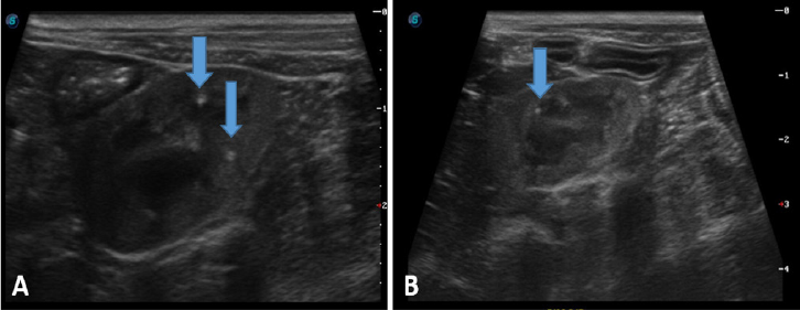
After almost 17 months of the first hospitalization (520th day), the owner reported that the patient had been experiencing weight loss and loss of appetite for a month. One day before the appointment, the patient began to experience vomiting and tremors. Physical examination revealed pale mucous membranes, tachycardia (160 bpm), and popliteal lymph nodes lymphadenomegaly. Body condition score was 3/9, with significant cachexia and severe periodontitis. Other parameters were within normal limits. Additional tests were requested, including CBC, ALT, creatinine, urea, phosphorus, and abdominal ultrasound. Results showed that the patient had severe normochromic normocytic anemia, with creatinine value of 7.7 mg/dL (0.5 to 1.5 mg/dL) and urea of 254 mg/dL 21.4 to 59.9 mg/dL) (14) (Table 1 and 2). Abdominal ultrasound showed both kidneys with reduced size, irregular contour, diffuse hyperechogenicity of the cortical region, and homogeneous echotexture, with slight loss of definition and alteration of the corticomedullary relationship, presence of a discrete echogenic halo in the medullary region parallel to the corticomedullary junction (medullary sign). Discreet hyperechogenic foci were noted in the topography of pelvic recesses (mineralizations) (Figure 1). In addition, signs of gastric hypersecretion/inflammatory gastropathy and prostatic hyperplasia were observed. The dog was diagnosed with stage four CKD according to IRIS, 2023 (14). The veterinary team informed the owner of the severity of the case and the need for additional tests, including blood gas analysis and urinalysis, as well as hospitalization and a consultation with a specialized nephrologist. The owner denied the request.

Abdominal ultrasound showed both kidneys with reduced size, irregular contour, diffuse hyperechogenicity of the cortical region, and homogeneous echotexture, with slight loss of definition and alteration of the corticomedullary relationship, presence of a discrete echogenic halo in the medullary region parallel to the corticomedullary junction (medullary sign). Discreet hyperechogenic foci were noted in the topography of pelvic recesses (mineralizations; arrow).
Seven days after the second consultation (527th day) the patient presented clinical worsening, including inappetence, hypocolored mucosa, cachexia, anorexia, adipsia, oliguria, purulent secretion in both eyes, pain on abdominal palpation, 8% dehydration, uremic breath with visible oral ulcers and instability of the incisor teeth in the jaw. The patient was hospitalized and underwent follow-up in the nephrology sector. Blood samples were evaluated and findings included hypochromic normocytic anemia (Table 1), severe hyperphosphatemia, and uremia (Table 3). Blood gas analysis revealed metabolic acidosis without significant electrolyte changes. The recommended treatment included fluid replacement, antiemetic, phosphorus chelator, analgesia, and insertion of a nasogastric tube for feeding. In addition, a urethral probe was inserted to measure urinary output.
Due to severe anemia, a blood transfusion with packed red blood cells was performed. The procedure occurred without complications and on the 545th day, new tests were carried out which showed an increase in hematocrit (Table 1) and persistence of uremia (Table 2). Erythropoietin and lubricating eye drops were added to the prescription, however, on day 546 the patient died.
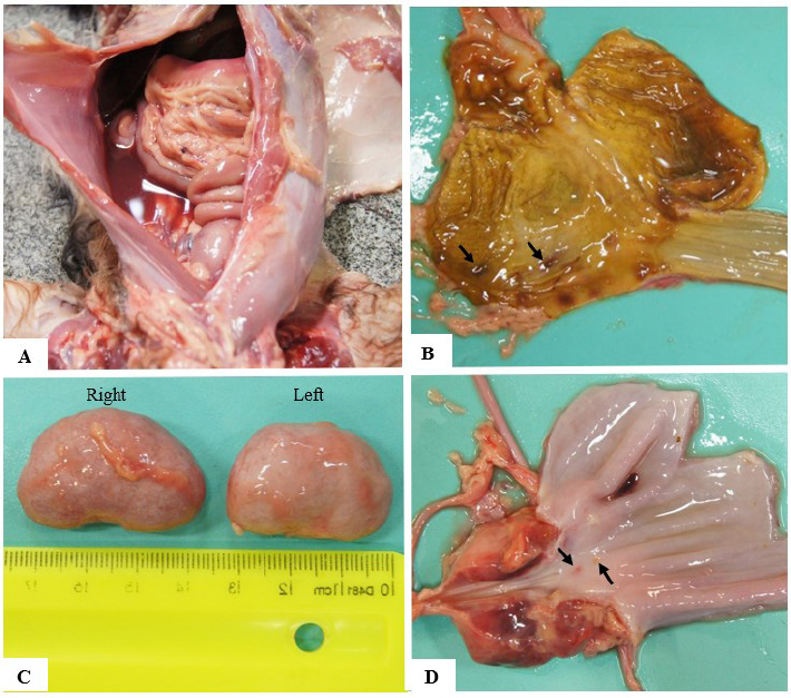
During the necropsy, the body condition score was 2/5. Ocular and oral mucous membranes were pale and there were ulcers in the latter. The tongue also presented ulceration in the ventral and lateral regions. When the abdominal cavity was opened, there was serosanguineous fluid, the liver was enlarged and the gastric mucosa contained multiple ulcers. Kidneys had marked hypotrophy, and the urinary bladder had an ulcerated focus on the mucosa. Lungs had diffuse congestion and discrete multifocal hemorrhages, in addition to the presence of foamy fluid in the bronchi and trachea, indicating pulmonary edema. Heart’s left ventricle showed concentric hypertrophy, congestion, and hemorrhagic areas (Figura 2).

Macroscopic images from the necropsy of a dog with suspected ethylene glycol poisoning. During the necropsy, the body condition score was 2/5. A. Presence of serosanguineous fluid in the abdominal cavity; B. Multiple ulcers in the gastric mucosa (black arrow). C. Kidneys had marked hypotrophy and irregular capsule. D. urinary bladder had an ulcerated focus on the mucosa (black arrows).
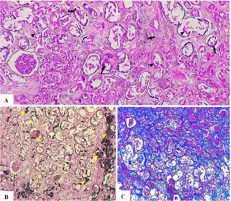
Histopathological analysis allowed us to confirm the macroscopic findings of the digestive and respiratory systems. Furthermore, foci of mineralization were observed in several organs, such as the heart, liver, stomach, intestine, pancreas, spleen, meninges, and especially the kidney. In the kidneys, it was still possible to notice moderate multifocal congestion, extensive fibroplasia, and moderate multifocal infiltrate of lymphocytes and plasma cells; as well as mineralization in collecting, proximal and distal tubules, and often basement membranes. Birefringent oxalate crystals were also found in the tubules. Furthermore, there was a desquamation of epithelial cells towards the luminal interior associated with tubular necrosis and a marked number of sclerotic glomeruli (Figure 3).
Photomicrograph of kidney from a dog poisoned by ethylene glycol. A) desquamation and necrosis of tubular epithelial cells (arrowhead) and sclerotic glomeruli (yellow asterisk). Birefringent oxalate crystals (arrow) in collecting, proximal and distal tubules, and basement membranes. HE, 10x. B) Oxalate crystals deposition (yellow arrowhead), which is shown in black, in proximal and distal tubules, and basement membranes. Von Kossa, 10x. C) Extensive fibroplasia in renal parenchyma (cortex renal), shown in blue. Masson’s trichrome, 10x.
3. Discussion
The present work describes detailed clinical and pathological findings of chronic kidney failure following EG poisoning in a dog in Brazil, after a serious contamination of treats. Once ingested, EG goes through an extensive metabolization process that results mainly in oxalic acid, the major nephrotoxic metabolite (19). Poisoning in dogs can cause three stages of clinical presentations. Stage I occurs between 30 min and 12 hours of the exposure, with ataxia, vomiting, polyuria, polydipsia, and hypothermia. Laboratory tests indicate metabolic acidosis, hyperosmolality, high osmolar gap, high anion gap (AG), prerenal azotemia, and calcium oxalate crystaluria. Stage II has cardiopulmonary manifestations that usually appear between 12 and 24 h, but are rarely recognized in dogs. Stage III has acute kidney injury or oligoanuric acute renal failure, with renal azotemia and hyperkalemia, that are manifested after 24 to 72 h (8). The patient in the present study was hospitalized with clinical signs and laboratory findings compatible with stage III of the poisoning. In the present case, the most prominent findings were vomiting and azotemia.
Acute kidney injury was confirmed by a combination of exams, including high azotemia, high phosphorus, urinalysis alterations, and abdominal ultrasound. It is important to highlight the relevance of urinalysis in this diagnosis, with a remarkable presence of oxalate crystals that are typical of EG poisoning (20). These crystals are the result of liver metabolism of oxalic acid and cause direct toxicity to kidney cell mitochondria.
Initially, the patient was diagnosed with stage four acute kidney injury (12), without probable cause. Only after the outbreak was reported by Brazilian officials, that a suspicion of EG was considered. This fact can be associated with the uniqueness of the contamination that is under investigation, with a possible replacement of propylene glycol with diethylene glycol, and the difficulty in establishing a diagnosis (8). Several reports claim the importance of early diagnosis, highlighting decontamination and antidotal therapy with fomepizole (21). Due to delayed diagnosis, antidotes were not prescribed in the present case and only symptomatic treatments were performed.
Despite recommendations for continuous monitoring, the patient only returned to veterinary care after 17 months. Clinical signs included tremors, loss of appetite, vomiting, pale mucous membranes, periodontitis, and cachexia, all commonly described in chronic diseases, including kidney failure (3). Further exams confirmed severe normochromic normocytic anemia, azotemia, and ultrasound findings compatible with stage four CKD (14). It is important to highlight the relevance of clinical monitoring since CKD is diagnosed.
The severity of anemia in the present case was remarkable, and treatment with blood transfusion was only partially successful. Anemia in patients with chronic kidney disease is multifactorial and includes decreased erythroid precursor cells, gastrointestinal bleeding, nutritional imbalance, iron deficiency, and systemic inflammation. It is believed the critical failing kidneys produce insufficient amounts of erythropoietin (12), with increased concentrations of parathormone and tumor necrosis factor-α (TNFα), that altogether induce erythroid hypoplasia (22).
An important continuous alteration was azotemia. Urea and creatinine are known biomarkers for kidney disease, increasing in cases of filtration insufficiency. However, it is important to highlight the fact that creatinine reached 11.59 mg/dL in a dog with severe cachexia, indicating this value is even more critical. Creatinine is mostly produced by muscle cells and atrophy found in cachexia can decrease blood levels (14). This indicates that the kidney dysfunction in our patient was most likely terminal. Clinical signs worsened during the last days of monitoring, with adipsia, oliguria dehydration, and oral erosions, compatible with uremia.
Abdominal ultrasound also confirmed the progression of kidney diseases. The most common findings in dogs are increased cortical echogenicity, abnormal cortical/medullar junction, and pyelectasis (23). Similar alterations were diagnosed in the present case, with the addition of discrete hyperechogenic foci in the topography of pelvic recesses, later diagnosed by histological analysis as mineralization areas. Necropsy findings included significant kidney alterations with consequences to other tissues, including the tongue, stomach, and heart. Tubular necrosis and desquamation of epithelial cells, leading to the formation of cellular casts, are common alterations described in previous reports (20). The presence of birefringent oxalate crystals is an important indication of EG poisoning (6) and corroborates with previous urinalysis results. It is also important to highlight that the degree of renal necrosis is not necessarily associated with the number of oxalate crystals present in the organ, since it is believed that kidney injury is also caused by the direct cytotoxic effect of metabolites of hippuric or glyoxylic acid.
Sclerotic glomeruli are an unusual lesion found in the present case (24) and may be directly related to interstitial injury to the kidney.
Systemic reverberation of kidney disease included serosanguineous fluid (25) and tongue necrosis, common in cases of chronic renal failure due to uremia, which promotes fibrinoid necrosis of the blood vessel wall, causing thrombosis, ischemia, and consequently ulceration of the affected organ (25). Circulatory changes, such as congestion and hemorrhagic foci in various internal organs, are also a consequence of EG poisoning (24). As noted, oxalate crystals can precipitate in practically all tissues of the animal's body (24) and accompany the mineralization of tissues; these findings have also been observed in other cases of EG poisoning in animals and humans (26).
4. Conclusion
This is the first report of EG poisoning in a dog in Brazil and one rare case that possibly evolved into CKD. It is important to highlight that other unknown factors might be involved in the pathogenesis of CKD. Through comprehensive clinical monitoring and laboratory, ultrasound, and anatomopathological examinations, it was possible to document the progression from severe acute kidney injury to chronic kidney disease, highlighting the severity and complexity of EG poisoning in dogs. This report aims to contribute to the understanding of the pathogenesis of EG intoxication, to further explore veterinary diagnosis, and the importance of clinical pathology monitoring. We would also like to highlight the relevance of quality control regarding animal feed to avoid such critical contaminations.
Acknowledgements
We thank the following colleagues for tube contributions regarding clinical management and pathology: Laís Di Paulie Taborda Prado, Fernanda Martins Paixão, Caroline Cavalcante Rodrigues, Nelcimara Mirley de Souza Correia, Nadjanaira Barbosa Abrão, Luana de Sousa Ribeiro, Heloisa Alvim Rodrigues Fraga, Amanda Lopes Ribeiro, Washington Cleyber de Oliveira Júnior, Filipe Augusto Vieira dos Santos, Bruna Siqueira Gomes de Lima.
Data availability statement
The complete dataset supporting the results of this study is available on request from the corresponding author.
References
-
1 Centers for Disease Control and Prevention (CDC). Toxic substances portal – Ethylene glycol. Atlanta (GA): US Department of Health and Human Services; 2021 [cited 2024 May 1]. Available from: https://wwwn.cdc.gov/TSP/substances/ToxSubstance.aspx?toxid=21
» https://wwwn.cdc.gov/TSP/substances/ToxSubstance.aspx?toxid=21 -
2 Davis DP, Bramwell KJ, Hamilton RS, Williams SR. Ethylene glycol poisoning: a case report of a record-high level and a review. J Emerg Med. 1997;15(5):653-657. doi: https://10.1016/S0736-4679(97)00145-5
» https://10.1016/S0736-4679(97)00145-5 -
3 Schweighauser A, Francey T. Ethylene glycol poisoning in three dogs: importance of early diagnosis and role of hemodialysis as a treatment option. Schweiz Arch Tierheilkd. 2016;158(2):109-14. doi: https://10.17236/sat00051
» https://10.17236/sat00051 -
4 Tart KM, Powell LL. 4-Methylpyrazole as a treatment in naturally occurring ethylene glycol intoxication in cats. J Vet Emerg Crit Care (San Antonio). 2011;21(3):268-72. doi: https://10.1111/j.1476-4431.2011.00638.x
» https://10.1111/j.1476-4431.2011.00638.x - 5 Hutchison TW, Dykeman JC. Presumptive ethylene glycol poisoning in chickens. Can Vet J. 1997;38(10):647. PMCID: PMC1576855.
-
6 Agerholm JS, Hansen KS, Voogd HL, Krogh AKH. Ethylene glycol toxicosis in milk-fed dairy calves. Acta Vet Scand. 2022;64(1):7. doi: https://10.1186/s13028-022-00626-1
» https://10.1186/s13028-022-00626-1 - 7 Thrall MA, Connally HE, Grauer GF, Hamar DW. Ethylene glycol. In: Peterson ME, Talcott PA, editors. Small animal toxicology. 3rd ed. St. Louis (MO): Elsevier Saunders; 2013. p. 551–567.
-
8 Scherk JR, Brainard BM, Collicutt NB, Bush SE, Almy FS, Koenig A. Preliminary evaluation of a quantitative ethylene glycol test in dogs and cats. J Vet Diagn Invest. 2013;25(2):219-25. doi: https://10.1177/1040638713477644
» https://10.1177/1040638713477644 -
9 Ghannoum M, Gosselin S, Hoffman RS, Lavergne V, Mégarbane B, Hassanian-Moghaddam H, et al. Extracorporeal treatment for ethylene glycol poisoning: systematic review and recommendations from the EXTRIP workgroup. Crit Care. 2023;27(1):56. doi: https://10.1186/s13054-022-04227-2
» https://10.1186/s13054-022-04227-2 -
10 Pivetta M, Beltran E, Stewart J, Dennis R. MRI findings of diffuse polioencephalopathy secondary to ethylene glycol intoxication in a dog. Vet Rec Case Rep. 2014;2(1):e000045. doi: https://10.1136/vetreccr-2014-000045
» https://10.1136/vetreccr-2014-000045 - 11 Kaneko JJ, Harhey IW, Bruss ML. Clinical biochemistry of domestic animals. 6th ed. San Diego (CA): Academic Press; 2008. p. 1–928. ISBN: 9780080568829.
-
12 Segev G, Cortellini S, Foster JD, Francey T, Langston C, Londoño L, et al. International Renal Interest Society best practice consensus guidelines for the diagnosis and management of acute kidney injury in cats and dogs. Vet J. 2024;6:106068. doi: https://10.1016/j.tvjl.2024.106068
» https://10.1016/j.tvjl.2024.106068 - 13 Jain NC. Essentials of veterinary hematology. Philadelphia (PA): Lea & Febiger; 1993.
-
14 International Renal Interest Society (IRIS). IRIS staging of CKD [Internet]. [cited 2024 Apr 25]. Available from: http://www.iris-kidney.com/education/guidelines/staging.html
» http://www.iris-kidney.com/education/guidelines/staging.html -
15 International Renal Interest Society (IRIS). IRIS treatment recommendations for CKD in dogs. 2023 [cited 2024 May 1]. Available from: https://iris-kidney.com/pdf/IRIS-DOG-Treatment_Recommendations_2023.pdf
» https://iris-kidney.com/pdf/IRIS-DOG-Treatment_Recommendations_2023.pdf - 16 Silverstein DC, Hopper K. Small animal critical care medicine. St. Louis (MO): Elsevier; 2015. p. 829–834.
- 17 Sink CA, Weinstein NM. Practical veterinary urinalysis. Hoboken (NJ): John Wiley & Sons; 2012.
-
18 Agência Nacional de Vigilância Sanitária (ANVISA). Resolução RE nº 3.008, de 9 de setembro de 2022 [Internet]. [cited 2024 Apr 25]. Available from: https://www.in.gov.br/en/web/dou/-/resolucao-re-n-3.008-de-9-de-setembro-de-2022-428657844
» https://www.in.gov.br/en/web/dou/-/resolucao-re-n-3.008-de-9-de-setembro-de-2022-428657844 - 19 Beauchamp GA, Valento M. Toxic alcohol ingestion: prompt recognition and management in the emergency department. Emerg Med Pract. 2016;18(9):1-20. PMID: 27538060.
-
20 Pomara C, Fiore C, D'Errico S, Riezzo I, Fineschi V. Calcium oxalate crystals in acute ethylene glycol poisoning: a confocal laser scanning microscope study in a fatal case. Clin Toxicol (Phila). 2008;46(4):322-4. doi: https://10.1080/15563650701419011
» https://10.1080/15563650701419011 -
21 McQuade DJ, Dargan PI, Wood DM. Challenges in the diagnosis of ethylene glycol poisoning. Ann Clin Biochem. 2014;51(2):167-78. doi: https://10.1177/0004563213506697
» https://10.1177/0004563213506697 -
22 Borin-Crivellenti S, Crivellenti LZ, Gilor C, Gilor S, Silva DG, Maia SR, et al. Anemia in canine chronic kidney disease is multifactorial and associated with decreased erythroid precursor cells, gastrointestinal bleeding, and systemic inflammation. Am J Vet Res. 2023;84(10):1-6. doi: https://10.2460/ajvr.23.05.0097
» https://10.2460/ajvr.23.05.0097 -
23 Perondi F, Lippi I, Marchetti V, Bruno B, Borrelli A, Citi S. How ultrasound can be useful for staging chronic kidney disease in dogs: ultrasound findings in 855 cases. Vet Sci. 2020;7(4):147. doi: https://10.3390/vetsci7040147
» https://10.3390/vetsci7040147 -
24 Leth PM, Gregersen M. Ethylene glycol poisoning. Forensic Sci Int. 2005;155:179-84. doi: https://10.1016/j. forsciint.2004.11.012
» https://10.1016/j. forsciint.2004.11.012 -
25 Barraza VCT, Marcus VB, Gonçalves PNC, Rosa EF, Flores MM. Nasal mucosal mineralization ulceration and epistaxis in a dog with uremia caused by chronic renal failure. Cienc Rural. 2023;53(2):e20210837. doi: https://10.1590/0103-8478cr20210837
» https://10.1590/0103-8478cr20210837 -
26 Hristov TS, Rusenov A, Simeonov R, Kalkanov I. Ethylene glycol intoxication in a pregnant cat and a tomcat: case report. Bulg J Vet Med. 2023;26(3):472-9. Available from: https://www.researchgate.net/publication/352178458_Case_report_ETHYLENE_ GLYCOL_INTOXICATION_IN_A_PREGNANT_CAT_AND_A_TOMCAT_CASE_REPORT
» https://www.researchgate.net/publication/352178458_Case_report_ETHYLENE_ GLYCOL_INTOXICATION_IN_A_PREGNANT_CAT_AND_A_TOMCAT_CASE_REPORT
Edited by
-
Editor:
Luiz Augusto B. Brito