Reuse of progesterone device did not affect embryo yield in locally adapted Brazilian sheep
Abstract
The reuse of progesterone devices is safe, efficient, and economically viable in small ruminants, supporting the evaluation of extended reuse protocols in superovulated ewes. Thus, the objective of this study was to test the efficiency of progesterone device reuse in superovulated ewes. Morada Nova (MN, n = 20) and Santa Inês (SI, n = 20) ewes were equally assigned to G-New (n = 20) and G-Used (n = 20) treatments. Ultrasound examinations in B- and Color Doppler modes were performed on D0 (device insertion), D7, D9 (device removal), D11, and D15 to assess ovarian dynamics. Superovulation consisted of six decreasing doses of p FSH (133 mg total) administered intramuscularly at 12 h intervals, starting 60 h before device removal. On D16, nonsurgical embryo recovery (NSER) was performed in ewes presenting at least one corpus luteum (CL). The number of follicles after p FSH treatment was greater (P < 0.05) in SI ewes. A breed-by-treatment interaction (P < 0.05) was observed for the interval to estrus, number of corpora lutea, and luteinized structures. Notably, a slight delay in estrus onset and poorer superovulatory responses occurred only in G-New MN ewes (60 % with CL and 4.7 ± 0.7 CL). However, they presented satisfactory numbers of CL and viable embryos per ewe flushed (7.8 ± 0.9 and 6.6 ± 1.1, respectively). The average total number of viable embryos in this trial was 9.2 ± 1.7. Positive correlations (P < 0.05) were found between ovarian blood perfusion on D15 and the number of luteinized structures/CL in G-New (r = 0.79) and G-Used (r = 0.54). However, correlations with recovered structures (r = 0.63) and viable embryos (r = 0.63) were observed only in G-New. In conclusion, reusing a progesterone device once for nine days (mid-term protocol) was as effective as using a new device in terms of embryo yield in locally adapted Brazilian ewes submitted to a superovulatory protocol and NSER.
Keywords:
nonsurgical; ovine; reused; superovulation; ultrasound
Resumo
A reutilização de dispositivos de progesterona é segura, eficiente e economicamente viável em pequenos ruminantes, o que respalda a avaliação de protocolos de reutilização estendida em ovelhas superovuladas. Assim, o objetivo deste estudo foi avaliar a eficiência da reutilização de dispositivos de progesterona em ovelhas superovuladas. Ovelhas das raças Morada Nova (MN, n = 20) e Santa Inês (SI, n = 20) foram igualmente distribuídas nos tratamentos G-Novo (n = 20) e G-Usado (n = 20). Exames ultrassonográficos nos modos B e Doppler colorido foram realizados nos dias D0 (inserção do dispositivo), D7, D9 (remoção do dispositivo), D11 e D15 para avaliar a dinâmica ovariana. A superovulação consistiu na administração de seis doses decrescentes de p-FSH (133 mg no total), por via intramuscular, em intervalos de 12 h, iniciando-se 60 h antes da remoção do dispositivo. No D16, realizou-se a recuperação não cirúrgica de embriões (RNCE) nas ovelhas que apresentaram pelo menos um CL. O número de folículos após o tratamento com p-FSH foi maior (p < 0,05) na raça SI. Houve interação raça × tratamento (p < 0,05) para o intervalo até o estro, número de corpos lúteos e estruturas luteinizadas. Observou-se discreto atraso no início do estro e menor resposta superovulatória apenas nas ovelhas MN do grupo G-Novo (60 % com CL e 4,7 ± 0,7 CL), embora tenham apresentado números satisfatórios de CL e de embriões viáveis por ovelha submetida à lavagem uterina (7,8 ± 0,9 e 6,6 ± 1,1, respectivamente). O número médio total de embriões viáveis neste ensaio foi de 9,2 ± 1,7. Além disso, foram observadas correlações positivas (p < 0,05) entre a perfusão sanguínea ovariana no D15 e o número de estruturas luteinizadas/CL no G-Novo (r = 0,79) e no G-Usado (r = 0,54); contudo, apenas no G-Novo houve correlação com o número de estruturas recuperadas (r = 0,63) e de embriões viáveis (r = 0,63). Conclui-se que a reutilização do dispositivo de progesterona por nove dias (protocolo de médio prazo) apresentou eficácia equivalente à do dispositivo novo quanto à produção de embriões em ovelhas brasileiras localmente adaptadas submetidas a protocolo superovulatório e à recuperação não cirúrgica de embriões.
Palavras-chave:
não cirúrgico; ovino; reutilizado; superovulação; ultrassom
1. Introduction
Santa Inês (SI) and Morada Nova (MN) are locally adapted Brazilian sheep breeds raised under semi-arid Caatinga conditions and included in the Brazilian Conservation Germplasm Bank. These breeds represent valuable genetic resources for sustainable livestock production in challenging environments (1).
In vivo embryo production by superovulation has recently been performed in MN (2,4) and SI (3) breeds using a mid-term (9-day) protocol. The 9-day protocol developed in Lacaune ewes was based on the ovarian follicular pattern (5) prioritizing the initiation of the FSH regimen in the presence of a greater number of small follicles while avoiding larger ovarian follicles, as well as enabling stimulation of follicles from two ovarian waves (6). Despite promising embryo yields, there are no reports describing the use of this protocol with the reuse of the progesterone device.
It has been demonstrated that a progesterone device previously used for 12 days can maintain luteal plasma progesterone concentrations for an additional six days in a short-term estrus induction protocol in acyclic goats (7) totaling 18 days of functional progesterone release. Similarly, in Santa Inês ewes, devices reused for a second time (10 days total) or third time (15 days total) in 5-day protocols maintained supraluteal progesterone concentrations and resulted in ovulation and fertility rates comparable to those obtained with new devices (8). Considering that progesterone device reuse has been shown to be sanitary-safe in goats (7,9), efficient (8), and a beneficial and economically viable alternative in sheep (10), the 9-day protocol may provide an additional opportunity for device reuse for a further nine days, reinforcing the previously reported efficacy in MN (2,4) and SI ewes (3).
Material reuse represents an effective strategy for reducing input requirements and minimizing agricultural waste generation. This sustainable approach contributes to mitigating environmental impacts while aligning with clean, green, and ethical practices in animal agriculture (11). Furthermore, although intravaginal devices represent a smaller cost component compared to FSH in superovulation protocols, as previously reported (6,12) their reuse may provide additional economic benefits that enhance the commercial viability of this biotechnology.
Although low progesterone concentrations during FSH superstimulation may favour embryo recovery (13), limited information is available regarding progesterone device reuse in mid-term (9-day) protocols for sheep superovulation. Addressing this gap is important because device reuse may reduce costs and environmental waste while maintaining efficiency, which is critical for the economic viability and sustainability of embryo production in conservation programs.
Therefore, this study aimed to compare the efficiency of new versus once-used progesterone devices for superovulation in Santa Inês and Morada Nova ewes.
2. Materials and methods
2.1 Ethical and animal conditions
This study was approved by the Ethics Committee for the Use of Animals of Embrapa Goats and Sheep (Process ID: 006/2016) and conducted from October to November in Sobral, Ceará State, Brazil (latitude 3°4′58.4″S and longitude 40°16′50.5″W).
Forty multiparous ewes of locally adapted Brazilian breeds (Santa Inês, n = 20; Morada Nova, n = 20) were selected. The animals had a mean body weight (BW)of 40.6 ± 2.1 kg (mean ± SEM), a body condition score of 3.3 ± 0.3 (scale 0–5), a mean age of 5.2 ± 0.2 years, and a mean parity number of 3.7 ± 0.9.
All females were maintained under an intensive production system with free access to corn silage, mineral salt, and water ad libitum. A balanced concentrate (200 g/animal) was provided twice daily.
2.2 Experimental design: estrus synchronization and superovulation in donor ewes
Ewes of both breeds were randomly assigned to two experimental groups and subjected to one of two estrus synchronization protocols, initiated in the evening on a random day of the estrous cycle (D0).
Each ewe received either a new intravaginal progesterone device (G-New; n = 10 Santa Inês ewes and n = 10 Morada Nova ewes) or a used device (G-Used; previously used once for nine days; n = 10 Santa Inês ewes and n = 10 Morada Nova ewes). The devices (0.33 g progesterone; CIDR® Eazi-Breed®, Zoetis, New Zealand) were maintained in situ for nine days.
All ewes received the superovulatory treatment starting 60 h before device removal, consisting of six decreasing doses (25 %, 25 %, 15 %, 15 %, 10 %, and 10 %) of p-FSH (133 mg total; Folltropin V®, Vetoquinol, Canada) administered i.m. at 12 h intervals. On Day 9, two equal doses of d-cloprostenol (37.5 μg; Prolise®, Agener União, Brazil) were administered i.m. concurrently with the fifth and sixth p-FSH injections.
All ewes also received three i.m. administrations of flunixin meglumine (2.25 mg/kg; Banamine®, MSD, São Paulo, Brazil) on Days 12, 13, and 15 (Figure 1).
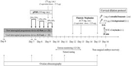
Schematic representation of the experimental procedures, including the estrus synchronization protocols followed by superovulatory treatment, and cervical dilation protocol in ewes subjected to nonsurgical embryo recovery (NSER) at 6.5 d after the device removal; pFSH: porcine follicle-stimulating hormone; i.m. intramuscular; i.v. intravenous; l.v.: latero-vulvar.
2.3 Estrus detection and natural mating
Ewes were kept with vasectomized rams equipped with ink-marked harnesses (ratio 10:1) and signs of estrus were observed twice daily (0800 and 1600) for 30 minutes, between 12 and 60 h after device removal. From the moment the females were first marked, they were mated three times at 12-hour intervals. Ewes were mated with rams (ratio 5:1) of the homologous breed, previously confirmed as fertile by breeding soundness examination. Rams underwent a comprehensive clinical examination with emphasis on reproductive tract evaluation. Libido was assessed during semen collection procedures and through behavioral observations. Semen quality was evaluated through mass motility (wave motion), progressive motility, sperm concentration, and morphological analysis of spermatozoa.
2.4 B- and Color Doppler mode ovarian ultrasonography
Ovarian ultrasonographic evaluations were performed on Days 0 (device insertion), 7 (first pFSH administration), 9 (sixth and final pFSH administration and device removal), 11 (36 h after device removal), and 15 (12 h before nonsurgical embryo recovery, NSER) (Figure 1). Examinations were conducted by an experienced technician using a Z5 Vet ultrasound system (Mindray®, Z5Vet, Digital Ultrasonic Diagnostic Imaging System, Brazil) equipped with a transrectal multifrequency (5–10 MHz) transducer. Standardized ultrasound settings were as follows: B-mode (depth 4.6 cm; gain 90 %; frame rate 55 frames/s; dynamic range 120 dB) and Color Doppler mode (frequency 5.7 MHz; gain 50 %; wall filter 183 Hz; pulse repetition frequency 1.1 kHz).
All visible antral follicles (≥ 2.0 mm) and detectable corpora lutea (CL) identified by B-mode ultrasonography were counted. Two perpendicular measurements were obtained to calculate the mean follicular diameters. Antral follicles were classified as small follicles (SF, ≤ 3.9 mm), medium follicles (MF, 4.0–5.9 mm), or large follicles (LF, ≥ 6.0 mm).
On Day 15, CL and luteinized anovulatory follicles (LAF) were quantified. LAF are defined as structures resulting from failed ovulation, measuring 5.0–10.0 mm in diameter (14). The total number of luteinized structures (CL + LAF) was calculated because ultrasonographic examination alone cannot definitively distinguish between ovulated and non-ovulated luteinized follicles without serial daily monitoring. This variable was considered an indicator of the total progesterone-producing capacity of the ovaries. The percentage of anovulatory luteinized follicles was calculated as (LAF/total luteinized structures × 100) to assess ovulatory efficiency in each experimental group.
Color Doppler ultrasonography was used to assess ovarian blood perfusion on Days 11 and 15. The mean percentage of ovarian blood perfusion area (Color Doppler signal area/ovarian cross-sectional area × 100) was calculated using ImageJ software (U.S. National Institutes of Health, Bethesda, MD, USA), based on three consecutive frames centered around the strongest Doppler signal.
2.5 Nonsurgical embryo recovery
All ewes in which corpora lutea were detected by ultrasonography were submitted to uterine flushing for NSER on Day 16 (6.5–7 days after progesterone intravaginal device removal).
A hormonal protocol for cervical dilation was performed using d-cloprostenol (37.5 μg) administered l.v. and estradiol benzoate (1 mg; Estrogin®, Biofarm, SP, Brazil) administered i.m., both 16 h before NSER. Oxytocin (50 IU; Oxytocin Forte®, UCB, SP, Brazil) was administered i.v. 20 min before uterine flushing.
The NSER procedure was conducted as previously described (15,16). Animals were subjected to the Embrapa analgesia and anesthesia protocol, which consisted of acepromazine maleate 1 % (0.1 mg/kg BW, i.m., 20 min before NSER; Aceproven®, Vencofarma, Londrina, Paraná, Brazil); dipyrone and n-butyl hyoscine bromide solution (5 mL i.v. and 5 mL i.m., 20 min before NSER; Buscofin Composto®, Agener União, Taboão da Serra, Brazil); and lidocaine 2 % (2 mL/animal epidural at S5–C1 and 5 mL intravaginally, administered 5 min and 3 min before NSER, respectively; Lidovet®, Bravet, Rio de Janeiro, Brazil).
The recovered fluid was filtered and examined under a stereomicroscope (Olympus SZ; Olympus Optical Co., Ltd., Tokyo, Japan) at 20–40× magnification. All retrieved structures were maintained in holding medium (Holding Plus, 0.4 % BSA; Embriocare, Cultilab, Brazil) and morphologically classified according to the criteria described in the 5th edition of the International Embryo Technology Society Manual (17). Embryos classified as Grade 1 to 3 were considered viable.
2.6 Variables and statistical analysis
The following parameters were calculated: follicular populations on Days 0, 7, 9, and 11 (total number of antral follicles, small follicles, medium follicles, and large follicles; diameter in mm of the largest antral follicle); estrus response (number of females in estrus/number of treated females × 100); interval to estrus (interval in hours from device removal to first estrus detection); behavioral estrus duration (interval in hours from the first to the last estrus detection); estrus duration (interval in hours between the first and last acceptance of mounting); percentage of ewes with CL one day before NSER; ovarian blood perfusion area (%) on Days 11 and 15 (Color Doppler signal area/ovarian cross-sectional area × 100); number of CL on Day 15; number of luteinized anovulatory follicles (LAF) on Day 15; percentage of ewes successfully penetrated and flushed during NSER; number of CL per ewe flushed; number of recovered structures; and number of viable embryos. The recovery rate was calculated as (number of recovered structures/number of corpora lutea) × 100. When the number of recovered structures exceeded the number of CL counted by ultrasonography, the recovery rate per ewe was capped at 100 %. The viability rate was calculated as (number of viable embryos / number of recovered structures) × 100. Ewes were also classified according to superovulatory response (< 10 CL or ≥ 10 CL one day before NSER) to evaluate the percentage of viable embryos obtained from different CL classes.
Data were analyzed using R software (Foundation for Statistical Computing, Vienna, Austria; Version 3.6.1, 2019). A 2 × 2 factorial arrangement was adopted in a completely randomized design (CRD) with time as a subdivided plot factor. Fixed effects included treatment, breed, day, and their interactions, according to the model proposed for a 2 × 2 factorial arrangement (18). Variables related to estrus, superovulatory response, and embryo production were tested for normality using ANODEV. When global tests were significant, Tukey’s test, chi-square test, or Fisher’s exact test was used for pairwise comparisons, as appropriate. Longitudinal discrete data related to ovarian follicular populations were analyzed by ANODEV using generalized linear mixed models with negative binomial or Poisson distributions and a logarithmic link function. Interactions involving day were evaluated using ANOVA followed by Tukey’s post hoc test for multiple comparisons. Pearson correlation coefficients were calculated between ovarian blood perfusion area (%) and estrus parameters, superovulatory response, and embryo production variables. Results are presented as least square means (LSMEANS) ± standard error of the mean (SEM). Differences were considered statistically significant at P < 0.05.
3. Results
3.1 Ovarian population
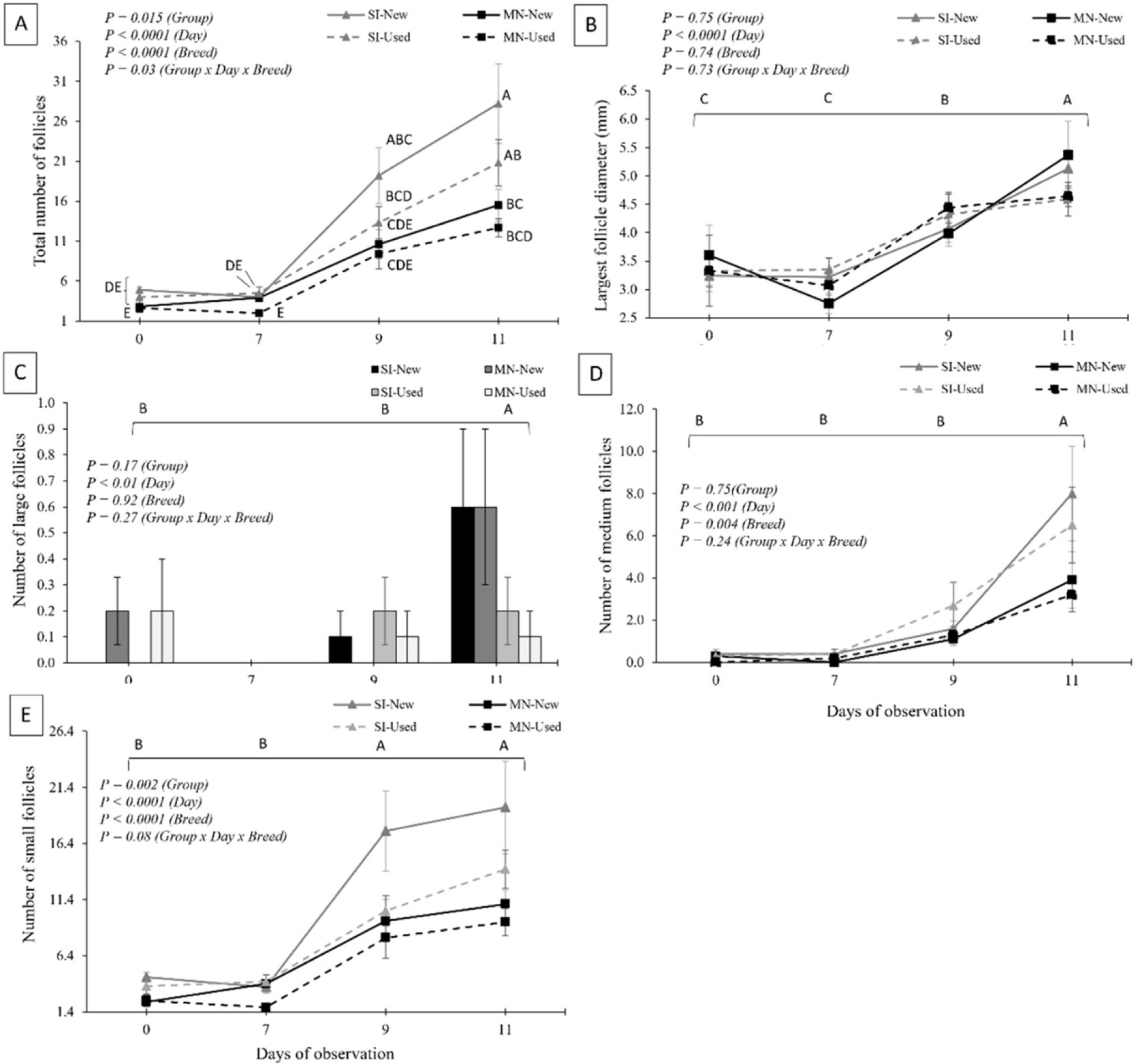
Ovarian follicular populations recorded for the treatment groups at progesterone intravaginal device insertion (Day 0), first (Day 7) and last (Day 9) p-FSH administrations, and 36 h after device removal (Day 11) are presented in Figure 2. A significant interaction among the main effects (treatment, breed, and day of evaluation) was observed only for the total number of follicles, which was greater (P < 0.05) at 36 h after device removal in Santa Inês ewes. A main effect of treatment (P < 0.05) was detected for the total number of follicles (G-New: 11.1 ± 1.2; G-Used: 8.7 ± 0.9) and the number of small follicles (G-New: 9.0 ± 1.0; G-Used: 6.7 ± 0.6). A breed effect (P < 0.05) was observed for the total number of follicles (SI: 12.4 ± 1.3; MN: 7.4 ± 0.7), number of small follicles (SI: 9.7 ± 1.0; MN: 6.0 ± 0.5), and number of medium follicles (SI: 2.5 ± 0.5; MN: 1.2 ± 0.2). A significant day effect (P < 0.05) was found for the total number of follicles, number of small, medium, and large follicles, and diameter of the largest follicle, with the highest values observed on Day 11 compared to Day 0.
Ovarian follicular populations (mean ± SEM) at progesterone intravaginal device insertion (Day 0 p.m.), first (Day 7 a.m.) and last (Day 9 p.m.) pFSH administration, and 36 h after (Day 11 a.m.) intravaginal device removal of locally adapted Brazilian (SI: Santa Inês; and MN: Morada Nova) ewes submitted to 9-days estrus synchronization protocol with a new (New) or a used (Used; previously used once for nine days) intravaginal progesterone device associated to superovulatory treatment, and nonsurgical embryo recovery. A,BDifferent superscripts indicate statistical difference (P < 0.05) by Tukey’s test.
3.2 Estrus, superovulatory response, and embryo recovery
Estrus behavior was not observed in any ewe at the time of device removal. The interval from device removal to estrus was less than 24 h in all groups, except in G-New MN ewes. Overall data related to estrus behavior, superovulatory response, and embryo recovery are presented in Table 1. NSER was successfully performed (i.e., successful cervical penetration and uterine flushing) in 93.7 % (15/16) of MN ewes and 89.5 % (17/19) of SI ewes.
Estrus data, superovulatory response, and in vivo embryo production (LSMEANS ± SEM) of locally adapted Brazilian ewes submitted to a 9-day estrus synchronization protocol with a new (G-New) or a used (G-Used) intravaginal progesterone device associated with superovulatory treatment and nonsurgical embryo recovery.
Significant treatment × breed interactions were observed for interval to estrus, number of corpora lutea (CL), and total luteinized structures (P < 0.05). In G-New MN ewes, the interval to estrus was longer (P < 0.05) compared to G-Used MN ewes. Lower superovulatory responses, reflected by reduced numbers of CL and total luteinized structures, were also observed in G-New MN. In contrast, greater SOV responses were observed in G-New SI ewes. The number of CL per ewe flushed, the number of recovered structures, and the number of viable embryos did not differ (P > 0.05) between treatment groups. The overall mean number of viable embryos per flushed ewe was 9.2 ± 1.7. The coefficient of variation for the number of viable embryos was 103 % overall (G-New MN: 92 %; G-New SI: 143 %; G-Used MN: 52 %; G-Used SI: 88 %).
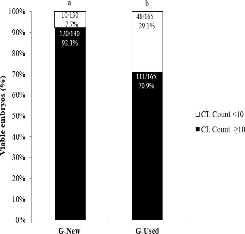
Ewes with a CL count <10 before NSER contributed to a greater percentage (P<0.05) to the production of total embryos in the G-New group compared to G-Used (Figure 3).
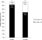
Percentages of viable embryos after nonsurgical embryo recovery from Morada Nova and Santa Inês ewes with <10 or ≥10 corpora lutea (CL) that underwent a protocol of 9-days estrus synchronization with a new (G-New) or a used (G-Used) intravaginal progesterone device associated to superovulatory treatment. A,B Comparison by Chi-square test, significant difference at P < 0.05.
Significant positive and negative correlations (P < 0.05) were observed between ovarian blood perfusion percentage on Day 11 and estrus characteristics, superovulatory response, and embryo recovery variables only in the G-Used group. On Day 15, significant correlations were detected in both treatment groups (Table 2).
Significant correlations of ovarian blood perfusion percentage on Day 11 and Day 15 with estrus responses, superovulatory responses, and embryo production of the locally adapted Brazilian ewes submitted to estrus synchronization protocol with a new (G-New) or a used (G-Used) intravaginal progesterone device associated with superovulatory treatment and nonsurgical embryo recovery.
4. Discussion
Estrus behavior was observed in all females of the G-New group, irrespective of breed, and only one Santa Inês ewe in G-Used failed to display estrus signs. This high synchronization rate suggests that progesterone concentrations were maintained at levels sufficient to prevent spontaneous ovulation and promote synchronous estrus (9,19). These findings are further supported by ultrasonographic monitoring of the ovarian follicular population. Although only slight differences in estrus behavior were observed between treatments, significant treatment × breed interactions were detected for superovulatory response, as reflected by total corpora lutea (CL) and total luteinized structures.
The total number of follicles was lower in Morada Nova compared to Santa Inês ewes. Breed effects are known to influence superovulatory responses (20), and some authors report that breed-related differences may account for approximately 30 % of the variability in embryo yield (21). The antral follicular count (AFC) is one of the principal determinants of superovulatory response and embryo production in sheep and has been suggested as a criterion for donor selection, thereby avoiding stressful procedures in animals with limited superovulatory potential (22, 23).
Interestingly, 40 % of G-New Morada Nova ewes did not exhibit CL on ultrasonography performed prior to embryo recovery. The proportion of ewes with absent or limited ovulation represents a major source of variability in MOET programs, with reported incidences of approximately 20–30 % (24, 25). One factor that may reduce ovulation rates is the presence of dominant follicles at the time of the first pFSH administration, as subordinate follicles may grow to preovulatory size but fail to ovulate (26).
The classification of large follicles (≥ 6.0 mm) was effective for confirming the absence of dominant follicles at treatment initiation (Day 7), which is critical for superovulatory success (24,25,26). The presence of large follicles (≥ 6 mm) at FSH onset has been associated with reduced ovulatory responses (20,25,26). However, this cutoff may be less appropriate for identifying preovulatory follicles on Day 11, when follicular maturation is advanced, and estrus is evident in most ewes. Other studies have defined large or preovulatory follicles using lower thresholds (≥ 5.0 mm) (4,27), which may provide greater sensitivity for detecting mature follicles. Nevertheless, this limitation did not affect the primary objective of comparing embryo yields between new and reused devices. Ovulatory failure and persistent anovulatory follicles are also reported even under the “Day 0 Protocol,” which is designed to initiate superstimulation in the absence of dominant follicles (27).
Embryo yield in the present study was greater than that reported in studies using similar protocols in Lacaune ewes (12) and/or comparable pFSH doses in Santa Inês ewes (28), despite lower reported percentages of LAF in those studies (7 % and 3.1–13.7 %, respectively). When satisfactory embryo yields (~7 viable embryos) are achieved, ovulatory failure rates of 15 % (6) and 30 % (29) have also been documented. In the present study, reuse of the progesterone device did not appear to influence the formation of anovulatory follicles.
In the G-New group, ovarian blood perfusion was a strong predictor of superovulatory response, correlating positively with CL number, total luteinized structures, and viable embryos on Days 11 and 15. In G-Used, moderate positive correlations were observed between CL count, total luteinized structures, and ovarian blood perfusion on Day 15. Moderate negative correlations were detected between the interval to estrus and ovarian blood perfusion on Day 11 (G-New) and Day 15 (G-Used).
Because correlation indicates association rather than causation, these findings must be interpreted cautiously. The greater variability in superovulatory responses observed in G-New may have contributed to the negative correlation observed in this group on Day 11. A rapid increase in estradiol secretion by preovulatory follicles would be expected to trigger estrus signs promptly, resulting in a shorter interval from device removal to estrus. Collectively, these data suggest that improved ovarian perfusion is associated with shorter estrus intervals, stronger superovulatory responses, and enhanced embryo production.
The observed correlations between ovarian blood perfusion and estrous behavior, superovulatory response, and embryo yield are consistent with previous studies evaluating the utility of Color Doppler ultrasonography for assessing luteal function (30) and embryo recovery potential (6,14,31). Although CL counting using Color Doppler may slightly decrease in accuracy as CL number increases (32), the overall estimate remains clinically valuable. Ewes presenting ≥ 10 CL contribute approximately 90 % of total embryo production, whereas those with < 10 CL contribute only about 10 % (4). Therefore, in cases of uncertainty in CL enumeration, assessment of total ovarian blood perfusion area—given its strong correlation with viable embryo numbers—may enhance decision-making regarding NSER through non-invasive ultrasonographic evaluation.
Mean viable embryo yield did not differ among treatment × breed groups. However, although not statistically significant, new progesterone devices yielded both the lowest and highest values, with the greatest coefficients of variation observed across genetic groups (Morada Nova and Santa Inês, respectively). Moreover, G-Used ewes with lower ovulatory responses (< 10 CL) contributed a higher proportion of viable embryos compared to G-New, suggesting more homogeneous responses. Under the conditions of the present study, device reuse appeared to reduce individual variability, a desirable characteristic in MOET programs (20).
In addition to supporting highly viable embryo production, the mid-term protocol effectively permitted reuse of intravaginal devices, totaling 18 days of device permanence. In commercial or experimental (cross-over) settings, donors are commonly superovulated twice at 30-day (12) to 60-day intervals (6). Reuse of an intravaginal device within the same donor during sequential superovulatory protocols may therefore be feasible. Given the maintained embryo yields, device reuse represents a practical strategy for reducing superovulatory costs and minimizing environmental waste, aligning with Green, Clean, and Ethical principles for controlled reproduction in sheep (13).
This study is the first to report successful reuse of intravaginal progesterone devices in superovulated Santa Inês and Morada Nova sheep, two locally adapted Brazilian breeds. Reuse has previously been described in estrus synchronization protocols for Toggenburg goats after autoclaving (7,9) and in Santa Inês sheep following washing and air-drying (8). In Toggenburg goats, devices used for 12 days were subsequently effective for an additional 6-day estrus induction protocol (7), indicating maintenance of plasma progesterone concentrations above 1 ng/mL for up to 18 days. In Santa Inês sheep, circulating progesterone concentrations did not differ between new and reused devices (used for ten days) from day 3 to the end of the 5-day synchronization protocol (8). Despite differences in superovulatory response following p-FSH treatment, high embryo yields per flushed ewe were observed across treatments. Total AFC appears to be the principal factor underlying breed differences. Notably, device reuse reduced individual response variability while maintaining high embryo production efficiency.
5. Conclusion
Reuse of an intravaginal device previously used for nine days resulted in embryo yields comparable to those obtained with a new device, demonstrating that mid-term (9-day) protocols can effectively support the reuse of intravaginal progesterone devices for superovulation in ewes. When combined with appropriate FSH dosing and administration strategies, protocols using either new or reused devices elicited robust ovarian responses, which, together with efficient natural mating and NSER, resulted in high numbers of viable embryos recovered from locally adapted Brazilian sheep.
Data availability statement
The data will be made available upon request to the corresponding author.
-
Generative AI use statementThe authors did not use generative artificial intelligence tools or technologies in creating or editing any part of this manuscript.
Acknowledgments
The authors would like to thank Embrapa Goats and Sheep collaborators for all their help and support during this experiment. This study was supported by Embrapa (Projects 11.15.02.003.01.00 and 22.13.06.026.00.04). Jeferson F. Fonseca and Maria E.F. Oliveira are CNPq fellows.
References
-
1. Fonseca JF, Silva KM. Brazilian Germplasm Bank: conservation of genetic resources of sheep and goats. (Documents 142) Sobral: Embrapa Goats & Sheep; Nov 2021. [cited 2025 Feb 06]. Available from: https://www.infoteca.cnptia.embrapa.br/infoteca/bitstream/doc/1137604/1/CNPC-2021-Art99.pdf
» https://www.infoteca.cnptia.embrapa.br/infoteca/bitstream/doc/1137604/1/CNPC-2021-Art99.pdf -
2. Arrais AM, Mello MRB, Vergani GB, Figueira LM, Esteves SN, Pereira VSA, Bartlewski PM, Oliveira MEF, Souza-Fabjan JMG, Fonseca JF. NonSurgical Embryo Recovery from Estrus Synchronized or Superovulated Morada Nova Ewes: A Feasible Strategy for Sheep Embryo Banking. Biopreserv Biobank. 2021;189:360–368. https://doi.org/10.1089/bio.2020.0125
» https://doi.org/10.1089/bio.2020.0125 -
3. Fonseca JF, Vergani GB, Lima MSD, Silva KM, Monteiro AWU, Ramos AF, Alves BRC, Souza-Fabjan JMG, Oliveira MEF, Batista RITP. Nonsurgical Embryo Recovery as a Feasible Tool for Supporting Embryo Biobanks of Locally Adapted Brazilian Sheep and Goats. Biopreserv Biobank. 2021;20:6. https://doi.org/10.1089/bio.2021.0066
» https://doi.org/10.1089/bio.2021.0066 -
4. Oliveira MEF, Arrais AM, Mello MRB, Vergani GB, Figueira LM, Esteves SN, Pereira VSAP, Garcia AR, Bartlewski PM, Fonseca, J.F. Study of the factors affecting embryo yields and quality in superovulated Morada Nova ewes that underwent non-surgical uterine flushing. Reprod Domest Anim. 2022;57(4):393-401. https://doi.org/10.1111/rda.14077
» https://doi.org/10.1111/rda.14077 -
5. Figueira LM. Alves NG, Souza-Fabjan JMG, Oliveira MEF, Lima RR, Souza GN, Fonseca JF. Preovulatory follicular dynamics, ovulatory response and embryo yield in Lacaune ewes subjected to synchronous estrus induction protocols and non-surgical embryo recovery. Theriogenology. 2020;145:238-246. https://doi.org/10.1016/j.theriogenology.2019.11.004
» https://doi.org/10.1016/j.theriogenology.2019.11.004 -
6. Figueira LM, Alves NG, Maia ALRS, Souza-Fabjan JMG., Batista RITP, Arrais AM, Lima RR, Oliveira MEF, Fonseca JF. In vivo embryo production and recovery in Lacaune ewes after imposing a superovulation treatment regimen is related to pFSH dose. Anim Reprod Sci. 2020; 223:106625. https://doi.org/10.1016/j.anireprosci.2020.106625
» https://doi.org/10.1016/j.anireprosci.2020.106625 -
7. Souza JMG, Torres CAA, Maia ALRS, Brandão FZ, Bruschi JH, Viana JHM, Oba E, Fonseca JF. Autoclaved, previously used intravaginal progesterone devices induces estrus and ovulation in anestrous Toggenburg goats. Anim Reprod Sci 2011; 129:50-55. https://doi.org/10.1016/j.anireprosci.2011.09.012
» https://doi.org/10.1016/j.anireprosci.2011.09.012 -
8. Pinna AE, Brandão FZ, Cavalcanti AS, Borges AM, Souza JMG, Fonseca JF. Reproductive parameters of Santa Inês ewes submitted to short-term treatment with re-used progesterone devices. Arq Bras Med Vet Zootec. 2012; 64:333-340. https://doi.org/10.1590/S0102-09352012000200012
» https://doi.org/10.1590/S0102-09352012000200012 -
9. Souza-Fabjan JMG, Torres CAA, Maia ALRS, Brandão FZ, Oba E, Bertoldo MJ, Fonseca JF. Re-used progesterone devices efficiently synchronise oestrus and ovulation after autoclaving process in Toggenburg goats during the breeding season. Anim. Reprod. Sci. 2014; 55: 818-822. https://doi.org/10.1071/AN14056
» https://doi.org/10.1071/AN14056 -
10. Swelum AA, Saadeldin IM, Moumen AF, Ali MA, Ba-Awadh H, Alowaimer AN, Efficacy of using previously used controlled internal drug release (CIDR®) insert on the reproductive performance, hormone profiles and economic measures of sheep. Reprod. Domest. Anim. 2018; 53:114-1122. https://doi.org/10.1111/rda.13212
» https://doi.org/10.1111/rda.13212 -
11. Martin BG, Kadokawa H. Clean, Green, and Ethical" Animal Production. Case Study: Reproductive Efficiency in Small Ruminants. J Reprod Develop. 2006; 52:145-152. https://doi.org/10.1262/jrd.17086-2
» https://doi.org/10.1262/jrd.17086-2 -
12. Figueira LM, Alves NG, Maia ALRS, Souza-Fabjan JMG, Batista RITP, Morais, MCC, Lima RR, Oliveira MEF, Fonseca JF. Embryo yield and quality are associated with progestogen treatment during superovulation protocol in lactating Lacaune ewes. Theriogenology. 2020; 155:132-138. https://doi.org/10.1016/j.theriogenology.2020.06.004
» https://doi.org/10.1016/j.theriogenology.2020.06.004 -
13. Marques JCS, Maciel JPO, Denis-Robichaud J, Conceicao RS, Bega AM, Moore S, Sirard MA, Baes CF, Cerri RLA. The effect of progesterone concentrations during superovulation of Holstein heifers in a randomized trial. J Dairy Sci. 2023 106:9677–9690. https://doi.org/10.3168/jds.2022-23065
» https://doi.org/10.3168/jds.2022-23065 -
14. Oliveira MEF, Ribeiro IF, Rodriguez MGK, Maciel GS, Fonseca JF, Brandão FZ, Bartlewski PM. Assessing the usefulness of B-mode and colour Doppler sonography, and measurements of circulating progesterone concentrations for determining ovarian responses in superovulated ewes. Reprod. Domest. Anim. 2018; 53:742e50. https://doi.org/10.1111/rda.13165
» https://doi.org/10.1111/rda.13165 -
15. Fonseca JF. Classificação de ovelhas para a colheita de embriões pela via transcervical de acordo com o grau de facilidade de transposição cervical. Circular Técnica 45 [Internet]. Sobral: Embrapa Caprinos e Ovinos; 2017 jul [cited 2025 Feb 06]. Available from http://www.infoteca.cnptia.embrapa.br/infoteca/handle/doc/1071938
» http://www.infoteca.cnptia.embrapa.br/infoteca/handle/doc/1071938 -
16. Fonseca JF, Oliveira MEF, Brandão FZ, Batista RITP, Garcia AR, Bartlewski PM, Souza-Fabjan JMG. Non-surgical embryo transfer in goats and sheep: the Brazilian experience. Reprod Fert Develop. 2019;31:17–26. https://doi.org/10.1071/RD18324
» https://doi.org/10.1071/RD18324 -
17. IETS 2024. Manual of International Embryo Technology Society (IETS), 5th edition [Internet] Champaign IL International Embryo Technology Society (IETS); [cited 2025 Feb 06] Available from: https://www.iets.org/Publications/IETS-Manual (accessed 15 May 2024).
» https://www.iets.org/Publications/IETS-Manual - 18. Kaps M, Lamberson WR. Biostatistics for Animal Science, 3rd Edition, Wallingford, United Kingdom: CABI Publishing; 2004. 459p.
-
19. Scaramuzzi RJ, Adams NR, Baird DT, Campbell BK, Downing JA, Findlay JK. A model for follicle selection and the determination of ovulation rate in the ewe. Reprod Fert Develop. 1993; 5:459-478. https://doi.org/10.1071/RD9930459
» https://doi.org/10.1071/RD9930459 -
20. Bartlewski PM, Seaton P, Oliveira MEF, Kridli RT, Murawski M, Schwarz T. Intrinsic determinants and predictors of superovulatory yields in sheep: Circulating concentrations of reproductive hormones, ovarian status, and antral follicular blood flow. Theriogenology. 2016; 86:130–143. https://doi.org/10.1016/j.theriogenology.2016.04.024
» https://doi.org/10.1016/j.theriogenology.2016.04.024 -
21. Khan SU, Jamal MA, Su, Y, Wei H-J, Qing Y, Cheng W. Towards Improving the Outcomes of Multiple Ovulation and Embryo Transfer in Sheep, with Particular Focus on Donor Superovulation. Vet Sci. 2022; 9:117. https://doi.org/10.3390/vetsci9030117
» https://doi.org/10.3390/vetsci9030117 -
22. Pinto PHN, Balaro MFA, Souza-Fabjan JMG, Ribeiro LS, Bragança GM, Leite CR, Arashiro EKN, Silva KM, Fonseca JF, Brandão FZ. Anti-Müllerian hormone and antral follicle count are more effective for selecting ewes with good potential for in vivo embryo production than the presence of FecGE mutation or eCG pre-selection tests. Theriogenology. 2018a.; 113:146-152. https://doi:org/10.1016/j.theriogenology.2018.02.018
» https://doi:org/10.1016/j.theriogenology.2018.02.018 -
23. Brasil OO, Moreira NH, Souto PLG, Silva CMG, Ramos AF. Ovarian assessment for pre-selection of embryo donor ewes. Small Rumin Res. 2022; 216:106803. https://doi.org/10.1016/j.smallrumres.2022.106803
» https://doi.org/10.1016/j.smallrumres.2022.106803 -
24. Cognie Y. State of the art in Sheep-Goat embryo transfer. Theriogenology. 1999; 51: 105–116. https://doi.org/10.1016/S0093-691X(98)00235-0
» https://doi.org/10.1016/S0093-691X(98)00235-0 -
25. Gonzalez-Bulnes A, Santiago-Moreno J, Cocero MJ, Lopez-Sebastian A. Effects of FSH commercial preparation and follicular status on follicular growth and superovulatory response in Spanish Merino ewes. Theriogenology. 2000; 54:1055–1064. https://doi.org/10.1016/s0093-691x(00)00414-3
» https://doi.org/10.1016/s0093-691x(00)00414-3 -
26. Veiga-Lopez A, Gonzalez-Bulnes A, Garcia-Garcia RM, Dominguez V, Cocero MJ The effects of previous ovarian status on ovulation rate and early embryo development in response to superovulatory FSH treatments in sheep. Theriogenology. 2005; 63:1973–1983. https://doi.org/10.1016/j.theriogenology.2004.09.055
» https://doi.org/10.1016/j.theriogenology.2004.09.055 -
27. Ribeiro ACS, Pinto PHN, Santos JDR, Ramalho NP, Laeber CCR, Balaro MFA, Batista RITP, Souza-Fabjan JMG, Fonseca JF, Ungerfeld R, Brandão FZ. The pFSH dose affects the efficiency of in vivo embryo production in Santa Inês ewes. Anim Reprod Sci. 2024; 264:107459. https://doi.org/10.1016/j.anireprosci.2024.107459
» https://doi.org/10.1016/j.anireprosci.2024.107459 -
28. Maciel GS, Rodriguez MGK, Santos VJC, Uscategui RAR, Nociti RP, Maronezi MC, Oliveira CS, Feliciano MAR, Vicente WRR, Fonseca JF, Oliveira MEF. Follicular dynamics and in vivo embryo production in Santa Inês ewes treated with small doses of pFSH. Anim Reprod Sci. 2019; 209:106137. https://doi.org/10.1016/j.anireprosci.2019.106137
» https://doi.org/10.1016/j.anireprosci.2019.106137 -
29. Veiga-Lopez A, Gonzalez-Bulnes A, Tresguerres JAF, Dominguez V, Ariznavarreta C, Cocero MJ. Causes, characteristics, and consequences of anovulatory follicles in superovulated sheep. Domest Anim Endocrinol. 2006; 30:76–87. https://doi.org/10.1016/j.domaniend.2005.06.001
» https://doi.org/10.1016/j.domaniend.2005.06.001 -
30. Figueira LM, Fonseca JF, Arashiro EKN, Souza-Fabjan JMG, Ribeiro ACS, Oba E, Viana, JHM, Brandão FZ. Colour Doppler ultrasonography as a tool to assess luteal function in Santa Inês ewes. Reprod Domest Anim. 2015; 50:643-650. https://doi.org/10.1111/rda.12543
» https://doi.org/10.1111/rda.12543 -
31. Oliveira MEF, Feliciano MAR, D’Amato CC, Oliveira LG, Bicudo SD, Fonseca JF. Vicente WRR, Visco E, Bartlewski PM. Correlations between ovarian follicular blood flow and superovulatory responses in ewes. Anim Reprod Sci. 2014; 144: 30-37. https://doi.org/10.1016/j.anireprosci.2013.10.012
» https://doi.org/10.1016/j.anireprosci.2013.10.012 -
32. Pinto PHN, Bragança GM, Balaro MFA, Arashiro EKN, dos Santos GB, Souza-Fabjan JMG, Fonseca JF, Brandão FZ. Colour-Doppler ultrasound imaging as a laparoscopy substitute to count corpora lutea in superovulated sheep. Reprod Domest Anim. 2018b; 53:266–269. https://doi.org/10.1111/RDA.13089
» https://doi.org/10.1111/RDA.13089