 Estrous cycle modulates the acute and persistent inflammatory muscle hyperalgesia in sedentary but not in exercised female mice
ABSTRACT
BACKGROUND AND OBJECTIVES Persistent pain is a significant public health issue, profoundly impacting quality of life. Women experience persistent pain more frequently and with greater intensity than men, which is attributed to sexual hormonal fluctuations. This study investigated the influence of the estrous cycle on the development and maintenance of acute and persistent inflammatory muscle hyperalgesia in sedentary and exercised female mice.
METHODS The study analyzed mechanical muscle hyperalgesia in female Swiss mice that underwent a model of acute and persistent inflammatory muscle hyperalgesia, during each of the four estrous cycle phases (proestrus, estrus, metestrus or diestrus). Swimming exercises were performed before the induction of acute and persistent inflammatory muscle hyperalgesia.
RESULTS The inflammatory stimulus of carrageenan during the proestrus phase induced the most intense acute muscle hyperalgesia. Conversely, in the estrus phase, the inflammatory stimulus induced the most intense persistent muscle hyperalgesia. The maintenance of persistent muscle hyperalgesia was not modulated by estrous cycle. Regular swimming exercise prevented the acute and persistent muscle hyperalgesia, regardless of the estrous cycle.
CONCLUSION These findings suggest that the estrous cycle phase during which an inflammatory insult occurs is critical for the development of acute inflammatory muscle hyperalgesia and its transition to the persistent phase. This effect was observed in sedentary, but not in exercised female mice, suggesting that regular physical activity may provide analgesic benefits regardless estrous cycle phase or the associated fluctuations in gender hormones. Further research is necessary to elucidate the mechanisms underlying the development of inflammatory muscle pain in females.
Keywords:
Estrous cycle; Female mice; Persistent hyperalgesia; Physical exercise; Muscle
HIGHLIGHTS
The estrous cycle, in which an inflammatory insult occurred, was determinant in the development of acute and persistent inflammatory muscle hyperalgesia
The inflammatory insult to the musculoskeletal muscle during the proestrus phase triggered the highest acute muscle hyperalgesia
The inflammatory insult to the musculoskeletal muscle during the estrus phase triggered the highest persistent muscle hyperalgesia
Regular swimming exercise prevented the acute and persistent muscle hyperalgesia, regardless of the estrous cycle phase
RESUMO
JUSTIFICATIVA E OBJETIVOS Dor persistente é um problema de saúde pública que impacta significativamente a qualidade de vida. Mulheres relatam sentir dores persistentes com maior intensidade e frequência do que homens, o que é atribuído às flutuações dos hormônios sexuais. Neste estudo, investigou-se a influência do ciclo estral no desenvolvimento e manutenção da hiperalgesia muscular inflamatória aguda e persistente em fêmeas sedentárias e exercitadas.
MÉTODOS A hiperalgesia muscular mecânica foi avaliada em camundongos fêmeas, da linhagem Swiss, submetidos ao modelo de hiperalgesia muscular inflamatória aguda e persistente, durante as quatro fases do ciclo estral (proestro, estro, metaestro ou diestro). O exercício foi realizado através da natação previamente à indução da hiperalgesia muscular.
RESULTADOS Quando o estímulo inflamatório da carragenina ocorre na fase proestro, desencadeia-se a hiperalgesia muscular aguda de maior intensidade. Por outro lado, durante a fase estro, o estímulo inflamatório desencadeia a hiperalgesia muscular persistente de maior intensidade. A duração da hiperalgesia muscular persistente não é modulada pelos ciclos estrais. Exercícios de natação regulares preveniram a hiperalgesia muscular aguda e persistente independentemente do ciclo estral.
CONCLUSÃO A fase do ciclo estral em que ocorreu o estímulo inflamatório é determinante para o desenvolvimento da hiperalgesia muscular aguda e sua transição para a fase persistente. Esse efeito foi observado em fêmeas sedentárias, mas não em exercitadas, sugerindo que a atividade física regular pode promover benefícios analgésicos independentemente da fase do ciclo estral ou das flutuações hormonais associadas. Pesquisas adicionais são necessárias para elucidar os mecanismos subjacentes ao desenvolvimento da hiperalgesia muscular inflamatória em fêmeas.
Descritores:
Camundongos fêmeas; Ciclo estral; Exercício físico; Hiperalgesia persistente; Músculo
DESTAQUES
O ciclo estral, no qual ocorreu o estímulo inflamatório, foi determinante para o desenvolvimento da hiperalgesia muscular inflamatória aguda e persistente
O estímulo inflamatório no músculo esquelético durante a fase proestro provocou a hiperalgesia muscular aguda mais intensa
O estímulo inflamatório no músculo esquelético durante a fase estro provocou a hiperalgesia muscular persistente mais elevada
A prática regular de natação preveniu a hiperalgesia muscular aguda e persistente, independentemente da fase do ciclo estral
INTRODUCTION
Chronic pain is a significant global health issue with a high socioeconomic impact, affecting millions worldwide1-3. It's more common in women, typically increases after puberty, and fluctuates throughout menopause4-6. For instance, the prevalence of back pain, headache, stomach pain and temporomandibular joint pain increases with pubertal development in girls7,8 and musculoskeletal pain, headaches or migraines, and vulvovaginal pain have a high incidence in the menopause period6. This strongly suggests that ovarian hormones have a significant influence on pain sensitivity.
While the impact of ovarian hormones on sensitivity to various pain conditions is clear, their role is remarkably complex. The two most important ovarian hormones are estrogen and progesterone. Several studies agree that fluctuations in estrogen levels significantly impact pain perception. These hormonal shifts lead to increased pain, while stable hormone levels offer a protective mechanism against nociception in females9-12. Progesterone, on the other hand, seems to have a pain-reducing effect13-15. Despite this complexity, most preclinical studies are performed using male subjects, which can lead to inefficient strategies to control chronic pain in women.
Regular physical exercise is a well-known strategy to reduce many chronic pain conditions16-18. Moreover, physically inactive individuals are more likely to develop chronic pain throughout their lives19-21. It was recently demonstrated that regular swimming exercise prevents the acute and persistent inflammatory muscle hyperalgesia in male mice by a mechanism dependent on macrophages22. However, little is known about the preventive effect of exercise on transition to chronic muscle hyperalgesia in female mice.
Given the significant implications of gender-related differences in pain processing, this study investigated the influence of estrous cycle phases on the development and maintenance of acute and persistent inflammatory muscle hyperalgesia in female mice. To this end, the authors used a model of acute and persistent inflammatory muscle hyperalgesia to analyze the behavioral nociceptive responses within each of the four estrous cycle phases in both sedentary and exercised female mice.
METHODS
Ethics and study design
A total of 100 female Swiss mice (Mus musculus), 60 days old, provided by Multidisciplinary Center for Biological Research (CEMIB) from UNICAMP, were used. The experimental procedure was approved by the local institutional Ethics Committee for animal’s use (CEUA-UNICAMP, protocol number 5234-1/2019), following the guidelines of the National Council for the Control of Animal Experimentation (CONCEA, Brazil) research, Brazilian Federal Law 11,794/2008 (Arouca Law) and ethics committee of the International Association for the Study of Pain in Conscious Animals. The animals were kept in plastic cages (five per cage) containing wood shavings and plastic tubes, under light/dark cycles of 12 h (light switched on at 06:00 a.m.) with water and food ad libitum, except during muscle injections, exercise sessions, and mechanical muscle hyperalgesia tests. Experimental sessions were conducted during the light phase, from 7:00 a.m., in a quiet room with a controlled temperature (±23 °C). The researcher responsible for assigning the animals to groups was aware of the group allocations. All behavioral experiments were conducted with women who were blinded to group allocations. After the behavioral testing period, all animals were euthanized, following the approved ethical protocol.
This study was planned based on previously published experiments. A detailed protocol, including the research question, key design features, and analysis plan, was prepared before the study.
Determining the stage of the estrous cycle
The estrous cycle was determined indirectly by using the non-invasive vaginal lavage method23. Initially, to check whether the female mice were cycling regularly, 4 days before carrageenan injection or exercise, estrous cycle was analyzed. This procedure was always performed at 7:30 h a.m. The vaginal cells were flushed by introducing 15µL of saline through a pipette and placing a few drops of cell suspension on a glass slide for microscopic examination. The visual analysis of the cells was used to determine the estrous cycle as follows: proestrus (predominance of nucleated epithelial cells), estrus (predominance of cornified cells), metestrus (predominance of leukocytes and presence of some cornified cells) and diestrus (leukocyte predominance)23. Female animals were selected for each experiment based on the stage of the estrous cycle. Factors that could confound the results were controlled: females were grouped by estrous cycle phase and housed in separate cages during each experiment.
The model of acute and persistent inflammatory muscle hyperalgesia
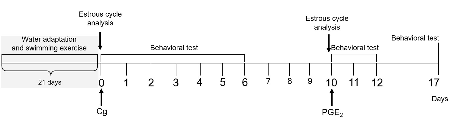
In this study, a model of persistent inflammatory muscle hyperalgesia24 was used, standardized in mice22,25. Immediately after determining the estrous cycle, λ – carrageenan (Cg, 100 µg/muscle) was injected into the belly of the gastrocnemius muscle to induce the acute inflammatory muscle hyperalgesia, after 10 days, when the nociceptive threshold was at baseline levels, Prostaglandin E2 (PGE2; 1 µg/muscle) was injected at the same site to trigger the persistent muscle hyperalgesia. The measurement of the hyperalgesic threshold was performed at the following time-points (Figure 1): 1) Immediately before the muscle injection (baseline); 2) after 1h, 3h and 6h of the injection at day 0; 3) daily until the 10 th day; 4) immediately before the injection of PGE2 (10 th day); 5) after 1 h and 4 h of the injection of PGE2 (10 th day); 6) at days 11, 12 and 1722,25. In control groups, isotonic saline (0.9% NaCl) was administered instead of carrageenan. Swimming exercise was performed before the first injection, and the estrous cycle was determined before carrageenan or PGE2 (Figure 1). All animals assigned to each experimental group were included in the analysis.
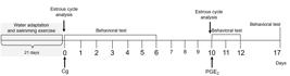
Experimental procedures in the model of persistent muscle hyperalgesia. Carrageenan (Cg) or saline (Sal) was administered into the gastrocnemius muscle. Behavioral test was applied from day 0 to 6 after the injection. At the 10th day, PGE2 was administered and behavioral test was applied at days 10, 11, 12 and 17. Estrous cycle analysis was determined before Cg or PGE2 injection. Swimming exercise procedures (gray square) were performed before Cg/Sal injections, with an interval of 48 h before the baseline behavioral test at day 0. Adapted from a reference study from 202122.
Mechanical muscle nociceptive threshold test
The mechanical muscle nociceptive threshold in mice was assayed through the Randall-Selitto digital algesimeter (Insight, Brazil)22,25. The device applies linear mechanical pressure to the gastrocnemius muscle through a rounded tip with 2 mm to evoke the nociceptive threshold of deep tissues26. Three measurements, at intervals of 5 minutes each, were performed to get the nociceptive threshold22,25. Baseline levels were measured two days after the end of the physical exercise period or equivalent to the sedentary group22. Mechanical muscle hyperalgesia was calculated as the difference (in grams) between the baseline measurement and the values obtained at each time point following carrageenan (or saline) and PGE2 injections. Mechanical muscle hyperalgesia was represented in the y-axis by increasing values22,25,27.
Intramuscular injection, drugs and doses
The drugs or their vehicles were injected into the mice's gastrocnemius muscle using a 30-gauge needle28, connected to a polyethylene catheter and a Hamilton syringe (50μL). The final volume was 20μl. The following drugs were used: λ – carrageenan (100 μg/muscle) and prostaglandin E2 (PGE2, 1μg/muscle) 22,24,25. All drugs were purchased from Sigma-Aldrich (St. Louis, MO, USA), and they were dissolved in isotonic saline (0.9% NaCl) to obtain the working solution. The stock solution of PGE2 was dissolved in ethanol, according to the datasheet, and re-suspended in saline.
Swimming exercise protocol
Swimming was performed as regular physical exercise. The water temperature was maintained at 31 ± 1 ºC and all procedures were carried out in individual cylindrical tanks with a smooth surface22,29. To minimize stress, mice were progressively adapted to the liquid environment, with a progressive increase in water depth, for 6 days22,30. Before starting the swimming exercise protocol, the animals remained at rest for 24 hours. The swimming protocol was performed for 15 days, split into three periods of 5 days each, with daily sessions of 50 minutes. To allow physiological adaptation and minimize the risk of fatigue or injury, a protocol with progressive increase in intensity was employed23. In the first period, mice performed swimming sessions with 4-minute passive pauses. In the second period, interval sessions of a 3-minute pause were used, and in the last period, mice swam 50 minutes without pauses. Mice were withdrawn from the water when inappropriate movements, such as floating, climbing, diving and bobbing, were observed31. For control groups, mice were maintained in individual tanks with shallow water for 10 minutes22. All animals from each experimental group were considered in the analysis.
Measurement of serum corticosterone levels
Before blood sampling, animals were habituated for one hour in a quiet room. Blood collection was carried out without the use of anesthesia or restraint. Blood samples were collected 24 hours before starting the exercise (baseline) and 24 hours after the end of the exercise protocol22. A small cut at the distal end of the animal's tail was made and 25 μL of blood was collected using non-heparinized glass capillaries. After 30 min, the samples were centrifuged, and the supernatant was collected and stored at -20◦C until analysis. Corticosterone was measured using the Enzyme-Linked ImmunoSorbent Assay (ELISA) test, according to the manufacturer's instructions (Kit DetectX - Arbor Assays).
Statistical analysis
According to the Kolmogorov-Smirnov test, all data followed a normal Gaussian’s distribution and allowed application of parametric tests. Quantitative data were analyzed by one-way ANOVA, two-way ANOVA, Mixed-effects Analysis or Student’s t test (unpaired). Following one-way and two-way ANOVA, post hoc contrasts were performed using the Tukey test. Area Under the Curve (AUC) analysis was performed for behavioral experiments to evaluate general effects of the interventions in acute and chronic hyperalgesia. AUC from acute period was considered from day 0 to day 4 after carrageenan injection (AUC Δ, g × 4 days) and AUC from chronic period was considered from day 10 to 17 (AUC Δ, g × 10–17 days). The sample size for continuous variables was calculated based on the estimated population standard deviation and the expected mean difference between groups, using the formula “n=1+[2C*(s/d)2]”, where “C=(zα+zβ)2”. Parameters were set at α=0.05 (two-sided), power = 80%, s=0.2, and d=0.532. Final values are reported in the figure legends. These values are expressed as means ± standard error of mean (SEM). All data were analyzed by GraphPad Prism 7.0 software. The outlier calculator tool from GraphPad website was applied to each group. For all tests, significance was set at p<0.05.
RESULTS
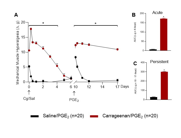
First, the authors analyzed the development of acute and persistent inflammatory muscle hyperalgesia in female mice independent of their estrous cycle. Administration of carrageenan into gastrocnemius muscle induced acute muscle hyperalgesia (n=20, p<0.05, Two Way ANOVA, Tukey post-test Figure 2A, AUC, Student t test, Figure 2B) when compared to saline control group (n=20). Administration of PGE2 into gastrocnemius muscle previously sensitized by carrageenan showed the persistent muscle hyperalgesia (n=20, p<0.05, Two Way ANOVA, Tukey post-test Figure 2A; AUC, Student T test, Figure 2C) when compared to saline control group (n=20).

Sexual dimorphism in acute and persistent inflammatory muscle hyperalgesia. (A) Behavioral nociceptive responses at different time points. AUC of the acute (B) and persistent (C) muscle hyperalgesia. The symbol “*” indicates differences for the saline control group.
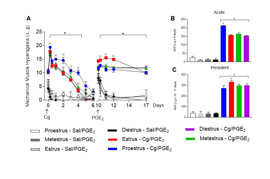
To investigate the influence of the estrous cycle on the development of acute and persistent inflammatory muscle hyperalgesia, carrageenan was administered at each of the four-cycle phases (proestrus, estrus, metestrus or diestrus). The estrous cycle phase in which PGE2 was injected was not considered. When carrageenan was injected during the proestrus (n=5), estrus (n=5), metestrus (n=5) or diestrus (n=5) phases, there were development of acute and persistent muscle hyperalgesia when compared to the respective saline control groups (p<0.05, Mixed-effects Analysis test, Tukey post-test, Figure 3A; AUC, One Way ANOVA, Tukey post-test, Figure 3B).
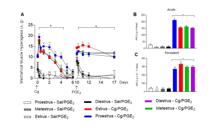
Estrous cycle modulates both acute and persistent muscle hyperalgesia. (A) Behavioral nociceptive responses in proestrus, estrus, metestrus and diestrus phases. AUC of the acute (B) and persistent (C) muscle hyperalgesia. The symbol “*” indicates differences for their respective saline control groups, “x” indicates differences for other groups in the acute or persistent phase.
A comparison across the different estrous cycle phases revealed that the carrageenan injection during the proestrus phase elicited a greater acute muscle hyperalgesia than that observed in the other phases (p<0.05, Mixed-effects Analysis test, Tukey post-test, Figure 3A; AUC, One Way ANOVA, Tukey post-test, Figure 3C). In addition, the carrageenan injection during the estrus phase elicited a greater persistent muscle hyperalgesia than that observed in the other phases (p<0.05, Mixed-effects Analysis, Tukey post-test, Figure 3A; AUC, One Way ANOVA, Tukey post-test, Figure 3C).
The estrous cycle phase at the time of PGE2 injection did not modulate the maintenance of persistent muscle hyperalgesia
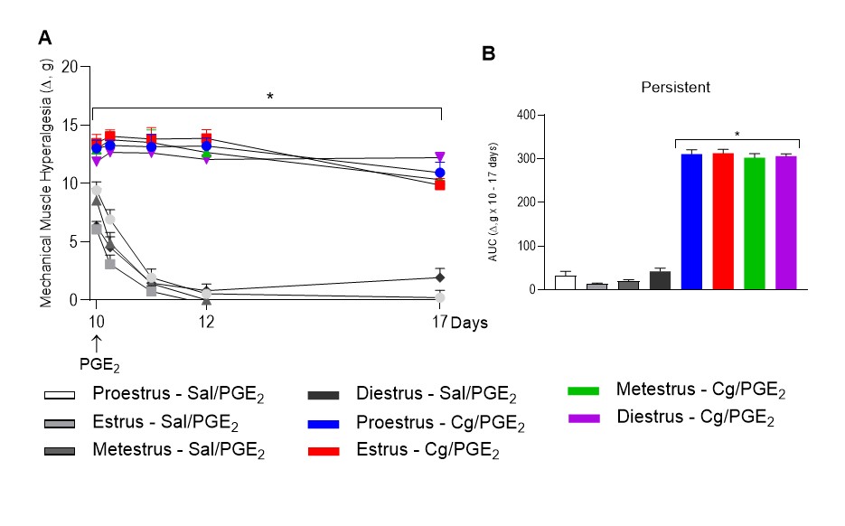
At this moment an analysis was performed to determine whether the estrous cycle phase, at the time of PGE2 injection in mice previously sensitized by carrageenan, would modulate the maintenance of the persistent muscle hyperalgesia. It was observed that the maintenance of this phase was not different among the estrous cycle phases (n=5, p>0.05, Mixed-effects Analysis, Figure 4A; AUC, One Way ANOVA, Figure 4B).

- Estrous cycle did not modulate persistent muscle hyperalgesia. (A) Behavioral nociceptive responses in proestrus, estrus, metestrus and diestrus phases. B) AUC of the persistent muscle hyperalgesia. The symbol “*” indicates differences for their respective saline control groups.
The estrous cycle did not modulate the regular physical exercise-prevented acute and persistent muscle hyperalgesia
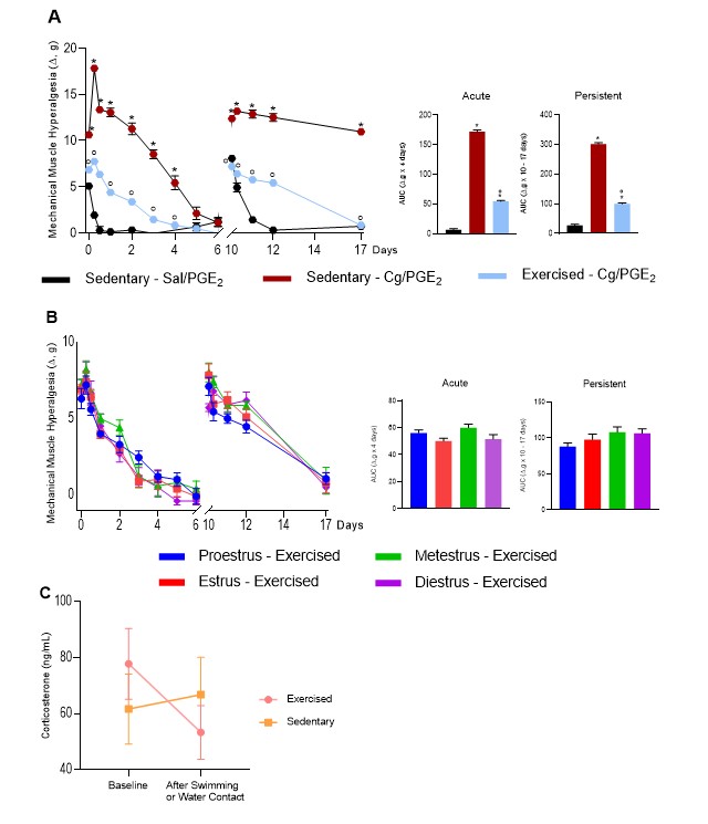
It was previously shown that regular physical exercise by swimming prevents acute and persistent inflammatory muscle hyperalgesia in male mice22. After that, the authors analyzed whether the estrous cycle would modulate this prevention in female mice. First, they observed that swimming was effective in preventing acute and persistent muscle hyperalgesia in females (n=20, p<0.05, Mixed-effects Analysis, Tukey post-test, Figure 5A; AUC, One Way ANOVA, Tukey post-test, Figure 5A). When the different estrous cycle phases were compared, there were no differences in behavioral responses (n=5, p>0.05, Mixed-effects Analysis, Figure 5B; AUC, One Way ANOVA, Figure 5B).

- Physical exercise-induced prevention of acute and persistent muscle hyperalgesia is independent of the estrous cycle. Behavioral nociceptive responses independent of the estrous cycle. (A) and in different phases of estrous cycle (B). Analysis of corticosterone levels (C). The symbol “*” indicates differences for their respective saline control groups and “°” indicates differences for the sedentary Cg/PGE2 group.
To analyze whether stress modulated the exercise-induced prevention of muscle hyperalgesia, the plasma levels of corticosterone before the start and 24 h after the end of the swimming protocol were quantified. There was no increase in plasma levels of corticosterone in exercised female mice (n=16, p>0.05, Two Way ANOVA, Figure 5C).
DISCUSSION
This study demonstrated that the estrous cycle, in which an inflammatory insult occurred, is determinant in the development of acute inflammatory muscle hyperalgesia and the consequent transition to the persistent stage, but not in its maintenance. In addition, in animals previously exercised, the estrous cycle, in which an inflammatory insult occurred, did not modulate the exercise-prevention of muscle hyperalgesia.
The study showed that intramuscular injection of carrageenan into the gastrocnemius muscle of female mice induced acute and persistent inflammatory muscle hyperalgesia throughout all phases of the estrous cycle. Interestingly, this inflammatory insult differentially affected the intensity of acute and persistent muscle hyperalgesia across various phases of the estrous cycle. Specifically, in the proestrus phase, carrageenan induced the most intense acute inflammatory muscle hyperalgesia, while in the estrus phase, the most intense persistent muscle hyperalgesia. These data highlight the impact of the estrous cycle on the outcome of acute and persistent muscle hyperalgesia triggered by an inflammatory insult.
Usually, women or female rodents show more pain than men across several noxious modalities, including mechanical-, electrical-, thermal- and chemical-induced pain33. In addition, women are more likely than men to report different pain conditions34,35. This evidence is, in part, attributed to sexual hormonal fluctuations36-40.
Estrogen41 and progesterone are steroid hormones secreted from the ovaries that play important roles in numerous tissues42, including in the metabolism of the musculoskeletal system43-45. Although there is a robust number of preclinical and clinical studies on the role of sex hormones in pain, the results are still unclear. There seem to be differences in the influence of female hormones based on the stimulus that triggered the pain condition43,46-50. Nevertheless, a consensus in the literature suggests a significant influence of estrogen fluctuations on pain sensitivity. Specifically, variations in hormone levels are associated with an increase in pain sensitivity, whereas stable hormonal profiles appear to confer a protective effect against nociception in females36-40. Progesterone has also been shown to be involved in pain processing, showing a protective role against pain39. Interestingly, a clinical pain model showed that increased pain associated with high progesterone levels was reduced by the increase in estradiol levels, suggesting an interaction between both hormones in pain perception51.
It is well known that in female mice, estradiol levels begin to increase at metestrus, reaching peak levels during proestrus and returning to baseline at estrus. Progesterone secretion also increases during metestrus and diestrus, with a decrease afterwards. Then the progesterone value rises to reach its second peak towards the end of proestrus52,53. Considering these oscillations, it is possible to hypothesize that the higher progesterone levels and the lower estradiol levels at the end of proestrus phase may be related to the highest acute muscle hyperalgesia induced by the inflammatory insult in the proestrus phase. In another way, the lower levels of estrogens and progesterone may have contributed to the highest persistent muscle hyperalgesia induced by the inflammatory insult in the estrus phase. Finally, the protective role of the increasing levels of estrogens and progesterone may have contributed to the lower intensity of persistent muscle hyperalgesia induced by the inflammatory insult in the proestrus and metestrus phases, respectively. The mechanisms underlying the protective role of estrogen in inflammatory muscle hyperalgesia are still under investigation. However, evidence shows that estrogens downregulate proinflammatory cytokines54-57, interact with adrenergic and serotoninergic systems58, and inhibit the signaling of NFkappaB59.
It was previously shown that regular swimming prevents the onset of acute inflammatory muscle hyperalgesia and its transition to the persistent stage in male mice22. Here, the authors demonstrated that females exhibit similar exercise-induced behavioral responses. Interestingly, the phase of the estrous cycle during the inflammatory insult did not affect the exercise-induced prevention of acute or persistent muscle hyperalgesia. Regular physical exercise of low or moderate intensity modulates the peripheral and central neuroimmune systems related to signaling pain pathways60 Therefore, they are efficient non-drug strategies for the treatment and prevention of chronic painful conditions61,62. Although the study has not evaluated the mechanisms underlying the exercise-prevention of acute and persistent muscle hyperalgesia in female mice, it showed that the exercise protocol is also effective in females, independent of the estrous cycle phase.
A limitation of this study is that sex hormones were not directly measured; instead, estrous cycle phases were determined using the non-invasive vaginal lavage method. Although this technique does not provide exact hormone levels, it is widely used and reliable for identifying cycle stages. Therefore, the interpretations regarding hormonal influences were based on indirect evidence.
CONCLUSION
The estrous cycle phase during which an inflammatory insult occurs is critical for the development of acute inflammatory muscle hyperalgesia and its transition to the persistent phase. Therefore, they should be taken into account in the clinical management of such conditions. Although changes in sex hormones throughout the cycle are known to influence pain sensitivity, the present findings suggest that well-designed exercise interventions can effectively prevent the progression from acute to chronic muscle pain. These findings underscore the potential of integrating individualized exercise programs into pain management strategies for women, suggesting that regular physical activity may provide analgesic benefits regardless menstrual cycle phase or the associated fluctuations in sex hormones.
-
Sponsoring sources:
FAPESP: processes 2017/17919-8 and 2021/02921-2. CNPQ: 306413/2022-1 Coordination for the Improvement of Higher Education Personnel - Brazil (CAPES): Grant 001.
-
Data availability
The data that support the findings of this study are available from the corresponding author upon reasonable request.
References
-
1 Gerdle B, Björk J, Cöster L, Henriksson K, Henriksson C, Bengtsson A. Prevalence of widespread pain and associations with work status: a population study. BMC Musculoskelet Disord. 2008;9(1):102. http://doi.org/10.1186/1471-2474-9-102 PMid:18627605.
» http://doi.org/10.1186/1471-2474-9-102 -
2 Rice ASC, Smith BH, Blyth FM. Pain and the global burden of disease. Pain. 2016;157(4):791-6. http://doi.org/10.1097/j.pain.0000000000000454 PMid:26670465.
» http://doi.org/10.1097/j.pain.0000000000000454 -
3 Treede RD, Rief W, Barke A, Aziz Q, Bennett MI, Benoliel R, Cohen M, Evers S, Finnerup NB, First MB, Giamberardino MA, Kaasa S, Korwisi B, Kosek E, Lavand’homme P, Nicholas M, Perrot S, Scholz J, Schug S, Smith BH, Svensson P, Vlaeyen JWS, Wang SJ. Chronic pain as a symptom or a disease: the IASP Classification of Chronic Pain for the International Classification of Diseases (ICD-11). Pain. 2019;160(1):19-27. http://doi.org/10.1097/j.pain.0000000000001384 PMid:30586067.
» http://doi.org/10.1097/j.pain.0000000000001384 -
4 Lund CI, Rosseland LA, Steingrímsdóttir ÓA, Engdahl BL, Stubhaug A, Furberg AS, Nielsen CS. How is age at menopause and reproductive lifespan associated with chronic pain outcomes in postmenopausal women? Pain. 2025;166(1):144-52. http://doi.org/10.1097/j.pain.0000000000003333 PMid:39058956.
» http://doi.org/10.1097/j.pain.0000000000003333 -
5 Gulati M, Dursun E, Vincent K, Watt FE. The influence of sex hormones on musculoskeletal pain and osteoarthritis. Lancet Rheumatol. 2023;5(4):e225-38. http://doi.org/10.1016/S2665-9913(23)00060-7 PMid:38251525.
» http://doi.org/10.1016/S2665-9913(23)00060-7 -
6 Strand NH, D’Souza RS, Gomez DA, Whitney MA, Attanti S, Anderson MA, Moeschler SM, Chadwick AL, Maloney JA. Pain during menopause. Maturitas. 2025;191:108135. http://doi.org/10.1016/j.maturitas.2024.108135 PMid:39500125.
» http://doi.org/10.1016/j.maturitas.2024.108135 -
7 Lund CI, Engdahl B, Rosseland LA, Stubhaug A, Grimnes G, Furberg AS, Steingrímsdóttir ÓA, Nielsen CS. The association between age at menarche and chronic pain outcomes in women: the Tromsø Study, 2007 to 2016. Pain. 2022;163(9):1790-9. http://doi.org/10.1097/j.pain.0000000000002579 PMid:35239542.
» http://doi.org/10.1097/j.pain.0000000000002579 -
8 LeResche L, Mancl LA, Drangsholt MT, Saunders K, Von Korff M. Relationship of pain and symptoms to pubertal development in adolescents. Pain. 2005;118(1-2):201-9. http://doi.org/10.1016/j.pain.2005.08.011 PMid:16213087.
» http://doi.org/10.1016/j.pain.2005.08.011 -
9 Lenert ME, Avona A, Garner KM, Barron LR, Burton MD. Sensory neurons, neuroimmunity, and pain modulation by sex hormones. Endocrinology. 2021;162(8):109. http://doi.org/10.1210/endocr/bqab109 PMid:34049389.
» http://doi.org/10.1210/endocr/bqab109 -
10 Gupta S, McCarson KE, Welch KMA, Berman NEJ. Mechanisms of pain modulation by sex hormones in migraine. Headache. 2011;51(6):905-22. http://doi.org/10.1111/j.1526-4610.2011.01908.x PMid:21631476.
» http://doi.org/10.1111/j.1526-4610.2011.01908.x -
11 Maurer AJ, Lissounov A, Knezevic I, Candido KD, Knezevic NN. Pain and sex hormones: a review of current understanding. Pain Manag. 2016;6(3):285-96. http://doi.org/10.2217/pmt-2015-0002 PMid:26983893.
» http://doi.org/10.2217/pmt-2015-0002 -
12 Hernandez-Leon A, De la Luz-Cuellar YE, Granados-Soto V, González-Trujano ME, Fernández-Guasti A. Sex differences and estradiol involvement in hyperalgesia and allodynia in an experimental model of fibromyalgia. Horm Behav. 2018;97:39-46. http://doi.org/10.1016/j.yhbeh.2017.10.011 PMid:29080671.
» http://doi.org/10.1016/j.yhbeh.2017.10.011 -
13 Coronel MF, Labombarda F, Roig P, Villar MJ, De Nicola AF, González SL. Progesterone prevents nerve injury-induced allodynia and spinal NMDA receptor upregulation in rats. Pain Med. 2011;12(8):1249-61. http://doi.org/10.1111/j.1526-4637.2011.01178.x PMid:21714841.
» http://doi.org/10.1111/j.1526-4637.2011.01178.x -
14 Frye CA, Duncan JE. Progesterone metabolites, effective at the GABAA receptor complex, attenuate pain sensitivity in rats. Brain Res. 1994;643(1–2):194-203. http://doi.org/10.1016/0006-8993(94)90025-6 PMid:8032914.
» http://doi.org/10.1016/0006-8993(94)90025-6 -
15 Gupta S, McCarson KE, Welch KMA, Berman NEJ. Mechanisms of pain modulation by sex hormones in migraine. Headache. 2011;51(6):905-22. http://doi.org/10.1111/j.1526-4610.2011.01908.x PMid:21631476.
» http://doi.org/10.1111/j.1526-4610.2011.01908.x -
16 Sluka KA, Frey-Law L, Hoeger Bement M. Exercise-induced pain and analgesia? Underlying mechanisms and clinical translation. Pain. 2018;159(Suppl 1):S91-7. http://doi.org/10.1097/j.pain.0000000000001235 PMid:30113953.
» http://doi.org/10.1097/j.pain.0000000000001235 -
17 Ikeda S, Tamura Y, Kakehi S, Takeno K, Kawaguchi M, Watanabe T, Sato F, Ogihara T, Kanazawa A, Fujitani Y, Kawamori R, Watada H. Exercise-induced enhancement of insulin sensitivity is associated with accumulation of M2-polarized macrophages in mouse skeletal muscle. Biochem Biophys Res Commun. 2013;441(1):36-41. http://doi.org/10.1016/j.bbrc.2013.10.005 PMid:24120496.
» http://doi.org/10.1016/j.bbrc.2013.10.005 -
18 Yoon H, Thakur V, Isham D, Fayad M, Chattopadhyay M. Moderate exercise training attenuates inflammatory mediators in DRG of Type 1 diabetic rats. Exp Neurol. 2015;267:107-14. http://doi.org/10.1016/j.expneurol.2015.03.006 PMid:25783659.
» http://doi.org/10.1016/j.expneurol.2015.03.006 -
19 Landmark T, Romundstad PR, Borchgrevink PC, Kaasa S, Dale O. Longitudinal associations between exercise and pain in the general population: the HUNT pain study. PLoS One. 2013;8(6):e65279. http://doi.org/10.1371/journal.pone.0065279 PMid:23776464.
» http://doi.org/10.1371/journal.pone.0065279 -
20 Landmark T, Romundstad P, Borchgrevink PC, Kaasa S, Dale O. Associations between recreational exercise and chronic pain in the general population: evidence from the HUNT 3 study. Pain. 2011;152(10):2241-7. http://doi.org/10.1016/j.pain.2011.04.029 PMid:21601986.
» http://doi.org/10.1016/j.pain.2011.04.029 -
21 Sluka KA, O’Donnell JM, Danielson J, Rasmussen LA. Regular physical activity prevents development of chronic pain and activation of central neurons. J Appl Physiol. 2013;114(6):725-33. http://doi.org/10.1152/japplphysiol.01317.2012 PMid:23271699.
» http://doi.org/10.1152/japplphysiol.01317.2012 -
22 de Azambuja G, Jorge CO, Gomes BB, Lourenço HR, Simabuco FM, Oliveira-Fusaro MCG. Regular swimming exercise prevented the acute and persistent mechanical muscle hyperalgesia by modulation of macrophages phenotypes and inflammatory cytokines via PPARγ receptors. Brain Behav Immun. 2021;95:462-76. http://doi.org/10.1016/j.bbi.2021.05.002 PMid:33964434.
» http://doi.org/10.1016/j.bbi.2021.05.002 -
23 Saito T, Ciobotaru A, Bopassa JC, Toro L, Stefani E, Eghbali M. Estrogen contributes to gender differences in mouse ventricular repolarization. Circ Res. 2009;105(4):343-52. http://doi.org/10.1161/CIRCRESAHA.108.190041 PMid:19608983.
» http://doi.org/10.1161/CIRCRESAHA.108.190041 -
24 Dina OA, Levine JD, Green PG. Muscle inflammation induces a protein kinase Cε–dependent chronic-latent muscle pain. J Pain. 2008;9(5):457-62. http://doi.org/10.1016/j.jpain.2008.01.328 PMid:18342576.
» http://doi.org/10.1016/j.jpain.2008.01.328 -
25 Jorge CO, de Azambuja G, Gomes BB, Rodrigues HL, Luchessi AD, de Oliveira-Fusaro MCG. P2X3 receptors contribute to transition from acute to chronic muscle pain. Purinergic Signal. 2020;16(3):403-14. http://doi.org/10.1007/s11302-020-09718-x PMid:32766958.
» http://doi.org/10.1007/s11302-020-09718-x -
26 Takahashi K, Taguchi T, Itoh K, Okada K, Kawakita K, Mizumura K. Influence of surface anesthesia on the pressure pain threshold measured with different-sized probes. Somatosens Mot Res. 2005;22(4):299-305. http://doi.org/10.1080/08990220500420475 PMid:16503582.
» http://doi.org/10.1080/08990220500420475 -
27 Santos DFS, Aquino BM, Jorge CO, Azambuja G, Schiavuzzo JG, Krimon S, Neves JS, Parada CA, Oliveira-Fusaro MCG. Muscle pain induced by static contraction in rats is modulated by peripheral inflammatory mechanisms. Neuroscience. 2017;358:58-69. http://doi.org/10.1016/j.neuroscience.2017.06.041 PMid:28673715.
» http://doi.org/10.1016/j.neuroscience.2017.06.041 -
28 Schiavuzzo JG, Teixeira JM, Melo B, da Silva dos Santos DF, Jorge CO, Oliveira-Fusaro MC, Parada CA. Muscle hyperalgesia induced by peripheral P2X3 receptors is modulated by inflammatory mediators. Neuroscience. 2015;285:24-33. http://doi.org/10.1016/j.neuroscience.2014.11.020 PMid:25446353.
» http://doi.org/10.1016/j.neuroscience.2014.11.020 -
29 Scariot PPM, Manchado-Gobatto FB, Torsoni AS, dos Reis IGM, Beck WR, Gobatto CA. Continuous aerobic training in individualized intensity avoids spontaneous physical activity decline and improves MCT1 expression in oxidative muscle of swimming rats. Front Physiol. 2016;7:132. http://doi.org/10.3389/fphys.2016.00132 PMid:27148071.
» http://doi.org/10.3389/fphys.2016.00132 -
30 Lima AA, Gobatto CA, Messias LHD, Scariot PPM, Forte LDM, Santin JO, Manchado-Gobatto FB. Two water environment adaptation models enhance motor behavior and improve the success of the lactate minimum test in swimming rats. Motriz. 2017;23(spe):e101607. http://doi.org/10.1590/s1980-6574201700si0009
» http://doi.org/10.1590/s1980-6574201700si0009 -
31 Kregel KC, Allen DL, Booth FW, Flesher MR, Henriksen EJ, Musch TI, O’ Leary DS, Parks CM, Poole, DC, Ra’anan AW, Sheriff DD, Toth LA. Resource book for the design of animal exercise protocols. Am J Vet Res. 2007;68(6):583. http://doi.org/10.2460/ajvr.68.6.583
» http://doi.org/10.2460/ajvr.68.6.583 -
32 Dell RB, Holleran S, Ramakrishnan R. Sample size determination. ILAR J. 2002;43(4):207-13. http://doi.org/10.1093/ilar.43.4.207 PMid:12391396.
» http://doi.org/10.1093/ilar.43.4.207 -
33 Bartley EJ, Fillingim RB. Sex differences in pain: a brief review of clinical and experimental findings. Br J Anaesth. 2013;111(1):52-8. http://doi.org/10.1093/bja/aet127 PMid:23794645.
» http://doi.org/10.1093/bja/aet127 -
34 Bouhassira D, Lantéri-Minet M, Attal N, Laurent B, Touboul C. Prevalence of chronic pain with neuropathic characteristics in the general population. Pain. 2008;136(3):380-7. http://doi.org/10.1016/j.pain.2007.08.013 PMid:17888574.
» http://doi.org/10.1016/j.pain.2007.08.013 -
35 Chen G, Luo X, Qadri MY, Berta T, Ji RR. Sex-dependent glial signaling in pathological pain: distinct roles of spinal microglia and astrocytes. Neurosci Bull. 2018;34(1):98-108. http://doi.org/10.1007/s12264-017-0145-y PMid:28585113.
» http://doi.org/10.1007/s12264-017-0145-y -
36 Hernandez-Leon A, De la Luz-Cuellar YE, Granados-Soto V, González-Trujano ME, Fernández-Guasti A. Sex differences and estradiol involvement in hyperalgesia and allodynia in an experimental model of fibromyalgia. Horm Behav. 2018;97:39-46. http://doi.org/10.1016/j.yhbeh.2017.10.011 PMid:29080671.
» http://doi.org/10.1016/j.yhbeh.2017.10.011 -
37 Delaruelle Z, Ivanova TA, Khan S, Negro A, Ornello R, Raffaelli B, Terrin A, Mitsikostas DD, Reuter U, and the European Headache Federation School of Advanced Studies (EHF-SAS). Male and female sex hormones in primary headaches. J Headache Pain. 2018;19(1):117. http://doi.org/10.1186/s10194-018-0922-7 PMid:30497379.
» http://doi.org/10.1186/s10194-018-0922-7 -
38 Maurer AJ, Lissounov A, Knezevic I, Candido KD, Knezevic NN. Pain and sex hormones: a review of current understanding. Pain Manag. 2016;6(3):285-96. http://doi.org/10.2217/pmt-2015-0002 PMid:26983893.
» http://doi.org/10.2217/pmt-2015-0002 -
39 Gupta S, McCarson KE, Welch KMA, Berman NEJ. Mechanisms of pain modulation by sex hormones in migraine. Headache. 2011;51(6):905-22. http://doi.org/10.1111/j.1526-4610.2011.01908.x PMid:21631476.
» http://doi.org/10.1111/j.1526-4610.2011.01908.x -
40 Lenert ME, Avona A, Garner KM, Barron LR, Burton MD. sensory neurons, neuroimmunity, and pain modulation by sex hormones. Endocrinology. 2021;162(8):bqab109. http://doi.org/10.1210/endocr/bqab109 PMid:34049389.
» http://doi.org/10.1210/endocr/bqab109 -
41 Pang H, Chen S, Klyne DM, Harrich D, Ding W, Yang S, Han FY. Low back pain and osteoarthritis pain: a perspective of estrogen. Bone Res. 2023;11(1):42. http://doi.org/10.1038/s41413-023-00280-x PMid:37542028.
» http://doi.org/10.1038/s41413-023-00280-x -
42 Belachew EB, Sewasew DT. Corrigendum: molecular mechanisms of endocrine resistance in estrogen-receptor-positive breast cancer. Front Endocrinol. 2021;12:689705. http://doi.org/10.3389/fendo.2021.599586 PMid:34046016.
» http://doi.org/10.3389/fendo.2021.599586 -
43 Pöllänen E, Sipilä S, Alen M, Ronkainen PH, Ankarberg-Lindgren C, Puolakka J, Suominen H, Hämäläinen E, Turpeinen U, Konttinen YT, Kovanen V. Differential influence of peripheral and systemic sex steroids on skeletal muscle quality in pre‐ and postmenopausal women. Aging Cell. 2011;10(4):650-60. http://doi.org/10.1111/j.1474-9726.2011.00701.x PMid:21388496.
» http://doi.org/10.1111/j.1474-9726.2011.00701.x -
44 Nilsson S, Gustafsson JÅ. Estrogen receptors: therapies targeted to receptor subtypes. Clin Pharmacol Ther. 2011;89(1):44-55. http://doi.org/10.1038/clpt.2010.226 PMid:21124311.
» http://doi.org/10.1038/clpt.2010.226 -
45 Cauley JA. Estrogen and bone health in men and women. Steroids. 2015;99(Pt A):11-5. http://doi.org/10.1016/j.steroids.2014.12.010 PMid:25555470.
» http://doi.org/10.1016/j.steroids.2014.12.010 -
46 Gulati M, Dursun E, Vincent K, Watt FE. The influence of sex hormones on musculoskeletal pain and osteoarthritis. Lancet Rheumatol. 2023;5(4):e225-38. http://doi.org/10.1016/S2665-9913(23)00060-7 PMid:38251525.
» http://doi.org/10.1016/S2665-9913(23)00060-7 -
47 Amandusson Å, Blomqvist A. Estrogenic influences in pain processing. Front Neuroendocrinol. 2013;34(4):329-49. http://doi.org/10.1016/j.yfrne.2013.06.001 PMid:23817054.
» http://doi.org/10.1016/j.yfrne.2013.06.001 -
48 Athnaiel O, Cantillo S, Paredes S, Knezevic NN. The role of sex hormones in pain-related conditions. Int J Mol Sci. 2023;24(3):1866. http://doi.org/10.3390/ijms24031866 PMid:36768188.
» http://doi.org/10.3390/ijms24031866 -
49 Lesnak JB, Inoue S, Lima L, Rasmussen L, Sluka KA. Testosterone protects against the development of widespread muscle pain in mice. Pain. 2020;161(12):2898-908. http://doi.org/10.1097/j.pain.0000000000001985 PMid:32658149.
» http://doi.org/10.1097/j.pain.0000000000001985 -
50 Averitt DL, Hornung RS, Murphy AZ. Role of sex hormones on pain. In: Sherman SM, editor. Oxford research encyclopedia of neuroscience. Oxford: Oxford University Press; 2019. http://doi.org/10.1093/acrefore/9780190264086.013.247
» http://doi.org/10.1093/acrefore/9780190264086.013.247 -
51 Stening K, Eriksson O, Wahren L, Berg G, Hammar M, Blomqvist A. Pain sensations to the cold pressor test in normally menstruating women: comparison with men and relation to menstrual phase and serum sex steroid levels. Am J Physiol Regul Integr Comp Physiol. 2007;293(4):R1711-6. http://doi.org/10.1152/ajpregu.00127.2007 PMid:17652363.
» http://doi.org/10.1152/ajpregu.00127.2007 -
52 Smith MS, Freeman ME, Neill JD. The control of progesterone secretion during the estrous cycle and early pseudopregnancy in the rat: prolactin, gonadotropin and steroid levels associated with rescue of the corpus luteum of pseudopregnancy. Endocrinology. 1975;96(1):219-26. http://doi.org/10.1210/endo-96-1-219 PMid:1167352.
» http://doi.org/10.1210/endo-96-1-219 -
53 Spornitz UM, Socin CD, Dravid AA. Estrous stage determination in rats by means of scanning electron microscopic images of uterine surface epithelium. Anat Rec. 1999;254(1):116-26. http://doi.org/10.1002/(SICI)1097-0185(19990101)254:1<116::AID-AR15>3.0.CO;2-X PMid:9892425.
» http://doi.org/10.1002/(SICI)1097-0185(19990101)254:1<116::AID-AR15>3.0.CO;2-X -
54 Chen BL, Li YQ, Xie DH, He QL, Yang XX. Blocking TNF-α with infliximab alleviates ovariectomy induced mechanical and thermal hyperalgesia in rats. Neurol Sci. 2012;33(3):527-33. http://doi.org/10.1007/s10072-011-0743-9 PMid:21874299.
» http://doi.org/10.1007/s10072-011-0743-9 -
55 Masiukiewicz US, Mitnick M, Grey AB, Insogna KL. Estrogen modulates parathyroid hormone-induced interleukin-6 production in vivo and in vitro*. Endocrinology. 2000;141(7):2526-31. http://doi.org/10.1210/endo.141.7.7537 PMid:10875254.
» http://doi.org/10.1210/endo.141.7.7537 -
56 Liu H, Liu K, Bodenner DL. Estrogen receptor inhibits interleukin-6 gene expression by disruption of nuclear factor κB transactivation. Cytokine. 2005;31(4):251-7. http://doi.org/10.1016/j.cyto.2004.12.008 PMid:16043358.
» http://doi.org/10.1016/j.cyto.2004.12.008 -
57 Torres‐Chávez KE, Sanfins JM, Clemente‐Napimoga JT, Pelegrini-Da-Silva A, Parada CA, Fischer L, Tambeli CH. Effect of gonadal steroid hormones on formalin‐induced temporomandibular joint inflammation. Eur J Pain. 2012;16(2):204-16. http://doi.org/10.1016/j.ejpain.2011.06.007 PMid:22323373.
» http://doi.org/10.1016/j.ejpain.2011.06.007 -
58 Okuda K, Iwasaka H, Hagiwara S, Takeshima N, Takatani J, Uchino T, Noguchi T. The antinociceptive effects of estradiol on adjuvant-induced hyperalgesia in rats involve activation of adrenergic and serotonergic systems. J Anesth. 2011;25(3):392-7. http://doi.org/10.1007/s00540-011-1142-3 PMid:21528403.
» http://doi.org/10.1007/s00540-011-1142-3 -
59 Liu C, Lo J, Kuo C, Chu CH, Chen LM, Tsai FJ, Tsai CH, Tzang BS, Kuo WW, Huang CY. Akt mediates 17β‐estradiol and/or estrogen receptor‐α inhibition of LPS‐induced tumor necresis factor‐α expression and myocardial cell apoptosis by suppressing the JNK1/2‐NFκB pathway. J Cell Mol Med. 2009;13(9B):3655-67. http://doi.org/10.1111/j.1582-4934.2009.00669.x PMid:20196785.
» http://doi.org/10.1111/j.1582-4934.2009.00669.x -
60 Sluka KA, Frey-Law L, Hoeger Bement M. Exercise-induced pain and analgesia? Underlying mechanisms and clinical translation. Pain. 2018;159(Suppl 1):S91-7. http://doi.org/10.1097/j.pain.0000000000001235 PMid:30113953.
» http://doi.org/10.1097/j.pain.0000000000001235 -
61 Hassett AL, Williams DA. Non-pharmacological treatment of chronic widespread musculoskeletal pain. Best Pract Res Clin Rheumatol. 2011;25(2):299-309. http://doi.org/10.1016/j.berh.2011.01.005 PMid:22094203.
» http://doi.org/10.1016/j.berh.2011.01.005 -
62 Mannerkorpi K, Henriksson C. Non-pharmacological treatment of chronic widespread musculoskeletal pain. Best Pract Res Clin Rheumatol. 2007;21(3):513-34. http://doi.org/10.1016/j.berh.2007.04.001 PMid:17602997.
» http://doi.org/10.1016/j.berh.2007.04.001
Edited by
-
Associate editor in charge:
Camila Squarzoni Dale https://orcid.org/0000-0002-3421-7799
The data that support the findings of this study are available from the corresponding author upon reasonable request.