
 Brainstem Expansive Lesion Suggestive of Glioma in a Renal Transplant Patient
ABSTRACT
Introduction: Immunosuppression following kidney transplantation is associated with a significantly increased risk of mortality and the development of neoplasms, especially due to the continuous use of immunosuppressants, which increase susceptibility to opportunistic infections and the development of tumors. Among the possible neurological complications in this group, glial tumors of the brainstem represent a rare and difficult-to-diagnose condition, with a high rate of malignancy and a guarded prognosis, whose therapeutic management is limited and often palliative.
Objective: To report a case of an immunosuppressed patient with a kidney transplant who developed an expansive lesion in the brainstem suggestive of glioma, treated empirically with radiotherapy due to the infeasibility of biopsy.
Methods: Descriptive observational study and literature review in the PubMed and SciELO databases, covering the past 20 years.
Results: The patient showed progressive clinical improvement and a favorable radiological response to empirical radiotherapy, with no metastases or tumor recurrence identified in the follow-up of the last 4 years.
Conclusion: Despite the rarity and poor prognosis of brainstem gliomas in immunosuppressed patients, this case demonstrates that empirical radiotherapy, when biopsy is not feasible, can be considered an alternative treatment and presents favorable results. The report also reinforces the need for neurological surveillance in transplant recipients and expands knowledge about neoplastic complications of the central nervous system in the context of prolonged immunosuppression.
Descriptors
Glioma; Neoplasm; Radiotherapy; Immunosuppression
RESUMO
Introdução: A condição de imunossupressão decorrente do transplante renal está associada a risco significativamente aumentado de mortalidade e desenvolvimento de neoplasias, especialmente em decorrência do uso contínuo de imunossupressores que elevam a susceptibilidade a infecções oportunistas e ao desenvolvimento de tumores. Entre as possíveis complicações neurológicas nesse grupo, os tumores gliais do tronco encefálico representam uma condição rara e de difícil diagnóstico, com elevada taxa de malignidade e prognóstico reservado, cujo manejo terapêutico é limitado e frequentemente paliativo.
Objetivo: Relatar um caso de paciente imunossuprimida com transplante renal que desenvolveu lesão expansiva no tronco encefálico sugestiva de glioma, tratada empiricamente com radioterapia devido à inviabilidade de biópsia.
Métodos: Estudo observacional descritivo e revisão da literatura nas bases PubMed e SciELO, abrangendo os últimos 20 anos.
Resultados: A paciente evoluiu com melhora clínica progressiva e resposta radiológica favorável à radioterapia empírica, não sendo identificadas metástases ou recidiva tumoral no seguimento dos últimos 4 anos.
Conclusão: Apesar da raridade e do grave prognóstico dos gliomas de tronco encefálico em pacientes imunossuprimidos, este caso demonstra que a radioterapia empírica, diante da inviabilidade de biópsia, pode ser considerada uma alternativa ao tratamento e apresentar resultado favorável. O relato reforça ainda a necessidade de vigilância neurológica em transplantados e amplia o conhecimento sobre complicações neoplásicas do sistema nervoso central nesse contexto de imunossupressão prolongada.
Descritores
Glioma; Neoplasia; Radioterapia; Imunossupressão
INTRODUCTION
Immunosuppressed patients, such as kidney transplant recipients, have a significantly increased risk of mortality and development of neoplasms, with a cancer incidence approximately twice as high as that of the general population¹. Studies indicate that, after kidney transplantation, the incidence of neoplasms increases progressively over time, reaching approximately 14% after 10 years and up to 40% after 20 years². This susceptibility is largely attributed to the continuous use of immunosuppressants, which not only predispose to opportunistic infections but are also implicated in an increased risk of tumor development³.
In the context of central nervous system (CNS) lesions in transplant recipients, the main differential diagnoses include primary lymphomas, particularly post-transplant lymphoproliferative disorder (PTLD), whose incidence ranges from 0.05% to 0.30%⁴, in addition to glial tumors, infectious and parasitic diseases (with an incidence of approximately 0.53%), leukoencephalopathies, and metastases are also common. Among glial tumors, brainstem gliomas represent a rare and particularly challenging group, with an estimated annual incidence of five to six cases per 100,000 people⁵. Although they account for only about 2% of brain tumors in adults⁵,⁶, approximately 80% of these lesions are malignant. In transplant patients, the occurrence of brainstem glioma is even rarer, making clinical and therapeutic diagnosis a real challenge⁴.
The clinical presentation of these tumors is generally insidious and nonspecific, making early diagnosis difficult. Symptoms vary depending on the nearby anatomical structures involved. Due to the complexity of the brainstem, biopsy is a high-risk procedure, and the diagnosis is often presumed based on clinical and neuroimaging findings⁵. Radiotherapy is considered the first-line treatment, even in the absence of histological confirmation, although the therapeutic response is, in most cases, modest and palliative in nature⁵,6. Cases of significant remission or prolonged stabilization of the disease are extremely rare, especially when treatment is initiated empirically, without histopathological confirmation6.
In this context, the present case report aims to describe the remission of a lesion suggestive of brainstem glioma in a renal transplant patient treated with empirical radiotherapy, highlighting the importance of early clinical recognition and the potential for a favorable response even in atypical clinical situations.
Ethical considerations
This case report was approved by the Research Ethics Committee of the Health Sciences Education and Research Foundation, in compliance with the National Health Council Resolution No. 466, dated December 12, 2012. Informed consent was obtained from the patient for the publication of clinical data, ensuring anonymization and the omission of any personal identifiers.
Case Report
A 52-year-old female patient, born in Correntina, Bahia state, with a history of chronic kidney disease secondary to recurrent urinary tract infection. She underwent hemodialysis from 1995 to 2003 and underwent living-related donor kidney transplantation from her sister in 2003. She was on an immunosuppressive regimen of sirolimus 2 mg/day, mycophenolate sodium 1,080 mg/day, and prednisone 5 mg/day, and was under regular follow-up at the transplant outpatient clinic of the Hospital de Base do Distrito Federal (HBDF), presenting with excellent graft function (creatinine 1.4 mg/dL) and no complications during transplant follow-up.
In November 2020, she presented with an episode of headache, vertigo, diplopia, and paresthesia in the left hemiface and left lower limb. Brain magnetic resonance imaging was performed, revealing a sequelae ischemic lesion in the region of the left third ventricle and brainstem, with imaging suggestive of gliosis and/or microangiopathic foci. She initiated treatment for ischemic cerebrovascular disease with acetylsalicylic acid 100 mg/day, with partial improvement of the headache and neurological symptoms.
In February 2021, she returned to the emergency department with a new-onset headache, associated with neurological symptoms such as bilateral limb weakness, imbalance, difficulty walking, and dysphagia; therefore, hospital admission was chosen for further investigation. On physical examination, she presented with reduced muscle strength (2/4+), ataxia, and mild dysphagia for solids; the remaining findings were unremarkable at the time.
An infectious screening was initiated, including serologies for HIV, viral hepatitis, syphilis, toxoplasmosis, and cytomegalovirus (all negative), as well as a rapid test for cryptococcosis (negative) and an IGRA, which yielded an indeterminate result. The brain computed tomography scan showed no changes compared to previous examinations.
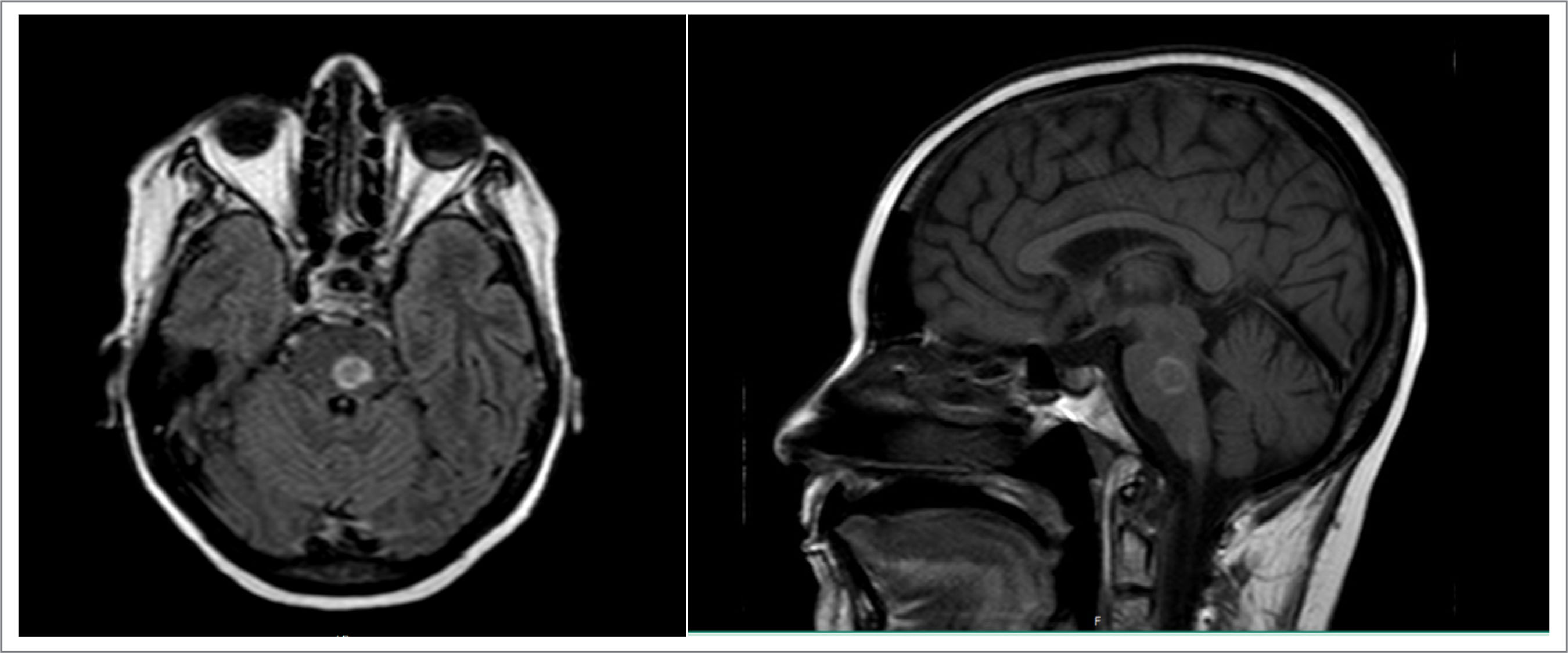
A cranial magnetic resonance imaging (MRI) scan was then performed, which showed expansive nodular lesions associated with significant edema involving the entire brainstem (Fig. 1), with the diagnostic hypotheses being infection or a CNS neoplasm.
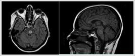
Given the suspicion of a neoplastic lesion, the decision was made to suspend mycophenolate and maintain sirolimus as the sole immunosuppressant. Furthermore, empirical treatment for neurotoxoplasmosis was initiated with pyrimethamine 75 mg/day + folinic acid 15 mg/day + clindamycin 600 mg every 8 hours, scheduled for 6 weeks; corticosteroid therapy with dexamethasone 4 mg every 6 hours to reduce cerebral edema; and biopsy or lumbar puncture was contraindicated by neurosurgery due to the risk of herniation. Despite the measures instituted, the patient progressed with worsening of her clinical and neurological status, presenting with dysphonia, diplopia, ataxia resulting in bed confinement, hemiplegia of the limbs, and progressive dysphagia, requiring the placement of a nasoenteric tube for feeding.
Given the worsening clinical and neurological condition, a multidisciplinary meeting was held (neurology, radiology, oncological neurosurgery, and neuroradiology), which, by consensus, suggested a diagnosis of brainstem glioma, without indicating a biopsy due to the high morbidity of the procedure and the unavailability of suitable material at the hospital.
After discussion with the family regarding the risks of empirical radiotherapy and the infeasibility of a biopsy, the decision was made to initiate treatment. On March 4, 2021, empirical oncological radiotherapy was initiated in a hypofractionated regimen. Eleven sessions were performed (267 cGy each), with a favorable outcome, improvement of the neurological status, and a follow-up magnetic resonance imaging revealing a 'marked reduction in edema and infiltration adjacent to the lesions in the pons and medulla oblongata, with a reduction in mass effect, as well as a reduction in perfusion and a pattern suggestive of necrosis within the lesions. The patient was discharged from the hospital on March 26, 2021, on a corticosteroid taper, maintaining outpatient follow-up with radiology, oncology, kidney transplantation, physiotherapy, and speech therapy, due to the motor and swallowing limitations still present.
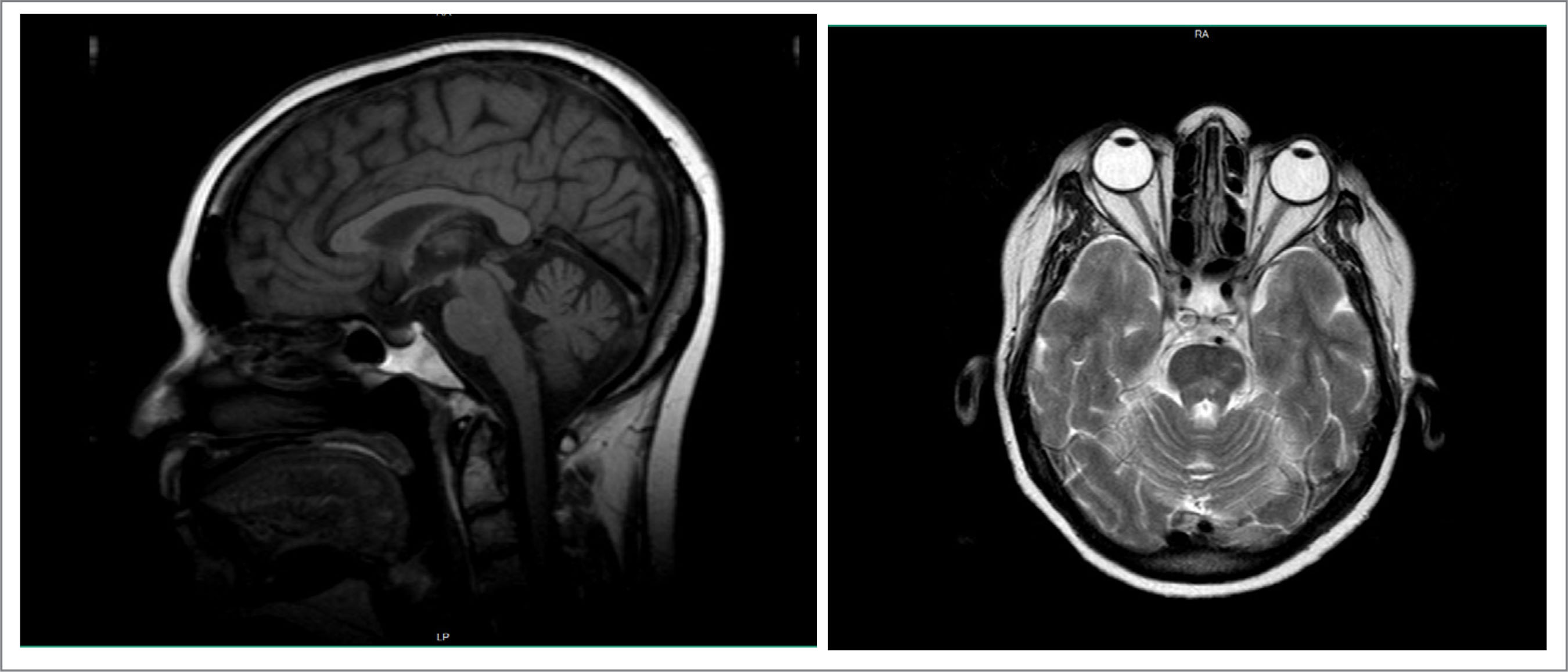
On April 14, 2021, she presented with a case of bilateral pulmonary thromboembolism, requiring hospitalization and full therapeutic anticoagulation. She was discharged from the hospital and maintained on anticoagulation for 1 year without further complications. The patient maintained outpatient follow-up with oncology, radiotherapy, and kidney transplantation, presenting with progressive improvement of the neurological deficit, with motor, speech, and swallowing recovery, though still presenting with ataxia, requiring assisted for ambulation, and occasional complaints of headache. Follow-up brain magnetic resonance imaging scans showed a progressive reduction in the tumor area until the most recent one (February 2025) (Fig. 2), which revealed the persistence of an area of encephalomalacia in the left half of the medulla oblongata and the central portion of the pons, with no signs of recurrence or tumor remnants. Currently, she maintains immunosuppressive treatment with sirolimus 2 mg/day and prednisolone 5 mg/day, with stable kidney function and no further complications.

DISCUSSION
This report describes an uncommon case of an expansive brainstem lesion suggestive of a glioma in a patient who underwent kidney transplantation, under prolonged immunosuppression, whose therapeutic approach was limited by the infeasibility of histopathological confirmation. Management based on a multidisciplinary evaluation, with immunosuppression adjustment and empirical radiotherapy, resulted in a sustained clinical and radiological response in a long-term follow-up, representing an atypical outcome in this context.
Kidney transplant recipients have an increased risk of developing neoplasms, directly related to the intensity and duration of immunosuppression. In addition to reducing antitumor immune surveillance, certain immunosuppressants exert direct pro-oncogenic effects. Calcineurin inhibitors are associated with increased expression of tumor growth factors and angiogenesis, while antiproliferative agents may contribute to persistent immune dysfunction. These mechanisms explain the higher incidence of solid and hematological malignancies observed in this population, with a cumulative risk increasing over the years post-transplant.
In the setting of neurological manifestations in transplant recipients, the differential diagnosis includes opportunistic infections, PTLD, and primary CNS neoplasms. Although PTLD is the primary CNS neoplasm associated with transplantation, glial tumors, especially in the brainstem, are rarely described. In adults, brainstem gliomas present with aggressive behavior, a presumptive diagnosis, and a guarded prognosis, with radiotherapy being the main therapeutic pillar, even in the absence of histological confirmation.
In addition to specific oncological treatment, the management of immunosuppression constitutes a central aspect in the approach to post-transplant neoplasms. Whenever possible, it is recommended to reduce the total immunosuppressive load, with special attention to suspending or reducing calcineurin inhibitors and antiproliferative agents. This strategy aims to partially restore immune surveillance, minimize tumor-stimulating stimuli, and avoid significant compromise of graft function.
In this context, mTOR pathway inhibitors, such as sirolimus and everolimus, play a significant role by combining immunosuppressive effects with antiproliferative, antiangiogenic, and potentially antineoplastic properties. Observational evidence and meta-analyses demonstrate a reduction in the incidence of post-transplant neoplasms in patients converted to mTOR-based regimens, particularly compared with continued calcineurin inhibitor therapy. In the case presented, suspending mycophenolate sodium and maintaining sirolimus as the primary immunosuppressive agent are in line with these recommendations and may have contributed to the observed tumor control.
The favorable response to empirical radiotherapy, with sustained lesion regression and progressive neurological recovery over a 4-year period, represents an unusual outcome for brainstem gliomas, especially in immunosuppressed patients. This finding reinforces the importance of individualized therapy, shared decision-making, and integrated action between nephrology, neurology, oncology, and radiotherapy.
This case further underscores the need for systematic neurological surveillance in the follow-up of transplant patients and the importance of a comprehensive diagnostic approach when progressive neurological symptoms arise. Although the absence of histopathological confirmation represents a limitation, the sustained clinical and radiological evolution strongly favors the presumptive diagnosis of glial neoplasia.
Finally, this report contributes to the literature by demonstrating that, in selected situations, careful adjustment of immunosuppression combined with empirical radiotherapy can result in prolonged disease control, while simultaneously preserving renal graft function and the patient's quality of life, which are central aspects of kidney transplant practice.
CONCLUSION
In patients who have undergone kidney transplantation under prolonged immunosuppression, the development of expansive CNS lesions represents a diagnostic and therapeutic challenge, especially when histopathological confirmation is infeasible. In this context, empirical radiotherapy, combined with judicious adjustment of immunosuppression by reducing the total immunosuppressive load and prioritizing regimens based on mTOR pathway inhibitors, may constitute a viable strategy, enabling disease control without compromising graft function. This case reinforces the importance of a multidisciplinary approach, continuous neurological surveillance, and individualized management in post-transplant neoplasms.
ACKNOWLEDGEMENT
To the nephrology, neurology, radiotherapy, and kidney transplant services of HBDF for their support and case discussions.
DECLARATION OF USE OF ARTIFICIAL INTELLIGENCE TOOLS
FUNDING
DATA AVAILABILITY STATEMENT
All dataset were generated or analyzed in the current study.
References
-
Wong G, Chapman JR, Craig JC, Kwan BCH, Chong KSC. De novo malignancies after kidney transplantation. Clin J Am Soc Nephrol, 2022; 17(3): 388-98. https://doi.org/10.2215/CJN.14570920
» https://doi.org/10.2215/CJN.14570920 -
Fuhrmann JD, Valkova K, von Moos S, Wüthrich RP, Müller TF, Schachtner T. Cancer among kidney transplant recipients >20 years after transplantation. Clin Kidney J, 2022; 15(6): 1152-9. https://doi.org/10.1093/ckj/sfac013
» https://doi.org/10.1093/ckj/sfac013 -
Huo Z, Zhao H, Xu H. Cancer risks in solid organ transplant recipients: results from a comprehensive analysis of 72 cohort studies. OncoImmunology, 2020; 9(1): 1848068. https://doi.org/10.1080/2162402X.2020.1848068
» https://doi.org/10.1080/2162402X.2020.1848068 -
White ML, Moore DW, Zhang Y, Mark KD, Greiner TC, Bierman PJ. Primary central nervous system post-transplant lymphoproliferative disorder: imaging appearances and differential. Insights Imaging, 2019; 10: 46. https://doi.org/10.1186/s13244-019-0726-6
» https://doi.org/10.1186/s13244-019-0726-6 -
Eisele SC, Reardon DA. Adult brainstem gliomas. Cancer, 2016; 122(18): 2847-54. https://doi.org/10.1002/cncr.29920
» https://doi.org/10.1002/cncr.29920 -
Liu H, Qin X, Zhao L, Zhao G, Wang Y. Epidemiology and survival of patients with brainstem gliomas: a population based study using the SEER database. Front Oncol, 2021;11: 692097. https://doi.org/10.3389/fonc.2021.692097
» https://doi.org/10.3389/fonc.2021.692097

 Source: Exam provided by the patient.
 Source: Exam provided by the patient.