
 Use of Aortohepatic Conduit in Liver Transplantation: Experience of Two Decades
ABSTRACT
Introduction: Aortohepatic conduits (AHC) are an alternative for graft revascularization when conventional arterial anastomosis is not feasible. They are usually made from the donor iliac artery and anastomosed to the infrarenal or supraceliac aorta. Although essential in some cases, the results in the literature are conflicting.
Objectives: To analyze the outcomes of the graft and patients undergoing primary liver transplantation (LTx) or retransplantation with aortohepatic conduits over 24 years. Methods: This is a retrospective and observational study analyzing medical records of patients who underwent LTx by a Brazilian team from 2000 to 2024. Patients aged 12 years or older who underwent revascularization using an aortohepatic conduit were included. Data on donors, recipients, surgical techniques, and intra- and postoperative outcomes were collected.
Results: 1.799 LTx procedures were performed during the study period, 43 used arterial conduits, and 41 were included in the final analysis. Most patients were male, with a mean age of 39.24 years. Among the 41 transplants, 17 were primary, and 24 were retransplants. All conduits were made with grafts from donor iliac and/or carotid arteries. The main indications for their use were arterial thrombosis and dissection or friability of the vascular intima. The mean hospital stay was 13 days, and the mean overall survival was 38.51 months. Postoperative complications included arterial thrombosis and biliary complications.
Conclusion: Aortohepatic conduits in LTx are a life-saving strategy in specific cases despite the increased risk of arterial thrombosis and biliary complications. Endovascular intervention shows promise, but prospective studies are needed to define the ideal anastomosis site, assess its impact on survival, and determine the role of antiplatelet therapy in this context.
Descriptors
Liver Transplantation; Vascular Graft; Vascular Surgical Procedures
RESUMO
Introdução: Os condutos aorto-hepáticos são uma alternativa para a revascularização do enxerto quando a anastomose arterial convencional não é viável. Geralmente, são confeccionados a partir da artéria ilíaca doadora e anastomosados na aorta infrarrenal ou supracelíaca. Embora essenciais em alguns casos, os resultados na literatura são conflitantes.
Objetivos: Analisar os desfechos do enxerto e dos pacientes submetidos a transplante hepático (TH) primário ou retransplante com condutos aorto-hepáticos ao longo de 24 anos.
Métodos: Trata-se de um estudo retrospectivo e observacional baseado na análise de prontuários de pacientes submetidos ao TH por uma equipe brasileira de 2000 a 2024. Incluíram-se pacientes com 12 anos ou mais cuja revascularização foi realizada com conduto aorto-hepático. Foram coletados dados sobre doador, receptor, técnica cirúrgica e desfechos intra e pós-operatórios.
Resultados: Foram realizados 1.799 TH no período estudado, com 43 utilizando condutos arteriais, sendo 41 incluídos na análise final. A maioria dos pacientes era do sexo masculino, com idade média de 39,24 anos. Entre os 41 transplantes, 17 foram primários e 24 retransplantes. Todos os condutos foram confeccionados com enxertos de artéria ilíaca e/ou carótida doadora. As principais indicações para seu uso foram trombose arterial e dissecção ou friabilidade da íntima vascular. O tempo médio de internação foi de 13 dias e a sobrevida global média foi de 38,51 meses. Complicações pós-operatórias incluíram trombose arterial e afecções biliares.
Conclusão: O uso de condutos aorto-hepáticos no TH é uma estratégia salvadora em casos específicos, apesar do risco aumentado de trombose arterial e complicações biliares. A intervenção endovascular mostra-se promissora, mas estudos prospectivos são necessários para definir o local ideal de anastomose, o impacto na sobrevida e o papel da antiagregação plaquetária.
Descritores
Transplante de Fígado; Enxerto Vascular; Procedimentos Cirúrgicos Vasculares
INTRODUCTION
In 1963, Starzl and his team performed the world's first liver transplant (LTx). Today, LTx is considered the only curative method for end-stage liver disease and a subset of primary liver tumors1,2. With advances in surgical and anesthetic techniques combined with effective immunosuppression regimens, the average survival of patients undergoing LTx reaches 90% in the first year1.
The success of LTx depends, among other factors, on a robust blood supply to the graft2,3. Under ideal conditions, arterial revascularization is performed through a conventional anastomosis of the donor hepatic artery (HA) to the recipient2,4. However, this is not always possible due to a multitude of factors, such as poor intraoperative arterial perfusion, diameter discrepancies between donor and recipient HAs, atherosclerosis, vascular intimal dissection or friability, anatomical variants, previous injuries, such as those caused by arterial chemoembolization, and the additional challenges of retransplantation3,5.
In these circumstances, using aortohepatic conduits emerges as an alternative for graft revascularization2,5. Usually, these arterial conduits (AHC) are made from the donor's iliac artery, obtained during the harvesting surgery. The most common anastomosis sites are the infrarenal aorta (88%) and the supraceliac aorta (8%) 4,6.
Although AHCs are a life-saving solution in many cases7, they are not without complications, and the results in the literature are conflicting. In a systematic review by Chatzizacharias et al.2, the use of aortohepatic conduits has been identified as an independent risk factor for HA thrombosis (HAT), in addition to being associated with increased operative time and intensive care unit (ICU) stay. In contrast, a single-center study with a 20-year follow-up found no statistically significant differences in graft survival, patient survival, or arterial or biliary complications associated with AHC6.
These discrepancies between the published results may reflect methodological differences between studies, variations in the protocols of each transplant center, and specific characteristics of the samples, such as comorbidities and underlying liver diseases.
Given this, this study aimed to analyze the outcomes of grafts and patients undergoing primary LTx or retransplantation with aortohepatic conduits over 24 years, performed by a transplant team in northeastern Brazil.
METHODS
Study design
The following is a documentary, observational, retrospective study based on the analysis of medical records of patients who underwent LTx, whose arterial reconstruction technique was performed using aortohepatic conduits. The period of analysis covers procedures performed from 2000 to 2024.
The research was conducted with patients transplanted by the team of the Liver Transplant Unit (LTU), whose home institution is the Oswaldo Cruz University Hospital (Hospital Universitário Oswaldo Cruz-HUOC), located in Recife, state of Pernambuco.
Population and eligibility criteria
Patients aged 12 years or older, of both genders, who underwent graft revascularization through an aortohepatic conduit, either supraceliac or infrarenal, were included.
Patients under 12 years of age, transplants with a living donor, and cases in which arterial revascularization techniques other than aortohepatic conduits were used were excluded.
Data collection and analysis procedures
Each patient transplanted by the LTU has a document with transplant records, which is also used for outpatient follow-up. Information was collected on the donor (age and cause of brain death), demographic and anthropometric data of the recipient [age, sex, body mass index (BMI)], medical history and comorbidities, indication for transplant, whether primary LTx or retransplantation, Model for End-Stage Liver Disease (MELD) score, as well as intra- and postoperative records.
The intraoperative parameters analyzed included transplant technique, ischemia times (total, cold, and warm), time for arterialization (interval between the portal and arterial revascularization), total surgery time, blood transfusion administration, presence of post-reperfusion syndrome, and complications.
Data on AHC included material used (vascular graft or prosthesis), the reason for choosing it over conventional anastomosis (examples: arterial thrombosis, dissection or friability of the intima, low flow, unfavorable anatomy, previous injury due to chemoembolization or atherosclerosis), and site of anastomosis (infrarenal or supraceliac).
In addition, postoperative records were analyzed, including length of hospital stay, survival time at 30 days, 6 months, and 1 year, overall survival time (from the date of transplantation to the last follow-up described in the medical records), and graft survival, as well as associated complications.
The local Research Ethics Committee approved the project – CAAE: 78677524.4.0000.5192.
RESULTS
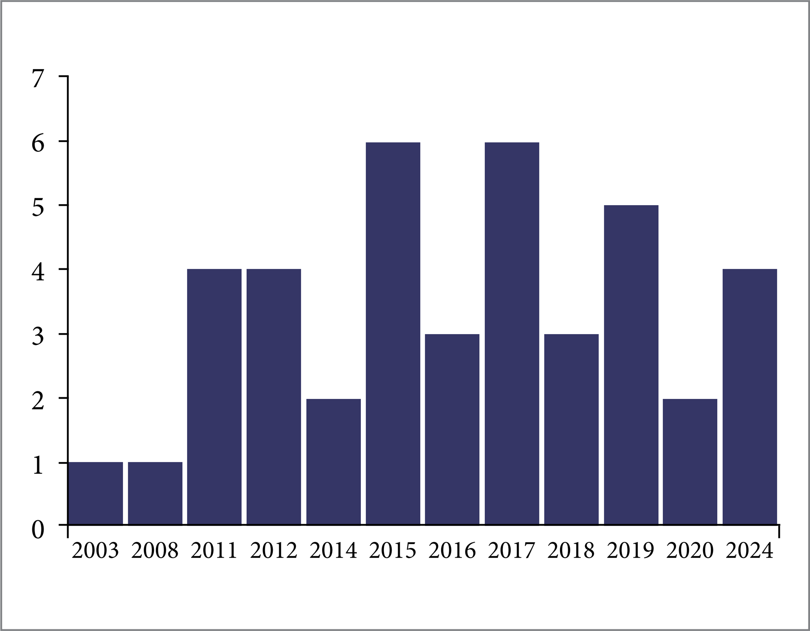
The team performed 1,799 LTx procedures from January 2000 to December 2024. Of these, 43 used AHC. Two were excluded because they were in patients under 12 years of age, resulting in a final sample of 41 transplants and 36 patients (excess justified by the need for retransplantation), corresponding to 2.28%.
Fig. 1 shows the annual distribution of transplants using aortohepatic conduits each year.

Population characteristics
Table 1 presents the general characteristics of the recipient sample studied. We considered n = 41 for the analysis since, except for biological sex, the other variables were likely to change between the primary transplant and retransplants, including cases of second and third retransplants.
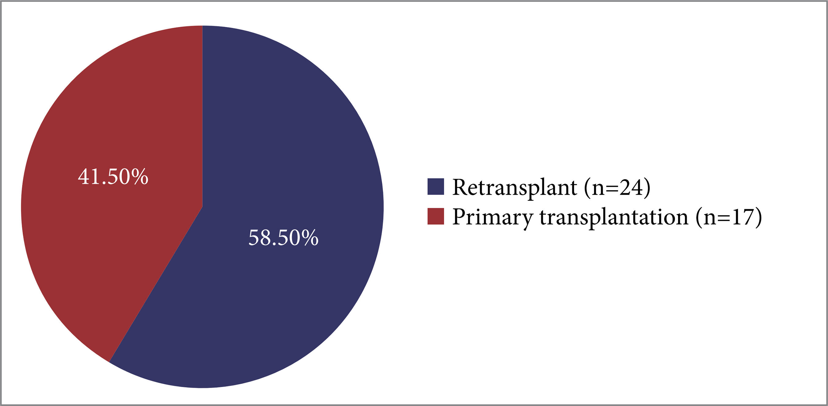
Seventeen patients underwent primary LTx, while 19 underwent retransplantation. Five required a second retransplantation, for a total of 24 retransplantation procedures.
The majority of transplants performed with AHC were retransplantation (Fig. 2). Among the 17 primary transplants, the main indications included autoimmune hepatitis (n = 6; 35.3%), cirrhosis of mixed etiology (n = 4; 23.5%), and chronic liver disease (CLD) secondary to hepatosplenic schistosomiasis (n = 3; 17.6%).

In the 24 retransplants, HAT was the main indication (n = 19; 79.2%), followed by primary graft dysfunction (n = 4; 16.7%). Only one patient required retransplantation due to the recurrence of primary sclerosing cholangitis.
The donors ranged in age from 8 to 77 years. Table 2 shows the mean age and the causes of brain death. The description of probable risk factors related to the donor, graft and/or harvesting was not equal among the transplants, so it was not scored.
Intraoperative data
Most transplants were performed using the conventional technique, with duct-to-duct biliary anastomosis and bile production during surgery (Table 3).
The time to arterialization, defined as the interval between portal and arterial revascularization, was recorded in 20 transplants, ranging from 14 minutes to 2 hours and 30 minutes, with a mean of 44 minutes. Four patients underwent simultaneous revascularization. In two other cases, conventional arterial anastomosis was initially performed without success. In the first case, the technique was modified to utilize AHC during the operation. In the second case, due to low flow in the HA identified by Doppler, the patient was re-approached on the 2nd postoperative day for AHC.
Post-reperfusion syndrome, as recorded in the individual documents for each transplant, was observed in 19 cases (46.3%) and characterized by a significant reduction in systolic blood pressure to less than 60 mmHg and/or a decrease of more than 30% in mean arterial pressure.
Intercurrences and complications during surgery were recorded in some cases, although not all medical records contain this information. Among the 41 transplants analyzed, the most frequently described events were hypotension in 12 cases (29.3%) and hypothermia in eight (19.5%). In addition, bradyarrhythmias were recorded in six (14.6%), metabolic acidosis in five (12.2%), urine output of less than 0.5 mL/kg/h in four (9.8%), and coagulopathy in three (7.3%).
Aspects related to AHC
All conduits were created using a vascular graft from the donor, and there is no record of using artificial prostheses. One transplant used the donor's carotid arteries as the conduit; in two other cases, the carotid artery was anastomosed to the iliac artery. In the remaining cases (n = 38; 92.7%), the vessel of choice was the donor's iliac artery.
Three cases involved conduits in the supraceliac aorta (7.3%), while 38 were in the infrarenal aorta (92.7%).
The main reasons for the need for AHC (Fig. 3) varied according to the indication for transplantation. In retransplants due to HA thrombosis (n = 19), the majority were related to arterial thrombosis itself (13/19; 68.4%), while in two cases, the indication was due to low arterial flow in the conventional anastomosis, as evidenced intraoperatively. Furthermore, arterial intimal friability was noted in one patient, while others denoted anatomical difficulties that made conventional vascular reconstruction unfeasible. The last patient had a history of transarterial chemoembolization due to hepatocarcinoma, a potential factor for endothelial injury. Although the anastomosis in the primary transplantation was initially conventional, the previous vascular injury compromised its viability, justifying the need for the conduit in the retransplantation.

In primary transplants for autoimmune hepatitis (n = 6), the primary justification was dissection or friability of the intima in 5 of 6 cases (83.3%). In one case, the need for a conduit was determined by low arterial flow detected on routine Doppler in the immediate postoperative period, requiring intervention to ensure adequate perfusion to the graft.
In cases of primary graft dysfunction (n = 4), the need for a conduit was exclusively associated with poor arterial perfusion. In patients with cirrhosis of mixed etiology (n = 4), two situations (50%) were related to vascular injury secondary to previous transarterial chemoembolization. At the same time, one case was attributed to pronounced atherosclerosis of the HA in an elderly patient and another to dissection of the HA intima.
In the three cases of CLD secondary to hepatosplenic schistosomiasis, two of primary sclerosing cholangitis, and the patient with secondary sclerosing cholangitis (due to a biliary complication after pancreaticoduodenectomy), the need for a conduit was attributed to dissection or friability of the recipient HA intima. The transplant patients with a previous diagnosis of schistosomiasis were older, had previously undergone splenectomies, and, at an earlier ultrasound examination, presented established vascular shunts and hepatofugal flow on Doppler.
In the case of Alagille syndrome, the indication for the conduit was motivated by local anatomical difficulty, and, finally, in the patient transplanted solely due to alcoholic cirrhosis, the presence of pronounced atherosclerosis was the determining factor for the conduit.
Postoperative results
Transplants were evaluated in two ways to analyze postoperative outcomes (Tables 4 and 5). Patient-related variables, such as overall survival, were analyzed considering the 36 recipients (n = 36), regardless of retransplantation. Graft-related outcomes, including graft survival and length of hospital stay for each transplant, were analyzed based on 41 Liver Transplants performed (n = 41).
To calculate the length of hospital stay, when there were two transplants in a single hospital stay, the period was counted in full from the initial admission for the first transplant. In the case of retransplantation, the time from the new surgery until hospital discharge or death during hospital stay was also considered.
Among the 16 patients who died during hospital stay, six (37.5%) died within 24 hours of the immediate postoperative period and four (25%) within 48 hours. Of the total, 10 cases (62.5%) were retransplants, while six (37.5%) were primary transplants. Regarding preoperative severity, half of the patients undergoing retransplantation were in serious condition, under mechanical ventilation and/or using vasoactive drugs.
The longest survival time observed was 12 years and 2 months among primary transplants and 11 years and 10 months among retransplants.
In addition to the need for retransplantation, some patients underwent reoperation during hospital stay (4/41; 9.76%), mainly due to hemoperitoneum.
Two cases described an endovascular approach. In the first case, after low arterial flow was identified on routine postoperative Doppler, arteriography with mechanical thrombectomy, heparin infusion into the arterial conduit, angioplasty, and implantation of a self-expandable metal stent in the proximal segment was performed. In the second case, arteriography was performed on the first postoperative day, followed by implantation of a stent in the conduit; this patient achieved a survival of 9 years and 1 month.
During outpatient follow-up, several patients were identified with late complications, including biliary and vascular complications, as well as other complications, in addition to hepatic complications. The most frequent biliary complications involved anastomotic stenosis and cholangitis, some requiring percutaneous drainage or a surgical approach. Vascular complications included arterial thrombosis, some progressing to retransplantation, and others being managed conservatively.
DISCUSSION
One of the primary challenges associated with LTx is the arterial reconstruction of the graft. The method of choice is direct anastomosis between the donor and recipient HA2,3. However, in certain situations, this approach may not be feasible; therefore, using AHC becomes an alternative to reestablish hepatic arterial perfusion.
The present is the second Brazilian study to analyze a series of patients undergoing LTx requiring arterial grafting. The first was conducted by Nunes et al.8, who evaluated 745 transplants performed from 1998 to 2015, of which 15 required arterialization conduits.
Like other centers, using aortohepatic conduits by the LTU transplant program is an exceptional procedure, accounting for 2,28% of transplants over the 24-year analysis period. Similarly, Vivarelli et al.³, in their multicenter study, also reported a rate of 2.8% for using conduits in transplants performed between 2003 and 2018. The study by Nunes et al.8 presented a percentage of 2.01%. The team opts for anastomosis in the infrarenal aorta instead of the supraceliac aorta when necessary. Patients undergo serial Doppler ultrasonography during hospital stay and outpatient follow-up.
The mean age of our sample was approximately 40 years, with a range of 13 to 69 years and a predominance of males. The mean BMI of 23.19 kg/m² indicated that most patients were as expected. More than 50% of the patients in the study by Nunes et al.8 were male; however, our sample consisted of younger patients. Most other centers did not identify age differences between patients with or without a conduit, and no statistically significant difference was determined concerning BMI2.
Liver retransplantation accounted for the majority of procedures in this study (24/41), highlighting the complexity of revascularization in these cases. This finding agrees with the literature, as demonstrated by Oberkofler et al.9, who observed a high proportion of retransplants (38%) in the cohort of patients undergoing arterial reconstruction with conduits. Furthermore, the systematic review by Chatzizacharias et al.2 confirms this trend, highlighting that AHCs were widely used in retransplantation procedures.
Thomas Starzl10, pioneer of LTx, already described, in 1984, the use of arterial grafts as a solution for complex revascularizations. AHCs are interposition grafts between the donor liver and the recipient's abdominal aorta9. These grafts can be allografts, typically obtained from the donor's iliac artery during harvesting, or be prosthetic. Our study performed all cases using allografts with donor iliac and/or carotid arteries. In the multicenter analysis by Oberkofler et al.9, of the 565 conduits, 98.6% were allografts (mainly iliac artery). At the same time, prosthetic grafts were used in eight cases (1.4%) and were associated with worse performance and a higher risk of occlusion, regardless of the anatomical site of implantation.
The main anastomotic sites are in the supraceliac or infrarenal aorta9. The literature is highly contentious regarding this choice, and there is no consensus on the most effective approach. It depends on numerous variables, including the patient's anatomy, the quality of the graft, and the surgical team's experience.
In our analysis, more than 90% of the conduits were infrarenal, reflecting the team's preference for this approach. This approach is technically more straightforward, with less challenging access, and has the benefit of uninterrupted renal arterial perfusion2,11.
However, this location has disadvantages due to the greater distance between the aorta and the hepatic hilum, which requires long conduits, making them more prone to thrombosis2. Furthermore, blood flow's angulation and caudocranial direction through the conduit can generate turbulence, increasing vascular resistance and compromising graft patency. This may result in early or late thrombosis, mainly due to the stress exerted on the vessel wall3,11.
In contrast, implanting conduits in the supraceliac aorta is technically more challenging but offers some advantages, such as the possibility of using shorter conduits, which reduces vascular resistance and can favor more physiological blood flow2,3,11. Furthermore, this location allows the arterial flow to be craniocaudal, which optimizes allograft perfusion, resembling natural flow3.
The study by Vivarelli et al.3 demonstrated that when AHC is necessary, supraceliac anastomosis significantly reduces the incidence of HA thrombosis and should, therefore, be recommended whenever possible. The authors evaluated 120 patients receiving LTx, and in this analysis, the use of infrarenal conduits was identified as the only independent risk factor for HAT (p = 0,009)3.
In contrast, Oberkofler et al.9 concluded that the site of AHC implantation had no impact on its patency. This was a large multicenter cohort with 428 (76%) infrarenal, 111 (20%) supraceliac, and less commonly other sites, including the common iliac artery (2.5%). There were no significant differences in early occlusion rates or occlusion-free survival regardless of the implantation site. Cox regression analysis indicated that the site of conduit placement was not an independent predictor of early occlusion [odds ratio (OR) 1.019; 95% confidence interval (95% CI) 0.402-2.583; p = 0.967] and occlusion-free survival [hazard ratio (HR) 1.040; 95% CI 0.579-1.869, p = 0.893]9.
The literature describes indications for the use of AHC in LTx, including HA thrombosis, intimal dissection or friability, low arterial flow, stenosis or occlusion of the celiac axis, anatomical variants, and vascular damage secondary to previous locoregional treatments3,5,9. Our findings align with these factors, with the primary indications observed being arterial thrombosis and dissection or friability of the vascular intima.
In the present study, we analyzed two cases in which, after the standard anastomosis failed, the technique was converted to AHC—one during the intraoperative period and the other re-approached on the second postoperative day.
Muiesan et al.11 described AHC as an alternative for emergency revascularization in cases of HAT and mycotic aneurysm of the HA, thus avoiding retransplantation.
We could also correlate the primary indication for transplantation with the justification for using the conduit. The cases of transplantation due to autoimmune hepatitis caught our attention; the primary rationale for implanting AHC was dissection or friability of the intima (83.3%). The autoimmune process and the hyperinflammatory state may compromise the integrity of the HA, as has already been demonstrated in studies on vasculopathy associated with autoimmune liver diseases12,13.
The mean surgical time in this study was 376 minutes; however, we did not compare this time with patients undergoing standard anastomosis.
Similarly, in the 20-year Denecke et al.14 analysis, the mean surgery time was 353 minutes ± 116.20. The present is a comparative study, and the authors pointed out that there was a longer operative time and a tendency towards prolonged ischemia times in the group of patients with conduit14. The Brazilian study by Nunes et al.8 reported an average surgical value of 420 minutes, slightly higher than the value presented here8.
In the review by Chatzizacharias et al.2, all studies reported longer operative times using AHC; however, this was not statistically significant in most cases. It is essential to note that, in our study, more than half of the cases involved retransplants, which is already associated with a longer surgical time.
In more than 90% of our cases, blood transfusion was necessary; most authors reported higher blood loss and transfusion requirements in their patients2,8.
Of the 16 patients who died during hospital stay, most died within the first 48 hours after surgery, and most were retransplant patients. Furthermore, many of these patients were already in critical condition preoperatively, requiring mechanical ventilation and/or support with vasoactive drugs. During surgery, hypotension, hypothermia, and bradyarrhythmias were frequent complications. Although the use of AHC is a point of discussion, the data suggest that the unfavorable evolution of some patients cannot be attributed exclusively to the arterial revascularization technique, as the previous severity and intraoperative complications also played a significant role.
The average hospital stay of our patients was approximately 13 days. Although some studies have indicated a more extended hospital stay for patients undergoing LTx with AHC, this difference was not statistically significant2,5. Furthermore, the need for ICU stay was slightly higher in this group2. Our sample did not have access to specific data on ICU admission.
Overall, 41.7% of our patients achieved a 1-year survival rate, and the longest overall survival time observed was 12 years and 2 months. Generally, 1-year survival after LTx using aortohepatic conduits has been reported to be 50 to 100%2, with better results for primary transplant cases9. Hibi et al.5 observed that the 5-year overall survival rates of patients were similar between the conduit group and the standard anastomosis group; it is worth noting that only patients undergoing primary LTx were included in the analysis.
Patients undergoing LTx with AHC may be more prone to graft loss due to HA thrombosis, acute rejection, and infection/sepsis5. HA thrombosis is one of the most serious complications after transplantation and is classified as early when it occurs up to 30 days after the procedure and late when it occurs later2,9. The incidence of HAT was higher in patients with AHC compared to those undergoing conventional anastomosis, especially late thrombosis2,5. Biliary complications, such as ischemic cholangiopathy, were also more frequent in this group2,5.
Oberkofler et al.9 described surgical and endovascular approaches to managing occlusive events. Retransplantation was the most commonly used surgical approach (65%) in cases of total occlusion. For partial occlusions, the endovascular approach was predominant (75%), primarily involving percutaneous transluminal angioplasty and stent implantation. Endovascular success was 82%, but a higher risk of reocclusion was observed in the first year following the intervention.
There is still little literature on the protective effect of antiplatelet therapy and anticoagulation in this population of transplant recipients. In our center, antiplatelet therapy is performed for a minimum period of 6 months, with long-term maintenance at the discretion of the attending surgeon.
Oberkofler et al.9 identified that using low-dose aspirin as prophylactic antiplatelet therapy improves arterial permeability and increases graft and patient survival in LTx with conduit. The analysis revealed that patients who received aspirin had significantly lower rates of early (3.7 vs. 12%) and overall (12 vs. 22%) arterial occlusion compared to those who did not receive aspirin (p < 0.001). In addition to the antiplatelet action, the authors highlighted the anti-inflammatory, antifibrotic and anticancer potential of the medication9. Hibi et al.5 stated that they have been prescribing aspirin prophylactically to their patients with aortohepatic conduits since the late 1990s, and, in those with high thromboembolic risk, clopidogrel5 is additionally administered.
This analysis expands knowledge on using AHC in LTx, offering new national evidence based on an extensive retrospective investigation spanning 24 years. In addition to evaluating the 41 cases from our center, we conducted a comprehensive literature review to contextualize our findings concerning previous studies and highlight the particularities of our sample. This study complements international findings, helping to understand the outcomes associated with this revascularization technique and contributing to guiding clinical decisions and the development of future research in the area.
This study is not without limitations. The small number of patients precluded robust statistical analysis, and the absence of a control group rendered direct comparisons challenging. In addition, the data collected spanned 24 years, during which changes occurred in immunosuppression, recipient characteristics, surgical technique, and perioperative care —factors that may have influenced the outcomes analyzed.
The retrospective design may have induced selection bias since patient selection was not random. Furthermore, this retrospective nature and the dependence on data available in medical records may have influenced the results, considering that the information was not always recorded homogeneously. Using physical medical records instead of digital ones made accessing and extracting data difficult, increasing the risk of errors.
We did not have access to clinical progress regarding the length of stay in the ICU and ward, which made it impossible to include important clinical and hemodynamic aspects that could have influenced early graft loss.
In addition, the length of hospital stay was considered for all patients, regardless of the hospital outcome, which may have reduced the average number of days in hospital stay since patients who died did not remain hospitalized for the same period as those who were discharged. However, this approach was chosen to ensure a more comprehensive and representative analysis of the study population, avoiding excluding cases with unfavorable outcomes.
CONCLUSION
Aortohepatic conduits in LTx are an exceptional measure; however, they become an essential strategy to ensure hepatic arterial perfusion in certain patients, especially in cases of thrombosis or dissection. However, this approach is associated with increased HA thrombosis and future biliary complications. Endovascular intervention for managing these complications shows promise. Future studies, preferably prospective and multicenter, are needed to clarify aspects such as the ideal anastomotic site, overall and graft survival rates, and the role of long-term antiplatelet therapy.
ACKNOWLEDGEMENT
The authors express their profound gratitude to the multidisciplinary team at UTF for their commitment and dedication to the patients. In particular, they thank Taciane, Rosângela, Mércia, Shirley, and Patrícia for their professionalism and essential contributions throughout the development of this research.
-
FUNDING
Not applicable.
DATA AVAILABILITY STATEMENT
All data were generated or analyzed during the study.
References
-
1 Meirelles Júnior RF, Salvalaggio P, Rezende MB, Evangelista A, Guardia B, Lourenço C, et al. Liver transplantation: history, outcomes and perspectives. Einstein, 2015; 13: 149. https://doi.org/10.1590/S1679-45082015RW3164
» https://doi.org/10.1590/S1679-45082015RW3164 -
2 Chatzizacharias NA, Aly M, Praseedom RK. The role of arterial conduits for revascularisation in adult orthotopic liver transplantation. Transplant Rev, 2017; 31: 121-66. https://doi.org/10.1016/j.trre.2016.10.008
» https://doi.org/10.1016/j.trre.2016.10.008 -
3 Vivarelli M, Benedetti Cacciaguerra A, Lerut J, Lanari J, Conte G, Pravisani R, et al. Infrarenal versus supraceliac aorto-hepatic arterial revascularisation in adult liver transplantation: multicentre retrospective study. Updates Surg, 2020; 72: 659-69. https://doi.org/10.1007/s13304-020-00839-x
» https://doi.org/10.1007/s13304-020-00839-x -
4 Reese T, Raptis DA, Oberkofler CE, Dutkowski P, Clavien P, Petrowsky H, et al. A systematic review and meta-analysis of rescue revascularization with arterial conduits in liver transplantation. Am J Transplant, 2019; 19: 551-63. https://doi.org/10.1111/ajt.15018
» https://doi.org/10.1111/ajt.15018 -
5 Hibi T, Nishida S, Levi DM, Daisuke S, Kyota F, Akin T, et al. Long-term deleterious effects of aortohepatic conduits in primary liver transplantation: proceed with caution. Liver Transpl, 2013; 19: 916-25. https://doi.org/10.1002/lt.23689
» https://doi.org/10.1002/lt.23689 -
6 Nikitin D, Jennings LW, Khan T, Sanchez E, Chinnakotla S, Randall H, et al. Twenty years of follow-up of aortohepatic conduits in liver transplantation. Liver Transpl, 2008; 14: 1486-90. https://doi.org/10.1002/lt.21575
» https://doi.org/10.1002/lt.21575 -
7 Jung DH, Park CS, Ha TY, Song FW, Park GC, Cho YP, et al. Placement of an aortohepatic conduit as an alternative to standard arterial anastomosis in liver transplantation. Ann Transplant, 2018; 23: 61. https://doi.org/10.12659/AOT.906307
» https://doi.org/10.12659/AOT.906307 -
8 Nunes RL, Cristina De Ataide E, Garcia MS, Perales S, Stucch R, et al. Interposição de enxerto ilíaco de doador para revascularização de implantes em transplante ortotópico de fígado: descrição da casuística em 745 transplantes realizados e revisão da literatura. Braz J Transpl, 2015; 18: 112-6. https://doi.org/10.53855/bjt.v18i4.134
» https://doi.org/10.53855/bjt.v18i4.134 -
9 Oberkofler CE, Raptis DA, Dinorcia J, Kaldas F, Muller P, Pita A, et al. How to handle arterial conduits in liver transplantation? Evidence from the first multicenter risk analysis. Ann Surg, 2021; 274: 1032-42. https://doi.org/10.1097/SLA.0000000000003753
» https://doi.org/10.1097/SLA.0000000000003753 - 10 Shaw BW, Iwatsuki S, Starzl TE. Alternative methods of arterialization of the hepatic graft. Surg Gynecol Obstet, 1984; 159: 490.
-
11 Muiesan P, Rela M, Nodari F, Melendez V, Smyrniotis V, Vougas V, et al. Use of infrarenal conduits for arterial revascularization in orthotopic liver transplantation. Liver Transpl Surg, 1998; 4: 232-5. https://doi.org/10.1002/lt.500040314
» https://doi.org/10.1002/lt.500040314 -
12 Tovoli F, Vannini A, Fusconi M, Frisoni M, Zauli D. Autoimmune liver disorders and small-vessel vasculitis: four case reports and review of the literature. Ann Hepatol, 2014; 13: 136-41. https://doi.org/10.1016/S1665-2681(19)30915-9
» https://doi.org/10.1016/S1665-2681(19)30915-9 -
13 Ruiz P, Sastre L, Crespo G, Blasé A, colmenero J, BAldecasas J, et al. Increased risk of portal vein thrombosis in patients with autoimmune hepatitis on the liver transplantation waiting list. Clin Transplant, 2017; 31: e13001. https://doi.org/10.1111/ctr.13001
» https://doi.org/10.1111/ctr.13001 -
14 Denecke C, Weiss S, Biebl M, Fritz J, Dziodzo T, Aigner F, et al. An arterial conduit is not a risk factor for survival following orthotropic liver transplantation: an analysis of 20 years of liver transplantation in Innsbruck. Ann Transplant, 2016; 21: 321-8. https://doi.org/10.12659/AOT.896659
» https://doi.org/10.12659/AOT.896659
Edited by
-
Seccion editor:
Ilka de Fátima Santana F. Boin https://orcid.org/0000-0002-1165-2149

 Source: Elaborated by the authors
 Source: Elaborated by the authors.
 Source: Elaborated by the authors