A retrospective analysis of maxillary sinus septa by cone beam computed tomography
Abstract
Aim To determine the prevalence, location, shape, size and base length of maxillary sinus septa using cone beam computed tomography, in order to provide critical anatomical information for safe and accurate surgical planning, particularly in procedures such as sinus lift surgeries.
Methods CT images of 198 patients were selected. The images were analyzed using XoranCat® software. All patients were classified according to sex, age and type of dentition. The existence and number of septa were investigated. If septa were present, their locations were determined and the height and length of their base were measured. The septa were classified as buccopalatal, sagittal or transverse. Statistical analysis was performed to evaluate the prevalence, location and morphology of the septum within the maxillary sinus according to the different variables. The Pearson Chi-square, Mann-Whitney and Kruskal-Wallis statistical tests were used, adopting a significance level of 5% (p<0.05).
Results Septa were found in 99 patients studied, thus indicating a prevalence of 50%. No significant differences (p<0.05) were found in the frequency of septa according to dentition type, gender and different age groups. Most of the septa found were in the middle region, with a prevalence of 52.5%, and the buccopalatal orientation was the most frequent (75.5%). The mean height and base length found in this study were 6.12 ± 3.32 and 6.61 ± 2.38 mm, respectively, with the highest measurements found in the middle region.
Conclusion Making a detailed anatomical study by tomographic images essential before sinus floor elevation surgeries and other maxillary procedures, including diagnosis and treatment planning in implantology and sinus pathology.
Keywords
Maxillary sinus; Sinus floor augmentation; Cone-Beam Computed Tomography
Introduction
Prosthetic rehabilitation using osseointegrated implants requires sufficient bone quantity and quality to promote implant stability1. However, the absence of maxillary molars and premolars can lead to a process of pneumatization of the maxillary sinus, gradually bringing it closer to the alveolar crest, reducing bone height and often making it impossible to place dental implants directly in this region2.
The proximity of the crest of the alveolar ridge to the floor of the maxillary sinus is challenging for the rehabilitation of the severely resorbed posterior maxilla, requiring prior surgeries for placement of bone grafts before the installation of osseointegrated implants3. The maxillary sinuses have bony septa inside them, with variable sizes, locations and thicknesses, making it necessary to detach the membrane from the floor of the sinus from the bone and create space for the placement of the graft4,5.
The prevalence of sinus septa is heterogeneous in the literature, with an average of 51.5% according to studies that used different imaging exams2,6. The identification of the maxillary septum through panoramic radiographs can only generate an approximate value of the size and location of the septum, since it is an examination that forms images with only two dimensions. However, cone beam computed tomography (CBCT) has become more suitable for implant planning, as it provides greater detail and greater precision in the anatomical evaluation of the maxillary sinus due to the generation of images with a three-dimensional nature7,8.
Therefore, this study aimed to determine the prevalence, location, morphology and orientation of the maxillary sinus septum through CBCT, allowing surgical planning of maxillary sinus lift with graft and preventing complications.
Matherials and Methods
Ethical considerations
This retrospective study analyzed archived cone beam computed tomography images from the database of a Private Clinic of Dental Radiology and Imaging, obtained from patients who underwent tomography for dental implant placement in the posterior maxilla between January 2010 and January 2014. The manuscript was prepared in accordance with the Strengthening of the Reporting of Observational Studies (STROBE). Participants had provided written informed consent for the use of their imaging data for research purposes, adhering to the principles outlined in the Declaration of Helsinki on clinical research. The study specifically involved the retrospective analysis of anonymized image data from a database, and no direct patient interaction or new imaging procedures were conducted for this research. The research ethics committee (Number 1.276.976) approved the use of human data for the study.
Sample
A total of 198 randomly selected cone beam computed tomography scans from patients were used, resulting in the analysis of 396 maxillary sinuses.
Inclusion criteria: Cone beam computed tomography images obtained from patients aged between 20 and 92 years, of both sexes, in whom the maxillary sinuses were within the normal anatomical aspects.
Exclusion criteria: Cone beam computed tomography images presenting maxillary sinus pathologies, fractures, perforations and/or previous surgical history, which would compromise the imaging evaluation.
Data collection
Clinical data on sex and age and the type of dentition present in the maxilla (totally edentulous; partially edentulous and fully dentate excluding third molars) were collected by reviewing patient records.
Image acquisition
Tomographic images of the maxilla were obtained using a cone beam computed tomography device I-Cat® Next Generation (Imaging Sciences International, Hatfield, Pennsylvania, EUA), using the image acquisition protocol with a voxel of 0.20 mm and a field of view (FOV) of 8 cm.
Image analysis and classifications
From the tomographic images of the maxilla, evaluations of the maxillary sinuses were made with the aim of verifying the existence of sinus septa and classifying and measuring them. These measurements correspond to the real size because the tomographic images do not have magnification (1:1). The entire process of identifying the septum and its measurements were performed by a single evaluator. For this purpose the XoranCat® (Xoran Technologies, Ann Arbor, Michigan, EUA – versão 3.1.62) software was used, with the possibility of manipulating brightness and contrast, and with the zoom tool. The program allows the evaluation of images in three planes, axial, sagittal, coronal, and also reconstructed panoramic images. The evaluations were performed in an environment with ideal luminescence conditions.
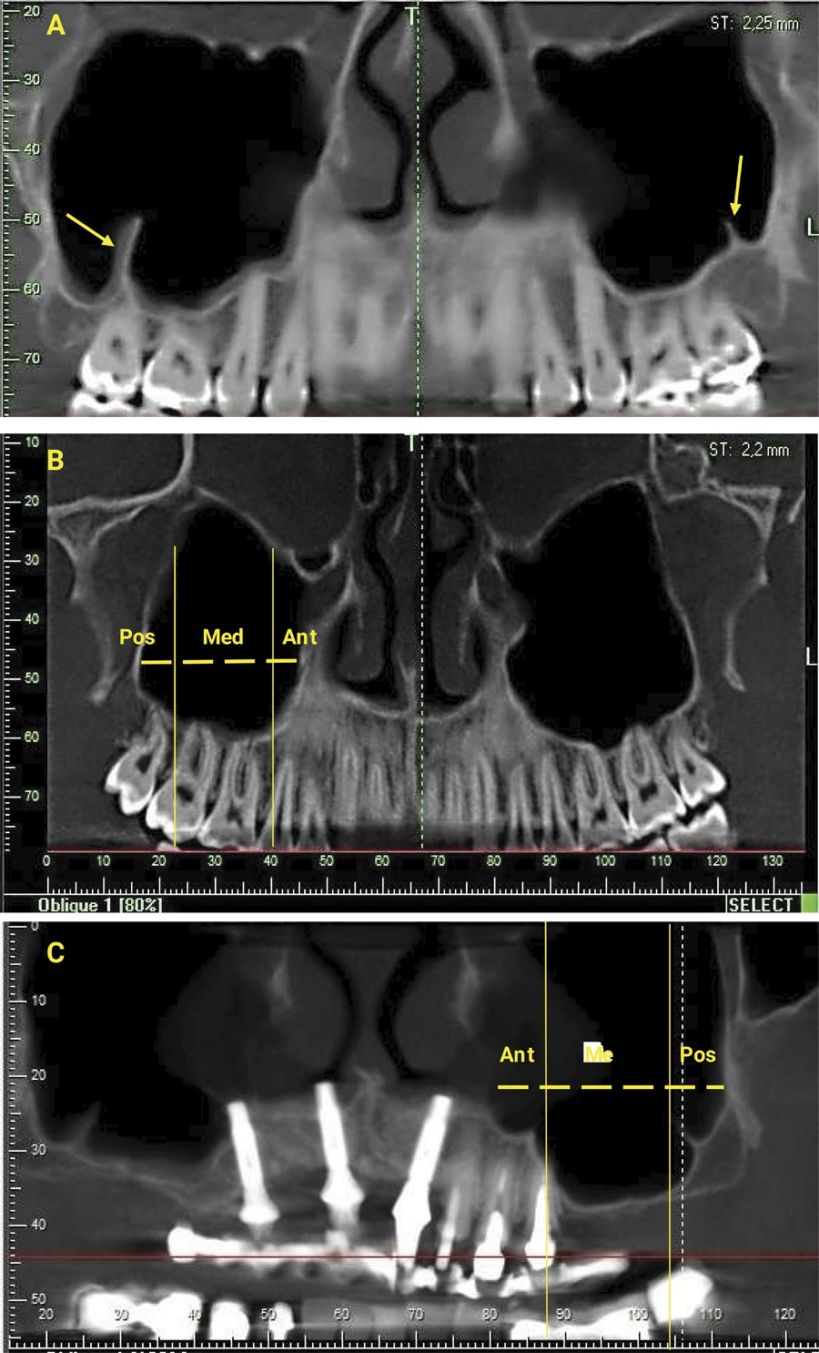
The existence and number of septa present were assessed through axial, coronal and sagittal sections, in addition to panoramic reconstructions of the tomographic image. Then, in the exams in which the presence of the septum was detected, their locations were specified through the panoramic image. According to the study by Kim et al. (2006)9, the septum located between the mesial region of the first premolar to the distal region of the second premolar was classified as anterior region, the septum between the mesial region of the first molar to the distal region of the second molar was classified as middle region, and the septum located distally to the region of the second molar was classified as posterior region.
In patients in whom teeth were absent, location was obtained following the method used by González-Santana et al.10 (2007), which divides the maxillary sinus into 3 parts. The maximum width is obtained from the anterior wall to the posterior wall of the cavity in the panoramic reconstruction and is then divided, maintaining the proportions of 1/2 of the width for the middle location and 1/4 of the width for the anterior and posterior portions.
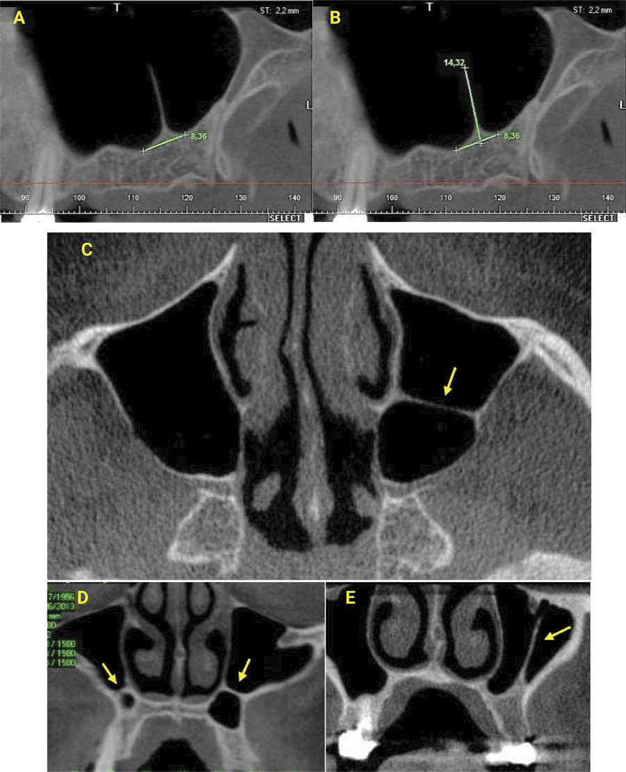
In the panoramic reconstructions, linear measurements of the septa were performed following the method used by van Zyl and van Heerden7 (2009). Initially, using the ruler tool, a line was drawn connecting the lowest points of the mesial and distal sides of the septum, thus determining its base. After this, another line was drawn from the apex of the septum to the base line, respecting the long axis of the septum. The measurements of the two lines were recorded, which resulted in the height of the septum and the length of its base, in millimeters. Only the largest height measurement presented by the septum along its trajectory was taken into consideration. Septa with less than 2.0 millimeters were not included in this study.
The orientation of the septum was classified as buccopalatal, sagittal or transverse. The buccopalatal septum exhibits a vertical orientation, connecting the walls of the maxillary sinus in a medial-lateral or lateral-medial manner, being oriented in a buccopalatal plane. Transverse septa were considered as those that have a horizontal orientation, parallel to the floor of the maxillary sinus, dividing the maxillary sinus into an upper and an lower portion. The sagittal type has an orientation parallel to the alveolar arch or the edentulous crest. For this classification, manipulation of the three available planes was allowed.
The septa were classified according to their shape and could be considered complete or incomplete, determined by manipulating the three available sections. The septum was only considered complete when it completely divided the sinus into two or more compartments. Figures 1 and 2 shows examples of the classifications used in the study.
A. Presence of septum observed in panoramic reconstruction. B. Classification of septa location as anterior, middle and posterior. C. Classification of septa location in case of absence of teeth.
Measurement in panoramic reconstruction: A. base of the septum, B. height and base of the septum. Classification of septal orientation, coronal view: C. buccopalatal, D. transverse and E. sagittal.
Statistical analysis
The data were tabulated in a spreadsheet using Microsoft Excel software and underwent statistical analysis using SPSS (Statistical Package for the Social Sciences). The results were expressed as percentages and statistical measures: mean, median and standard deviation. To assess the association between the variables defined by age group, sex and type of dentition, Pearson’s chi-square test or Fisher’s exact test were used when the condition for using the chi-square test was not met. Regarding the numerical variables, the Mann-Whitney statistical tests were used to compare two categories and, in the case of comparison between two or more categories, the Kruskal-Wallis statistical tests were used. The significance level adopted in this study was 5% (p<0.05).
Results
Results by patient
The age of the patients analyzed ranged from 20 to 92 years and had a mean of 49.89 years, with a standard deviation of 13.83 years and a median of 51 years. Of the 198 patients analyzed, 19 (9.6%) were between 20 and 29 years of age, 20 (10.1%) between 30 and 39 years of age, 56 (28.3%) between 40 and 49 years of age, 47 (23.7%) between 50 and 59 years of age and 56 (28.3%) were 60 years of age or older. Of the group analyzed, 73 (36.9%) were male and 125 (63.1%) were female.
It is noteworthy that the majority (77.8%) had partial dentition, followed by patients with complete dentition (13.1%) and completely edentulous patients (9.1%). Exactly half of the patients had septa, that is, a prevalence of septa of 50% was found when taking into account the septa found per patient. Among those who had septa, the lowest percentage (24.2%) corresponded to those who had three or more septa and the highest percentage (40.4%) to those who had two septa. Regarding the side, more than half (55.6%) had septa on both sides and those who had septa only on the right and left sides were 19.2% and 25.2% respectively.
Table 1 presents the study of the association between the presence of a septum and each of the variables: age group, sex and type of dentition. From this table, it is possible to calculate that the greatest percentage difference in those who had a septum occurred between the two age groups: 50 to 59 years and 20 to 29 years, being higher in those aged 50 to 59 years (55.3% x 36.8%). However, no significant association was found between the presence of a septum and any of the three variables (p > 0.05).
Results by septum
In total, 200 septa were found out of a total of 396 maxillary sinuses analyzed, thus finding a prevalence of 50.50% when the total number of septa was taken into account by the number of sinuses. Table 2 presents the distribution of the septa analyzed according to side, location, shape, height, width and orientation. There was no significant association between the type of dentition and any of the variables: location of the septum, side of the septum and orientation of the septum (p > 0.05), as can be seen in Table 3.
Distribution of the analyzed septa according to side, location, shape, height, width and orientation.
Table 4 shows the evaluation of the height and width of the septa found according to age group, sex, type of denture, location, side and orientation. It can be observed that there were significant differences (p < 0.05) between age groups and between locations in the variables of height and length of the base of the septa.
Discussion
The present study analyzed the prevalence, location, morphology and dimensions of maxillary sinus septa using cone beam computed tomography (CBCT), a method that provides three-dimensional accuracy essential for surgical planning in implant dentistry. Our findings revealed that sinus septa were present in 50.5% of the analyzed maxillary sinuses, a value that aligns with the variability found in the literature, where prevalence rates range from 9.5% to 69% depending on the population studied and imaging methodology employed11,12.
For instance, the Brazilian study by Furtado et al.13reported a prevalence of 26%, while Abesi et al.14found 37.9% in an Iranian population. It is important to note that differences in image acquisition protocols, such as voxel size and resolution, as well as the criteria adopted for classifying and measuring septa, may account for the variability in prevalence rates reported across studies. The higher prevalence in our study may be related to technical differences, including the use of thinner image slices, higher spatial resolution, and a larger sample size (396 sinuses), all of which may have facilitated the identification of finer septa not detected in studies using lower-resolution imaging.
When stratified by age, although no statistically significant differences were found, a trend toward increased prevalence was observed in older individuals, particularly those over 60 years. This may be attributed to progressive maxillary bone resorption associated with aging and tooth loss, which can contribute to the development of secondary septa, consistent with prior studies11,12,15.
Regarding the type of dentition present, no significant association was recorded between dentition and the presence of septum in this study, corroborating the publication of Jang et al.16. Furthermore, no significant association was recorded between the type of dentition and the number of septa present per patient. However, other authors reported an increased prevalence in totally edentulous patients than in patients with partial or complete dentition15,17.
In the present study, the location in which septa were most frequently found was the middle portion, with a prevalence of 52.5% of septa in this region, which is consistent with most of the data previously reported, according to a systematic review of Malec et al.18 and the study published by Al-Zahrani et al.11. It is assumed that the greater prevalence of septa in the middle region may be due to the formation of secondary septa occurring more frequently after the loss of a molar tooth than a premolar, since a greater pneumatization process is found after the extraction of a molar tooth. According to Sharan and Madjar19, the reason for this appears to be the large bone defect left by the extraction of a molar, which requires a long time to repair, thus allowing pneumatization of the sinus.
There was no significant association between the type of dentition and the location of the septum in the present study, although other authors reported an increased prevalence in edentulous patients. This finding is attributed to the development of secondary septa found in edentulous areas11.
Regarding orientation, the most frequent type of septum was the buccopalatal, with 151 septa being found out of 200 analyzed, demonstrating a prevalence of 75.5%. This form of septal orientation can be compared to an inverted Gothic arch14. Next, the most frequent septa were the transverse septa with a prevalence of 16.5% and the sagittal septa, which comprised 8% of the total septa analyzed.
The average height of the septa found in this study was 6.12 ± 3.32 mm, with a minimum size found of 2.01 mm and a maximum of 20.82 mm, a measurement close to that obtained by other authors12,15. Park et al.20 found an average of 7.78 mm and 7.89 mm in the right and left breasts, respectively, and Pommer et al.21 obtained an average septal size of 7.5 mm. Some authors report a significant difference in the size of the septa in relation to their dentition. Jang et al.16 found an average of 6.01 ± 2.21 mm and 5.30 ± 1.90 mm in dentate and edentulous patients respectively. Ranticelli et al.22 divided their measurement into primary and secondary septa, and found an average size of 5.5 mm and 7.8 mm respectively. However, as in the research by Al-Zahrani et al.11, no significant differences were found between the size of the septa and the type of dentition in our study.
A significant difference in the size of the septa is observed when the age group of the patient is taken into account, with the tallest patients being between 50 and 59 years old, with an average of 7.36 ± 3.89, followed by the age group of 20-29 years old, with 6.99 ± 2.85 mm. This fact possibly occurred due to the greater pneumatization process that follows the loss of a molar tooth, with the frequency of tooth loss in older patients being higher than in younger patients. In addition, many authors report an increase in the size of the septum in edentulous patients than in dentate patients11,13,14. Finally, no significant differences were found in septum sizes when each type of orientation was taken into account.
In the literature searched, no study was found that addressed the length of the base. In this study, the mean length of the base was 6.61 ± 2.38 mm. Significant differences were observed when the patient’s age group and location were taken into account. It is important to emphasize that the length of the base appears to be directly proportional to the size of the septum, since higher measurements were observed in the same age groups and in the same location for both variables. However, further studies on the subject are needed to prove this hypothesis.
Regarding location, as in the case of height, a higher mean base length was found in the middle portion compared to the anterior and posterior locations, with 7.15 ± 2.58 mm. As in the case of height, this finding appears to be due to the greater process of sinus pneumatization in the case of molar extractions, since a greater inferior projection of the recesses separated by septa results in a greater base length in the methodology used in this study.
Further research with a larger sample is needed to confirm the high prevalence of septa found in this study when compared to other publications on the subject. It is also necessary to evaluate the direction of the septa through the angle formed between the long axis of the septum in axial view and the median sagittal line, since it was not possible to provide this information in this study due to limitations of the software used.
Clinical implications are critical with the presence of septa complicates sinus lift procedures by limiting access and increasing the risk of Schneiderian membrane perforation, especially when septa are located in the surgical window pathway. Therefore, preoperative identification of the location, size, and orientation of septa is vital for surgical planning—guiding the choice between a single lateral window, double window, or transcrestal approach—and for the strategic placement of bone grafts.
Although the CBCT evaluation enabled detailed analysis, limitations include the lack of angular measurement of septa due to software restrictions. Future studies should incorporate angular assessments and larger multicentric samples to confirm our findings and refine surgical protocols.
In summary, the identification and characterization of maxillary sinus septa using CBCT are not merely anatomical observations—they are fundamental steps in reducing surgical risks and improving outcomes in maxillary bone grafting and implant rehabilitation. In the studied sample, maxillary sinus septa were detected in 50.5% of the sinuses, with higher frequency in the middle region. These anatomical variations, regardless of dentition status or degree of sinus pneumatization, reinforce the need for detailed tomographic evaluation before surgical procedures. The identification of septa through cone beam computed tomography is crucial in implant dentistry, particularly for sinus floor elevation surgeries. Knowledge of their location, dimensions, and orientation contributes to safer surgical planning, optimal bone graft placement, and reduced risk of Schneiderian membrane perforation, ultimately improving clinical outcomes in implant-supported rehabilitation.
Acknowledgments
The authors would like to thank the radiology clinic partner used in data collection.
References
-
1 Esfahrood ZR, Ahmadi L, Karami E, Asghari S. Short dental implants in the posterior maxilla: a review of the literature. J Korean Assoc Oral Maxillofac Surg. 2017 Apr;43(2):70-6. doi: 10.5125/jkaoms.2017.43.2.70.
» https://doi.org/10.5125/jkaoms.2017.43.2.70 -
2 Souza CF, Loures AO, Lopes DGF, Devito KL. Analysis of maxillary sinus septa by cone-beam computed tomography. Rev Odontol UNESP. 2019;48:e20190034. doi: 10.1590/1807-2577.03419.
» https://doi.org/10.1590/1807-2577.03419 -
3 Bacelar SMA, Guimarães Neto, UG. Sinus lift: Realização e técnicas cirúrgicas. Brazilian Journal of Implantology and Health Sciences. 2019;1(4):119-46. doi: 10.36557/2674-8169.2019v1n5p119.
» https://doi.org/10.36557/2674-8169.2019v1n5p119 -
4 Testori T, Scaini R, Deflorian M, Wallace SS. Sinus augmentation: the lateral approach. In: Artzi Z, editor. Bone augmentation by anatomical region: techniques and decision-making. Hoboken (NJ): John Wiley & Sons; 2020. Chapter 15, p.283-308. doi: 10.1002/9781119427926.ch15.
» https://doi.org/10.1002/9781119427926.ch15 -
5 Schiller LA, Barbu HM, Iancu SA, Brad S. Incidence, size and orientation of maxillary sinus septa-a retrospective clinical study. J Clin Med. 2022 Apr;11(9):2393. doi: 10.3390/jcm11092393.
» https://doi.org/10.3390/jcm11092393 -
6 Areal Neto PGE, Freitas GB, Silva RLB, Siqueira MV, Ciotti DL, Alvesand MAL, et al. [Evaluation of the prevalence of bony septa in maxillary sinuses by means of cone beam computed tomography (CBCT)], Int J Dev Res. 2020;10(4):35303-9. Portuguese. doi: 10.37118/ijdr.18617.04.2020.
» https://doi.org/10.37118/ijdr.18617.04.2020 -
7 van Zyl AW, van Heerden WF. A retrospective analysis of maxillary sinus septa on reformatted computerised tomography scans. Clin Oral Implants Res. 2009 Dec;20(12):1398-401. doi: 10.1111/j.1600-0501.2009.01802.x.
» https://doi.org/10.1111/j.1600-0501.2009.01802.x - 8 Neugebauer J, Ritter L, Mischkowski RA, Dreiseidler T, Scherer P, Ketterle M, et al. Evaluation of maxillary sinus anatomy by cone-beam CT prior to sinus floor elevation. Int J Oral Maxillofac Implants. 2010 Mar-Apr;25(2):258-65.
-
9 Kim MJ, Jung UW, Kim CS, Kim KD, Choi SH, Kim CK, et al. Maxillary sinus septa: prevalence, height, location, and morphology. A reformatted computed tomography scan analysis. J Periodontol. 2006 May;77(5):903-8. doi: 10.1902/jop.2006.050247.
» https://doi.org/10.1902/jop.2006.050247 -
10 González-Santana H, Peñarrocha-Diago M, Guarinos-Carbó J, Sorní-Bröker M. A study of the septa in the maxillary sinuses and the subantral alveolar processes in 30 patients. J Oral Implantol. 2007;33(6):340-3. doi: 10.1563/1548-1336(2007)33[340:ASOTSI]2.0.CO;2.
» https://doi.org/10.1563/1548-1336(2007)33[340:ASOTSI]2.0.CO;2 -
11 Al-Zahrani MS, Al-Ahmari MM, Al-Zahrani AA, Al-Mutairi KD, Zawawi KH. Prevalence and morphological variations of maxillary sinus septa in different age groups: a CBCT analysis. Ann Saudi Med. 2020 May-Jun;40(3):200-6. doi: 10.5144/0256-4947.2020.200.
» https://doi.org/10.5144/0256-4947.2020.200 -
12 Alhumaidan G, Eltahir MA, Shaikh SS. Retrospective analysis of maxillary sinus septa - A cone beam computed tomography study. Saudi Dent J. 2021 Nov;33(7):467-73. doi: 10.1016/j.sdentj.2020.11.001. Epub 2020 Nov 11.
» https://doi.org/10.1016/j.sdentj.2020.11.001 -
13 Furtado DM, Martins-Júnior PA, Alves TK, Santos RM, Coutinho DC, Aranha Neto IS, et al. Prevalence and characterization of maxillary sinus septa in a brazilian population. J Clin Exp Dent. 2021 Jul;13(7):e642-e647. doi: 10.4317/jced.56467.
» https://doi.org/10.4317/jced.56467 -
14 Abesi F, Motaharinia S, Moudi E, Haghanifar S, Khafri S. Prevalence and anatomical variations of maxillary sinus septa: A cone-beam computed tomography analysis. J Clin Exp Dent. 2022 Sep;14(9):e689-e693. doi: 10.4317/jced.59599.
» https://doi.org/10.4317/jced.59599 -
15 Orhan K, Kusakci Seker B, Aksoy S, Bayindir H, Berberoglu A, Seker E. Cone beam CT evaluation of maxillary sinus septa prevalence, height, location and morphology in children and an adult population. Med Princ Pract. 2013;22(1):47-53. doi: 10.1159/000339849. Epub 2012 Jul 24.
» https://doi.org/10.1159/000339849 -
16 Jang SY, Chung K, Jung S, Park HJ, Oh HK, Kook MS. Comparative study of the sinus septa between dentulous and edentulous patients by cone beam computed tomography. Implant Dent. 2014 Aug;23(4):477-81. doi: 10.1097/ID.0000000000000107.
» https://doi.org/10.1097/ID.0000000000000107 -
17 Poli O, Manzon L, Niglio T, Ettorre E, Vozza I. Masticatory force in relation with age in subjects with full permanent dentition: a cross-sectional study. Healthcare (Basel). 2021 Jun;9(6):700. doi: 10.3390/healthcare9060700.
» https://doi.org/10.3390/healthcare9060700 -
18 Malec M, Smektala T, Trybek G, Sporniak-Tutak K. Maxillary sinus septa: prevalence, morphology, diagnostics and implantological implications. Systematic review. Folia Morphol (Warsz). 2013;73(3):259-66. doi: 10.5603/FM.2014.0041.
» https://doi.org/10.5603/FM.2014.0041 - 19 Sharan A, Madjar D. Maxillary sinus pneumatization following extractions: a radiographic study. Int J Oral Maxillofac Implants. 2008 Jan-Feb;23(1):48-56.
-
20 Park YB, Jeon HS, Shim JS, Lee KW, Moon HS. Analysis of the anatomy of the maxillary sinus septum using 3-dimensional computed tomography. J Oral Maxillofac Surg. 2011 Apr;69(4):1070-8. doi: 10.1016/j.joms.2010.07.020.
» https://doi.org/10.1016/j.joms.2010.07.020 -
21 Pommer B, Ulm C, Lorenzoni M, Palmer R, Watzek G, Zechner W. Prevalence, location and morphology of maxillary sinus septa: systematic review and meta-analysis. J Clin Periodontol. 2012 Aug;39(8):769-73. doi: 10.1111/j.1600-051X.2012.01897.x.
» https://doi.org/10.1111/j.1600-051X.2012.01897.x -
22 Rancitelli D, Borgonovo AE, Cicciù M, Re D, Rizza F, Frigo AC, et al. Maxillary sinus septa and anatomic correlation with the schneiderian membrane. J Craniofac Surg. 2015 Jun;26(4):1394-8. doi: 10.1097/SCS.0000000000001725.
» https://doi.org/10.1097/SCS.0000000000001725
-
Data availability:
Datasets related to this article will be available upon request to the corresponding author.
Edited by
-
Editor:
Dr. Altair A. Del Bel Cury
Datasets related to this article will be available upon request to the corresponding author.