Antagonistic potential of Trichoderma koningiopsis and its effect on plant defense response
Abstract
The genus Trichoderma is the most studied and widely used in biological control of plant diseases worldwide, showing efficiency against phytopathogens in different agricultural crops. This study aimed to evaluate the antagonistic potential of four Trichoderma koningiopsis strains isolated from dystrophic red latosol in controlling the main phytopathogens associated with soybean diseases, namely Fusarium tucumaniae, Macrophomina phaseolina, Rhizoctonia solani, and Sclerotinia sclerotiorum, as well as their effect on the activation of soybean defense responses by assessing chitinase, β-1,3-glucanase, phenolic compounds, proteins, and phenylalanine ammonia-lyase (PAL). The disease control potential was evaluated in vitro using the direct culture pairing method, in which the mycelial growth of each pathogen was measured daily. To assess plant resistance induction, the T. koningiopsis strains were applied by spraying soybean plants at the V3 growth stage, with plant material collected over the following five days. Results showed that all four tested strains were effective in the biocontrol of phytopathogens, acting mainly through mycoparasitism and competition. In addition, they were also highlighted as inducers of plant resistance, increasing chitinase and β-1,3-glucanase concentrations in plant tissues.
Keywords:
biological control; competition; mycoparasitism; resistance induction
Resumo
O gênero Trichoderma é o mais estudado e utilizado no controle biológico de doenças no mundo, apresentando eficiência no controle de fitopatógenos de diferentes culturas agrícolas. O presente estudo, visa avaliar o potencial antagonista de quatro estirpes de Trichoderma koningiopsis prospectadas em latossolo vermelho distrófico, no controle dos principais fitopatógenos causadores de doenças na cultura da soja, sendo Fusarium tucumaniae, Macrophomina phaseolina, Rhizoctonia solani e Sclerotinia sclerotiorum, e seu efeito na ativação de resposta de defesa de plantas de soja, avaliando as enzimas Quitinase, β-1,3 Glucanase, compostos fenólicos, proteínas e fenilalanina amônia-liase (FAL). O potencial de controle de doenças, foi avaliado in vitro utilizando o médoto direto de pareamento de culturas, onde foi medido diariamente o crescimento micelial de cada patógeno. E para avaliação da indução de resistência de plantas, utilizou-se a aplicação das estirpes de T. koningiopsis via pulverização de plantas de soja no estádio V3, coletando-se material vegetal nos cinco dias posteriores. Os resultados mostram que as quatro estirpes testadas foram eficientes no biocontrole dos fitopatógenos, apresentando como mecanismo de ação o micoparasitismo e a competição. Destacando-se também como indutor de resistência em plantas, aumentando a concentração de quitinase e β-1,3 glucanase nos tecidos vegetais.
Palavras-chave:
controle biológico; competição; micoparasitismo; indução de resistência
1. Introduction
Soybean (Glycine max L. Merril) has a well-established market for grains and derivatives, gaining global importance due to its diverse applications in agribusiness, including animal feed production, vegetable oil, the food industry, as well as the chemical industry (Costa Neto et al., 2000).
Among the factors that limit high yield in this crop, soilborne diseases stand out, especially root rots caused by rhizospheric fungi (Correia and Michereff, 2018). The main phytopathogenic agents affecting soybean include Macrophomina phaseolina, the Fusarium spp. complex, Sclerotinia sclerotiorum, and Phomopsis longicolla.
Biological control of diseases has been consolidated as an effective and environmentally safe tool, reducing ecological risks. Rhizosphere-associated agents are particularly relevant, since the soil system tends to be a relatively stable environment, with less climatic fluctuation compared to the aerial part of plants. Consequently, the survival and activity of antagonistic organisms in this environment are dynamic and favorable (Bettiol and Morandi, 2009). Among these agents, the genus Trichoderma stands out worldwide, being recognized as plant symbionts and effective fungal mycoparasites, with several species demonstrating potential as commercial biofungicides (Schuster and Schmoll, 2010).
Trichoderma spp. is a saprophytic, cosmopolitan, soil-inhabiting fungus, belonging to the family Ascomycota. It has strong antagonistic potential in the control of bacteria, fungi, and nematodes, especially those causing root rots such as M. phaseolina, Fusarium spp., S. sclerotiorum, and P. longicolla. The most studied species include Trichoderma harzianum, T. virens, T. viride, and T. asperellum (Blaszczyk et al., 2014; Medeiros et al., 2022). In addition to colonizing plant roots, these microorganisms also inhabit the rhizosphere and the rhizoplane, exhibiting a complex mode of action: antibiosis through the production of metabolites toxic to phytopathogens; chitin degradation, an essential component of the fungal cell wall (favoring mycoparasitism); competition for nutrients and space; and induction of plant resistance. Together with plant growth promotion, siderophore production, and phosphorus solubilization, these characteristics make Trichoderma an excellent option for use in agriculture as a biocontrol agent (Tyśkiewicz et al., 2022; Gajera et al., 2013; Segarra et al., 2010; Borges-Chagas et al., 2015).
Therefore, the objective of this study was to evaluate the direct effect of Trichoderma koningiopsis strains in the control of Fusarium tucumaniae, Macrophomina phaseolina, Rhizoctonia solani, and Sclerotinia sclerotiorum, as well as their effect on the activation of defense responses in soybean plants, by assessing the enzymes chitinase, β-1,3-glucanase, phenolic compounds, proteins, and phenylalanine ammonia-lyase (PAL).
2. Material and Methods
2.1. Direct effect against the phytopathogen
Four strains of Trichoderma koningiopsis isolated from dystrophic red latosol were studied. The POP#GoG-010 method – Identification of microorganisms by DNA sequencing (ITS) – was used for species-level identification.
In a laminar flow chamber, Petri® dishes containing solidified PDA medium were prepared. A 7 mm mycelial disc of each phytopathogen was placed on the right side of the plate, at 1 cm from the edge. The phytopathogens used in the experiments were: Fusarium tucumaniae (CMES 25), Macrophomina phaseolina (CMES 1574), Rhizoctonia solani (CMES 1861), and Sclerotinia sclerotiorum (CMES 2131), obtained from the “Collection of Multifunctional Microorganisms” of Embrapa Soybean, Londrina, PR, Brazil.
On the left side of the same plate, at 1 cm from the edge, a 7 mm mycelial disc of the antagonist was added. The treatments consisted of four phytopathogens and four T. koningiopsis strains, plus the control.
Plates were sealed with plastic film and incubated in a BOD chamber at 25 °C with a 12-hour photoperiod. Mycelial growth of the phytopathogens was measured every 24 hours using a graduated ruler, until the control plates reached full growth.
The percentage of mycelial growth inhibition was calculated based on the Formula 1:
At the end of the experiment, light microscopy was performed to evaluate potential morphological damages to the hyphae of the phytopathogen. The experimental design adopted was completely randomized, with four replications per treatment. The data obtained were subjected to the Lilliefors normality test; since the experiment involved both qualitative and quantitative factors, ANOVA (Scott-Knott test at 5% probability) was conducted, followed by regression analysis using the Rbio software (Bhering, 2017).
2.2. Induction of resistance
2.2.1. Plant cultivation in a greenhouse
Soybean seeds of the cultivar Brasmax Zeus IPRO were sown in 8 L pots containing commercial substrate. The treatments consisted of four replicates of pots, each containing two soybean plants, totaling five treatments: the four T. koningiopsis strains (MS04, MS09, MS27, MS28) plus the control (without inoculum), arranged in a completely randomized design.
When soybean reached the V3 stage, T. koningiopsis was applied via foliar spray at a concentration of 1×10⁹ spores mL−1 for each tested strain. After 12 hours, the plants were challenge-inoculated with a powdery mildew spore suspension at a concentration of 1×10⁶ spores mL−1.
To quantify resistance-related variables over time, plant material for analyses was collected at 0, 24, 48, 72, and 96 hours after the mildew spores inoculation.
2.2.2. Preparation of the enzymatic extract
The methodology described by Guzzo and Martins (1996), was followed. For extract preparation, tissue samples (approximately 0.3 g) were homogenized in a mortar containing 0.2 g of glass microbeads, 0.1 g of Dowex resin, and 0.2 g of PVPP (polyvinylpolypyrrolidone). The homogenate was transferred to an Eppendorf tube, and 0.5 mL of sodium borate buffer was added. The mixture was allowed to react for 5 minutes. Subsequently, the samples were centrifuged at 20.000 g for 20 minutes at 4 °C, and the supernatant was collected into another labeled Eppendorf tube.
2.2.3. Determination of total proteins
Total protein content was quantified following the methodology of Bradford (1976). In numbered test tubes, 40 μL of the plant extract described in section 2.2.1 was mixed with 460 μL of distilled water and 1 mL of Bio-Rad reagent (Sigma). The mixture was then vortexed and absorbance was measured at 595 nm using a spectrophotometer.
2.2.4. Determination of chitinase activity
In Eppendorf tubes, 100 mg of chitinase substrate, 400 μL of 0.2 M sodium phosphate buffer, and 500 μL of the prepared plant extract were combined and incubated in a water bath at 50 ºC for 40 minutes. Subsequently, the mixture was centrifuged at 7,000 rpm for 10 minutes, and the supernatant was transferred to a new Eppendorf tube. Absorbance was then measured using a spectrophotometer at 595 nm.
2.2.5. Determination of β-1,3-glucanase activity
The methodology described by Guzzo and Martins (1996) was followed. In Eppendorf tubes, 200 μL of plant extract, 400 μL of 50 mM sodium acetate buffer (pH 5.0), and 200 μL of Curdlan substrate (Sigma) were added. The tubes were then incubated in a water bath at 40 ºC for 50 minutes. The reaction was stopped by adding 200 μL of 2N HCl, followed by cooling on ice for 10 minutes. Subsequently, the mixture was centrifuged at 10,000 × g for 5 minutes to remove insoluble, non-hydrolyzed substrate, and the absorbance of the supernatant was measured at 600 nm using a spectrophotometer.
2.2.6. Determination of Phenylalanine Ammonia-Lyase (PAL) activity
The methodology proposed by Rodrigues et al. (2006) was followed. A 0.5 g portion of plant material was weighed, transferred to crucibles, and ground with 4 mL of TRIS-HCl buffer (pH 8.0). The resulting material was transferred to an Eppendorf tube and centrifuged at 6,000 × g at 4 ºC for 10 minutes. An aliquot of 200 μL of the supernatant was transferred to a labeled test tube, and 3 mL of TRIS extraction buffer was added. The solution was then vortexed to obtain the enzymatic extract.
Subsequently, 1.5 mL of the enzymatic extract was transferred, supplemented with 1 mL of extraction buffer and 0.5 mL of phenylalanine, and incubated in a water bath at 40 ºC for 30 minutes. After incubation, the reaction was stopped by cooling on ice. Absorbance was measured at 290 nm using a quartz cuvette in a spectrophotometer.
2.2.7. Determination of total phenolic compounds
The analysis of total phenolic compounds was carried out in two steps, following the method adapted from Bieleski and Turner (1966). A 0.3 g portion of plant material was weighed, 5 mL of MCA solution (methanol, chloroform, water 6:2.5:1.5) was added, and the mixture was ground in a mortar. The resulting material was transferred to an Eppendorf tube and centrifuged at 6,000 rpm at 20 ºC for 20 minutes, and the supernatant was collected. The remaining solid residue was subjected to a second extraction by adding 4 mL of MCA, vortexing, and centrifuging again at 6,000 rpm at 20 ºC for 20 minutes. The supernatant from this second extraction was combined with the first, obtaining the MCA extract. To this extract, 1 mL of chloroform and 1.5 mL of deionized water were added, and the mixture was centrifuged at 6,000 rpm at 20 ºC for 15 minutes.
The second step involved the determination of total phenolics using the method adapted from Jennings (1981). An aliquot of 0.5 mL of the upper phase of the supernatant was taken, 0.5 mL of water and 0.5 mL of 1:10 diluted Folin–Ciocalteau reagent were added, and the solution was vortexed. After 15 minutes, 5 mL of alkaline reagent A (prepared from 2% sodium carbonate in 0.1 N sodium hydroxide solution) was added. The mixture was vortexed again, and after 50 minutes, absorbance was measured at 760 nm using a spectrophotometer.
2.2.8. Statistical analysis
The experimental design was completely randomized, with four replicates per treatment. The data were subjected to the Lilliefors test for normality. As this was a qualitative × quantitative experiment, analysis of variance (ANOVA) was performed using the Scott–Knott test at 5% probability, followed by regression analysis in the Rbio software (Bhering, 2017).
3. Results and discussion
As shown in Figure 1, for the control of M. phaseolina, three of the four strains (MS04, MS09, MS28) reduced radial growth of the phytopathogen by 76%, while strain MS27 reduced it by 54% compared to the control. In contrast, for F. tucumaniae, no significant differences were observed among the treatments with T. koningiopsis; however, all treatments differed from the control, with the strains under study promoting a 67% reduction in mycelial growth of F. tucumaniae.

Antagonistic effect of T. koningiopsis strains MS04, MS09, MS27, and MS28 on the mycelial growth (cm) of M. phaseolina, F. tucumaniae, P. longicolla, and S. sclerotiorum, respectively, over four days.
Mendoza et al. (2015) observed the same effect in the in vitro control, where eleven Trichoderma spp. isolates exhibited hyphal parasitism of M. phaseolina, and four showed antibiosis, forming a clear inhibition halo on the plate. Sridharan et al. (2021) reported that T. longibrachiatum reduced M. phaseolina growth by 58%, in addition to demonstrating its antibiosis effect.
The production of β-1,3-glucanase and N-acetylhexosaminidase, as well as mycoparasitism by Trichoderma spp. strains, was also observed in experiments with M. phaseolina isolates from bean and sorghum, which may explain their success in reducing mycelial growth (Larralde-Corona et al., 2008).
The same was reported by Pimentel et al. (2020), who found that the most effective isolate in controlling F. virguliforme, which affects soybean in the USA, was T. harzianum, capable of reducing the radial growth of the phytopathogen by 92%. The study also demonstrated mycoparasitic activity of T. harzianum, as well as the induction of plant resistance genes, such as β-1,6-glucan synthase, α-1,3-glucanase, chitinase, and endochitinase.
The production of secondary metabolites may also explain the advantage of Trichoderma spp. in dual culture assays. Mironenka et al. (2021) observed that, in addition to suppressing the growth of F. culmorum, the antagonist produced the metabolite zearalenone on the plate. However, metabolite production by Trichoderma spp. is broad, including alcohols, acids, esters, ketones, and others (Li et al., 2018).
Analyzing the results obtained with the four T. koningiopsis strains (Figure 1 and Table 1), an average reduction of 54–71% in mycelial growth was observed for Phomopsis longicolla and 44–63% for Sclerotinia sclerotiorum compared to the control, promoted by the Trichoderma spp. strains.
Regression equation and adjusted R2 for the mycelial growth of M. phaseolina, F. tucumaniae, P. longicolla, and S. sclerotiorum.
Das et al. (2014) reported a reduction of 84% and 78% in the growth of Phomopsis vexans when using T. viride and T.harzianum, respectively. In another in vitro study, Anita et al. (2012) achieved 100% inhibition of Phomopsis theae Petch growth when exposed to metabolites of T. atroviride. They also described the synthesis of defense-related enzymes, such as amylase, cellulase, and chitinase, when the organisms were placed in direct confrontation.
There are numerous studies involving Trichoderma spp. and Sclerotinia sclerotiorum. Zhang et al. (2016) observed that, in dual culture, the antagonist hyphae intertwined with S. sclerotiorum hyphae and formed a hook structure, evidencing mycoparasitism. The study reported a 51.2% reduction in the mycelial growth of the phytopathogen and also confirmed the growth-promoting effect exerted by Trichoderma spp.
Another study involving the co-inoculation of two strains, Trichoderma harzianum and Trichoderma asperellum, challenged with S. sclerotiorum, demonstrated effectiveness in inducing resistance responses in eggplant, with increased shikimic acid production after 72 hours, in addition to the expression of defense-related enzymes, such as phenylalanine ammonia-lyase and peroxidase (Pratap-Singh et al., 2021). Another reported mechanism that explains the antagonist’s success in suppressing S. sclerotiorum is the production of volatile organic compounds that inhibit the phytopathogen (Silva et al., 2021).
The mycoparasitism mechanism can be observed in Figures 2 and 3, where strain MS28 parasitizes the hyphae of M. phaseolina, F. tucumaniae, P. longicolla, and S. sclerotiorum.

Mycoparasitism by T. koningiopsis on: (a) M. phaseolina, (b) F. tucumaniae, (c) P. longicolla, and (d) S. sclerotiorum.

Under a light microscope, narrowing and coiling damage to M. phaseolina hyphae was observed in dual plates with T. koningiopsis strain MS27 (Figure 4B), in addition to mycoparasitism (Figure 4C).
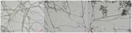
It can be observed that the four T. koningiopsis strains exhibited potential for the biological control of the studied phytopathogens, with mycoparasitism of the hyphae confirmed in Figure 4C.
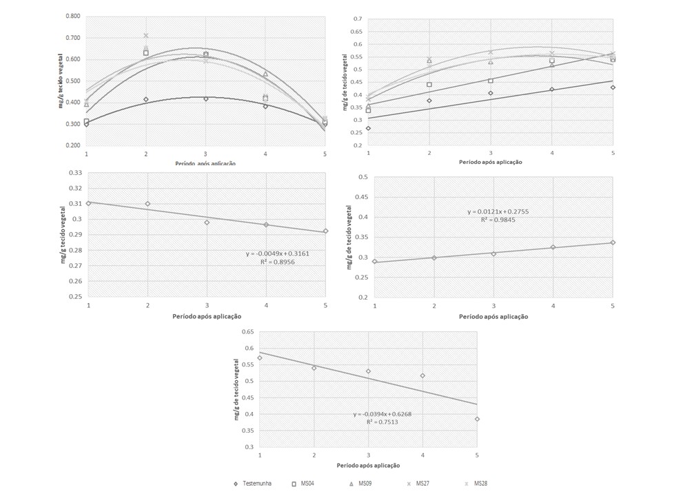
In the study evaluating the potential of strains MS07, MS09, MS27, and MS28 in inducing resistance in soybean plants, the activated enzymes were chitinase and β-1,3-glucanase (Figure 5 and Table 2). However, no significant differences were observed among treatments for phenolic compounds, proteins, or PAL activity.

Defense response of soybean plants inoculated with T. koningiopsis strains MS04, MS09, MS27, and MS28, showing the expression of chitinase, β-1,3-glucanase, phenolic compounds, proteins, and PAL, respectively.
Linear equation and adjusted R2 for the defense response of soybean plants inoculated with T. koningiopsis strains MS04, MS09, MS27, and MS28, regarding the expression of chitinase and β-1,3-glucanase.
When resistance is induced in plants, a large production of defense-related proteins is activated, known as pathogenesis-related proteins (PR-proteins), leading to an increase in their concentration in plant tissues. This includes enhanced levels of peroxidases, chitinases, β-1,3-glucanases, PAL, salicylic acid, and lignin deposition (Verbene et al., 2000).
The “priming” effect is a term used in the context of resistance induction, referring to the pre-conditioning of the plant against infection, which allows its defense system to be activated more rapidly (Pascholati and Dalio, 2018). Studies have demonstrated the direct role of Trichoderma spp. in activating priming, inducing faster immune responses in plants and increasing the effectiveness of defense. This leads to greater accumulation and activation of these otherwise inactive cellular proteins, making them readily available in response to pathogen attack (Morán-Diez et al., 2021).
Liu et al. (2021), studying the effect of Trichoderma spp. on cabbage plants infected with Botrytis cinerea, observed that application of the agent increased ascorbate peroxidase by 1.46-fold, while also controlling B. cinerea infection. Pratap-Singh et al. (2021) reported that treating eggplant plants with T. harzianum and T. asperellum challenged with S. sclerotiorum resulted in an increase in shikimic acid concentration, peaking 72 hours after application, whereas phenolic compounds showed no significant difference.
In the present study, the peak production of chitinase and β-1,3-glucanase occurred on the second and fifth day after application, respectively, with an average increase of 59% for chitinase and 27% for β-1,3-glucanase.
Gajera et al. (2015), when treating different peanut seedlings with T. viride challenged with Aspergillus niger, observed an increase on the third day after application of 3.5- and 2.3-fold for polyphenol oxidase and β-1,3-glucanase, respectively; and an increase of 1.6-fold for PAL on the sixth day, and 2.3- to 2.8-fold on the ninth day after application.
4. Conclusion
The four T. koningiopsis strains proved to be effective agents for the control of the studied phytopathogens, demonstrating their capacity for mycoparasitism and competition for space and nutrients. Strain MS27 showed the highest control of mycelial growth of M. phaseolina and S. sclerotiorum. The antagonist also proved efficient in activating defense responses in soybean plants, increasing the concentration of chitinase and β-1,3-glucanase in plant tissues.
Data Availability Statement
References
-
ANITA, S., PONMURUGAN, P. and GANESH BABU, R., 2012. Significance of secondary metabolites and enzymes secreted by Trichoderma atroviride isolates for the biological control of Phomopsis canker disease. African Journal of Biotechnology, vol. 11, no. 45, pp. 10350-10357. https://doi.org/10.5897/AJB12.599
» https://doi.org/10.5897/AJB12.599 - BETTIOL, W. and MORANDI, M.A.B., 2009. Biocontrole de doenças em plantas: usos e perspectivas Jaguariuna: Empresa Brasileira de Pesquisa Agropecuária, Embrapa Meio Ambiente; Brasília: Ministério da Agricultura, Pecuária e Abastecimento.
-
BHERING, L.L., 2017. Rbio: a tool for biometric and statistical analysis using the R Platform. Crop Breeding and Applied Biotechnology, vol. 17, no. 2, pp. 187-190. https://doi.org/10.1590/1984-70332017v17n2s29
» https://doi.org/10.1590/1984-70332017v17n2s29 -
BIELESKI, R.L. and TURNER, N.A., 1966. Separation and estimation of amino acids in crude plant extratcts by thin-layer electrophoresis and chomatograghy. Analytical Biochemistry, vol. 17, no. 2, pp. 278-293. https://doi.org/10.1016/0003-2697(66)90206-5 PMid:5971422.
» https://doi.org/10.1016/0003-2697(66)90206-5 -
BLASZCZYK, L., SIWULSKI, M., SOBIERALSKI, K., LISIECKA, J. and JĘDRYCZKA, M., 2014. Trichoderma spp.: application and prospects for use in organic farming and industry. Journal of Plant Protection Research, vol. 54, no. 4, pp. 309-317. https://doi.org/10.2478/jppr-2014-0047
» https://doi.org/10.2478/jppr-2014-0047 -
BORGES-CHAGAS, L.F., CHAGAS JUNIOR, A.F., RODRIGUES DE CARVALHO, M., DE OLIVEIRA MILLER, L, and OROZCO COLONIA, B.S., 2015. Evaluation of the phosphate solubilization potential of Trichoderma strains (Trichoplus JCO) and effects on rice biomass. Journal of Soil Science and Plant Nutrition, vol. 15, no. 3, pp. 794-804. https://doi.org/10.4067/S0718-95162015005000054
» https://doi.org/10.4067/S0718-95162015005000054 -
BRADFORD, M.M., 1976. A rapid and sensitive method for the quantification of microgram quantities of protein utilizing the principle of protein-dye binding. Analytical Biochemistry, vol. 72, no. 1-2, pp. 248-254. https://doi.org/10.1016/0003-2697(76)90527-3 PMid:942051.
» https://doi.org/10.1016/0003-2697(76)90527-3 - CORREIA, K.C. and MICHEREFF, S.J., 2018. Desafios do manejo de doenças radiculares causadas por fungos Recife: EDUFRPE, 208 p.
-
COSTA NETO, P.R., ROSSI, L.F.S., ZAGONEL, G.F. and RAMOS, L.P., 2000. Produção de biocombustível alternativo ao óleo diesel através da transesterificação de óleo de soja usado em fritura. Química Nova, vol. 23, no. 4, pp. 531-537. https://doi.org/10.1590/S0100-40422000000400017
» https://doi.org/10.1590/S0100-40422000000400017 - DAS, S.N., SARMA, T.C. and TAPADAR, S.A., 2014. In vitro evaluation of fungicides and two species of Trichoderma against Phomopsis vexans causing fruit rot of brinjal (Solanum melongena L.). International Journal of Scientific and Research Publications, vol. 4, no. 9, pp. 1-2.
- GAJERA, H., DOMADIYA, R., PATEL, S., KAPOPARA, M. and GOLAKIYA, B., 2013. Molecular mechanism of Trichoderma as bio-control agents against phytopathogen system: a review. Current Research in Microbiology and Biotechnology
-
GAJERA, H.P., SAVALIYA, D.D., PATEL, S.V. and GOLAKIYA, B.A., 2015. Trichoderma viride induces pathogenesis related defense response against rot pathogen infection in groundnut (Arachis hypogaea L.). Infection, Genetics and Evolution : Journal of Molecular Epidemiology and Evolutionary Genetics in Infectious Diseases, vol. 34, pp. 314-325. https://doi.org/10.1016/j.meegid.2015.07.003 PMid:26160540.
» https://doi.org/10.1016/j.meegid.2015.07.003 -
GUZZO, S.D. and MARTINS, E.M.F., 1996. Local and systemic induction of β 1,3 glucanase and chitinase in coffee leaves protected against hemileia vastatrix by Bacillus thuringiensis. Journal of Phytopathology, vol. 144, no. 9-10, p. 449-454. https://doi.org/10.1111/j.1439-0434.1996.tb00322.x
» https://doi.org/10.1111/j.1439-0434.1996.tb00322.x -
JENNINGS, A.C., 1981. The determination al dihydroxy phenolic compounds in extracts of plant tissues. Analytical Biochemistry, vol. 118, no. 2, pp. 396-398. https://doi.org/10.1016/0003-2697(81)90600-X
» https://doi.org/10.1016/0003-2697(81)90600-X -
LARRALDE-CORONA, C.P., SANTIAGO-MENA, M.R., SIFUENTES-RINCÓN, A.M., RODRÍGUEZ-LUNA, I.C., RODRÍGUEZ-PÉREZ, M.A., SHIRAI, K. and NARVÁEZ-ZAPATA, J.A., 2008. Biocontrol potential and polyphasic characterization of novel native Trichoderma strains against Macrophomina phaseolina isolated from sorghum and common bean. Applied Microbiology and Biotechnology, vol. 80, no. 1, pp. 167-177. https://doi.org/10.1007/s00253-008-1532-0 PMid:18523764.
» https://doi.org/10.1007/s00253-008-1532-0 -
LI, N., ALFIKY, A., WANG, W., ISLAM, M., NOUROLLAHI, K., LIU, X. and KANG, S., 2018. Reconhecimento e inibição mediados por compostos voláteis entre agentes de controle biológico de Trichoderma e Fusarium oxysporum Microbiol Frontal, vol. 9, pp. 2614. https://doi.org/10.3389/fmicb.2018.02614 PMid:30455673.
» https://doi.org/10.3389/fmicb.2018.02614 -
LIU, C.M., LIU, S.Y., LIAO, C.K., LO, C.T., LIN, K.C. and PENG, K.C., 2021. Cabbage defense response provoked by Trichoderma Th-LAAO. Archives of Microbiology, vol. 203, no. 4, pp. 1641-1647. https://doi.org/10.1007/s00203-020-02174-6 PMid:33432379.
» https://doi.org/10.1007/s00203-020-02174-6 - MEDEIROS, F.H.V., BAVIA, G.P. and SEIXAS, C.D.S., 2022. Manejo de doenças fúngicas radiculares da soja. In: M.C. MEYER, A.F. BUENO, S.M. MAZARO and J.C. SILVA. Bioinsumos na cultura da soja Brasília: Embrapa.
-
MENDOZA, J.L.H., PÉREZ, M.I.S., PRIETO, J.M.G., VELÁSQUEZ, J.D.Q., OLIVARES, J.G.G. and LANGARICA, H.R.G., 2015. Antibiose de cepas de Trichoderma spp nativas do nordeste do México contra o fungo patogênico Macrophomina phaseolina Brazilian Journal of Microbiology: Publication of the Brazilian Society for Microbiology, vol. 46, no. 4, pp. 1093-1101. https://doi.org/10.1590/S1517-838246420120177
» https://doi.org/10.1590/S1517-838246420120177 -
MIRONENKA, J., RÓŻALSKA, S., SOBOŃ, A. and BERNAT, P., 2021. Trichoderma harzianum metabolites disturb Fusarium culmorum metabolism: metabolomic and proteomic studies. Microbiological Research, vol. 249, pp. 126770. https://doi.org/10.1016/j.micres.2021.126770 PMid:33932742.
» https://doi.org/10.1016/j.micres.2021.126770 -
MORÁN-DIEZ, M.E., MARTÍNEZ DE ALBA, Á.E., RUBIO, M.B., HERMOSA, R. and MONTE, E., 2021. Trichoderma and the Plant Heritable Priming Responses. Journal of Fungi (Basel, Switzerland), vol. 7, no. 4, pp. 318. https://doi.org/10.3390/jof7040318 PMid:33921806.
» https://doi.org/10.3390/jof7040318 - PASCHOLATI, S.F. and DALIO, R.J.D., 2018. Fisiologia do parasitismo: como as plantas se defendem dos patógenos. In: L. AMORIM, J.A.M. REZENDE and A. BERGAMIM FILHO, eds. Manual de fitopatologia 5. ed. Ouro Fino: Ceres, pp. 424-450.
-
PIMENTEL, M.F., ARNÃO, E., WARNER, A.J., SUBEDI, A., ROCHA, L.F., SROUR, A., BOND, J.P. and FAKHOURY, A.M., 2020. Trichoderma isolates inhibit fusarium virguliforme growth, reduce root rot, and induce defense-related genes on soybean seedlings. Plant Disease, vol. 104, no. 7, pp. 1949-1959. https://doi.org/10.1094/PDIS-08-19-1676-RE PMid:32396055.
» https://doi.org/10.1094/PDIS-08-19-1676-RE -
PRATAP-SINGH, S., KESWANI, C., PRATAP SINGH, S., SANSINENEA, E. and XUAN HOAT, T., 2021. Trichoderma spp. mediated induction of systemic defense response in brinjal against Sclerotinia sclerotiorum. Current Research in Microbial Sciences, vol. 2, pp. 100051. https://doi.org/10.1016/j.crmicr.2021.100051 PMid:34841342.
» https://doi.org/10.1016/j.crmicr.2021.100051 -
RODRIGUES, A.A.C., BEZERRA NETO, E. and COELHO, R.S.B., 2006. Indução de resistência a Fusarium oxysporum f. sp. Tracheiphilum em Caupi: eficiência de indutores abióticos e atividade enzimática elicitada. Fitopatologia Brasileira, vol. 31, no. 5, pp. 492-499. https://doi.org/10.1590/S0100-41582006000500009
» https://doi.org/10.1590/S0100-41582006000500009 -
SCHUSTER, A. and SCHMOLL, M., 2010. Biology and biotechnology of Trichoderma. Applied Microbiology and Biotechnology, vol. 87, no. 3, pp. 787-799. https://doi.org/10.1007/s00253-010-2632-1 PMid:20461510.
» https://doi.org/10.1007/s00253-010-2632-1 -
SEGARRA, G., CASANOVA, E., AVILÉS, M. and TRILLAS, I., 2010. Trichoderma asperellum strain T34 controls Fusarium wilt disease in tomato plants in soilless culture through competition for iron. Microbial Ecology, vol. 59, no. 1, pp. 141-149. https://doi.org/10.1007/s00248-009-9545-5 PMid:19536588.
» https://doi.org/10.1007/s00248-009-9545-5 -
SILVA, L.R., MUNIZ, P.H.P.C., PEIXOTO, G.H.S., LUCCAS, B.E.G.D., SILVA, J.B.T. and MELLO, S.C.M., 2021. Mycelial Inhibition of Sclerotinia sclerotiorum by Trichoderma spp. volatile organic compounds in distinct stages of development. Pakistan Journal of Biological Sciences: PJBS, vol. 24, no. 4, pp. 527-536. https://doi.org/10.3923/pjbs.2021.527.536 PMid:34486312.
» https://doi.org/10.3923/pjbs.2021.527.536 -
SRIDHARAN, A.P., SUGITHA, T., KARTHIKEYAN, G., NAKKEERAN, S. and SIVAKUMAR, U., 2021. Metabolites of Trichoderma longibrachiatum EF5 inhibits soil borne pathogen, Macrophomina phaseolina by triggering amino sugar metabolism. Microbial Pathogenesis, vol. 150, pp. 104714. https://doi.org/10.1016/j.micpath.2020.104714 PMid:33383148.
» https://doi.org/10.1016/j.micpath.2020.104714 -
TYŚKIEWICZ, R., NOWAK, A., OZIMEK, E. and JAROSZUK-ŚCISEŁ, J., 2022. Trichoderma: the current status of its application in agriculture for the biocontrol of fungal phytopathogens and stimulation of plant growth. International Journal of Molecular Sciences, vol. 23, no. 4, pp. 2329. https://doi.org/10.3390/ijms23042329 PMid:35216444.
» https://doi.org/10.3390/ijms23042329 -
VERBENE, M.C., VERPOORTE, R., BOL, J.F., MERCADO-BLANCO, J. and LINTHORST, H.J.M., 2000. Overproduction of salicylic acid in plants by bacterial transgenes enhances pathogen resistance. Nature Biotechnology, vol. 18, no. 7, pp. 779-783. https://doi.org/10.1038/77347 PMid:10888849.
» https://doi.org/10.1038/77347 -
ZHANG, F., GE, H., ZHANG, F., GUO, N., WANG, Y., CHEN, L., JI, X. and LI, C., 2016. Biocontrol potential of Trichoderma harzianum isolate T-aloe against Sclerotinia sclerotiorum in soybean. Plant Physiology and Biochemistry : PPB, vol. 100, pp. 64-74. https://doi.org/10.1016/j.plaphy.2015.12.017 PMid:26774866.
» https://doi.org/10.1016/j.plaphy.2015.12.017
*
e-mail: maira.schuster@hotmail.comEditor: