Ameliorating effects of Ascorbic acid and Ammi Visnaga seeds on cigarette and water- pipe smoking Cytotoxicity in the lung and heart ventricle
Abstract
A current study investigated the effects of smoking (Cigarettes and Water-pipes) on the lung and cardiac ventricles by comparing the cytotoxic effects of smoking with certain natural antioxidants. 56 male albino rats were randomly assigned to 7 groups (N = 8 per group). Group 1 was a negative control that was exposed to fresh air; Group 2 was a positive group exposed to the most popular cigarette brands in the Jordanian market (red LM cigarettes) for 30 consecutive days, one cigarette per rat. Groups 3 and 4 were exposed to a cigarette smoking treatment with one of the natural antioxidants (vitamin C, Ammi Visnagia seed extract, respectively). Rats in group 5 were exposed to flavored water-pipe smoke resulting from the complete burning of 20 g from one coastal for 30 days, one session a day, and groups 6–7 were exposed to cigarette smoking with treatment with one of the selected natural antioxidants (Ascorbic acid vitamin C, Ammi Visnagia seed extract). According to immunohistochemistry investigations, smoking exposure has been linked to negative tissue consequences. Both types of smoking models induced the expression of the p53 protein in heart and lung tissues. However, p53 was mildly expressed in the heart and considerably in the lung. In conclusion, investigations were conducted on the impact of smoking on different levels, such as histological changes in the trachea, lung, and heart ventricle, as well as its influence on the expression of the p53 protein. The administration of Ammi Visnaga seed extract and Ascorbic acid exhibited protective effects against the detrimental effects of smoking through all the aforementioned methods.
Keywords:
Ammi Visnaga; vitamin C; ascorbic acid; cigarette; water-pipe smoking; cytotoxicity
Resumo
Um estudo atual investigou os efeitos do fumo (cigarros e narguilés) no pulmão e ventrículos cardíacos, comparando os efeitos citotóxicos do fumo com certos antioxidantes naturais. Um total de 56 ratos albinos machos foram aleatoriamente divididos em 7 grupos (N = 8 por grupo). O grupo 1 foi um controle negativo exposto ao ar fresco; o grupo 2 foi um grupo positivo exposto às marcas de cigarro mais populares no mercado jordaniano (cigarros LM vermelhos) por 30 dias consecutivos, um cigarro por rato. Os grupos 3 e 4 foram expostos a um tratamento de tabagismo com um dos antioxidantes naturais (vitamina C, extrato de semente de Ammi Visnaga, respectivamente). Os ratos do grupo 5 foram expostos à fumaça de narguilé aromatizada resultante da queima completa de 20 g de carvão por 30 dias, uma sessão por dia, e os grupos 6-7 foram expostos ao tabagismo com tratamento com um dos antioxidantes naturais selecionados (ácido ascórbico, vitamina C, extrato de semente de Ammi Visnaga). De acordo com investigações de imuno-histoquímica, a exposição ao tabagismo foi associada a consequências negativas nos tecidos. Ambos os tipos de modelos de tabagismo induziram a expressão da proteína p53 nos tecidos cardíaco e pulmonar. No entanto, o p53 foi levemente expresso no coração e consideravelmente no pulmão. Em conclusão, foram conduzidas investigações sobre o impacto do tabagismo em diferentes níveis, como alterações histológicas na traqueia, pulmão e ventrículo cardíaco, bem como sua influência na expressão da proteína p53. A administração de extrato de semente de Ammi Visnaga e ácido ascórbico exibiu efeitos protetores contra os efeitos prejudiciais do tabagismo por todos os métodos mencionados.
Palavras-chave:
Ammi Visnaga; vitamina c; ácido ascórbico; cigarro; fumo de narguilé; citotoxicidade
1. Introduction
Tobacco smoking continues to be a major preventable cause of illness and death around the world, accounting for more than 8 million deaths each year (Mustafa et al., 2023). The World Health Organization (WHO) estimates that smoking cigarettes causes the deaths of approximately 3.5 million people yearly; by 2030, this figure is expected to rise to 10 million (Addissouky et al., 2024). All forms of tobacco are harmful, including cigarette smoking, water-pipe smoking, e-cigarettes, and smokeless tobacco (Zakaria et al., 2021). Combustible cigarettes produce a complex mixture of about 7000 chemical elements upon breathing in, containing carcinogens, oxidants, and toxins, which can trigger inflammatory pathways that cause smoking-related disease (Kim et al., 2023). The most important chemical is Nicotine, which is the main reason to consume tobacco cigarettes, and can increase blood pressure and heart rate, and also provides protection against ulcerative colitis. The main causes of smoking-related morbidity and mortality include lung malignancies, atherosclerosis, stroke, and chronic obstructive pulmonary disease (COPD) (Mustafa et al., 2023; Addissouky et al., 2024). The Effects of nicotine consumption have been associated with a lower incidence of Parkinson’s disease and Alzheimer’s disease.
Smoking causes the death of around one in every ten people globally. Cigarettes have been linked to harmful poisons and chemicals. Smoking's negative impacts on human health, as well as interest in smoking-related disorders, have long been documented (Hashemi et al., 2023; Sharma, 2017). The components of tobacco smoke are complex, which makes the toxicological process very intricate. The majority of research has shown that smokers have lower-quality semen, malfunctioning reproductive hormone systems, and poor spermatogenesis, sperm maturation, and spermatozoa function when compared to nonsmokers (Hashemi et al., 2023; Esakky et al., 2015). About 1.3 billion adults smoke worldwide, according to the World Health Organization (WHO) for 2020(Djuartina et al., 2023; Sharareh et al., 2020).
Since nicotine creates addiction and encourages smoking, it is to blame for the rising number of smokers (Hashemi et al., 2023; Djuartina et al., 2023; Sharareh et al., 2020). More than 7,000 compounds, including 70 carcinogens and hundreds more harmful ones, are found in cigarette smoke. The dangerous effects of exposure to hazardous chemicals in cigarette smoke depend on the concentration and length of time spent exposed (Mendel et al., 2018; Bonnie et al., 2015). Oxidative stress and lung injury can be caused by a variety of chemicals and environmental reasons, including cigarette smoke(Chírico et al., 2018; Fischer et al., 2015). Additionally, lipid peroxidation, oxidative stress, inflammatory mediator production, and cellular recruitment are all brought on by cigarette smoke (Chírico et al., 2018; Fischer et al., 2015; Pena et al., 2016).
To counteract the oxidative damage induced by ROS, many organisms use an antioxidant defense mechanism that includes metal-chelating agents, different enzymatic and nonenzymatic antioxidants, and free radical scavenging (Rietjens et al., 2017). Particularly, various foods and medicinal plants—including fruits, vegetables, and grains—are high in naturally occurring antioxidants (Raeeszadeh et al., 2022). Furthermore, well-known antioxidants present in a wide variety of plants include phytoestrogen chemicals and different forms of vitamins (Asensi-Fabado and Munné-Bosch, 2010). Medicago sativa, also known as alfalfa, is a herb that is high in many vitamins, especially C, and phytoestrogenic compounds such as luteolin, coumestrol, quercetin, medicarpin, daidzein, and genistein (Raeeszadeh and Fallah, 2018). In addition to its role as an antioxidant, alfalfa has been shown to have positive effects on several illnesses, including hypercholesterolemia, diabetes, thalassemia, and several malignancies. Because of the aforementioned health benefits, this plant is processed and used in many different medical forms all over the world (Rietjens et al., 2017; Raeeszadeh et al., 2022; Moser and Chun, 2016).
Currently, with the growing trend of risk causes of human illnesses and people's changing attitudes in many nations, the global trend towards the treatment of various illnesses has led to the usage of medical herbs and natural foods (Moser and Chun, 2016; Petrović et al., 2023). Vitamin C (Ascorbic acid), is a crucial water-soluble antioxidant nutrient that decreases the oxidative properties of lethal substances(Abdel-Hamid, 2018), and also can scavenge free radicals and form a strong defense line in contradiction to the reactive oxygen species (ROS) that induce cellular damage(21). It provides a low-cost, safe, and promising therapeutic approach for improving clinical outcomes,(Petrović et al., 2023), (Samoh et al., 2024; Slavin and Lloyd, 2012). Certain fruits and vegetables are excellent sources of vitamin C, particularly citrus fruits, strawberries, green peppers, and white potatoes, which belong to various fruit and vegetable categories (Petrović et al., 2023; Slavin and Lloyd, 2012). These findings were validated by a Swedish cohort study that included 1725 males and discovered that fruits and vegetables have cardiovascular-protective properties (Moser & Chun, 2016; Petrović et al., 2023; Slavin and Lloyd, 2012).
Numerous investigations have been carried out about the antioxidant capability of the Apiaceae family (Umbelliferae), with favorable effects(Thiviya et al., 2021). Ammi visnaga L., a member of the Apiaceae, often known as Toothpick weed in England or Khella in Arab nations, has only lately been studied for its multiple medicinal characteristics, among which the antioxidants have been tackled by relatively little research (Kamal et al., 2022). This genus's primary active ingredients include furanochromone, flavonoids, coumarin, and its derivatives (Elgamal et al., 1993; Aourabi et al., 2019).
Ammi species contain flavonoids classified as flavonols (quercetin, kaempferol, and isorhamnetin) or flavones (apigenin, luteolin, and chrysoeriol). These flavonoids are powerful antioxidants and free radical scavengers (Aourabi et al., 2019). A. visnaga (AV) has demonstrated various pharmacological activities, including diuretic, nephroprotective, antioxidant, anti-inflammatory, cancer-fighting, enzymatic modulator, and antihypertensive properties (Nirumand et al., 2018; Mohamed and Abdel-Motaal, 2023). Many medical conditions can be treated with A. visnaga, including hyperlipidemia, diabetes, and kidney stones. It is also well-known for its anti-cardiovascular and anti-asthmatic effects (Nirumand et al., 2018; Mohamed and Abdel-Motaal, 2023). P53 primarily regulates the cell cycle, apoptosis, and DNA repair (Budanov, 2014). Additionally, it plays non-canonical roles in redox balance, glucose metabolism, and autophagy (White, 2016). p53 also has dual functions in oxidative stress, acting (Hong et al., 2024)as a prooxidant that induces damage and as an antioxidant that reduces stress (Budanov, 2014; Butler et al., 2023). The conflicting roles of p53 in regulating the redox state may be linked to the specific conditions of the cells, which might be either stressed or non-stressed. Clarifying the intricacies of p53 in regulating redox balance would enhance our comprehension of the mechanisms underlying oxidative stress-related clinical disorders, hence aiding in treating diseases associated with redox imbalance (Byrnes et al., 2020; Liu et al., 2020; Hong et al., 2024; Li et al., 2024).
To investigate the impact of water-pipe and cigarette smoke on certain antioxidant enzymes and blood biochemical assays. This study will additionally evaluate the immunohistochemistry of rats' lung and cardiac tissues during a period of exposure and sub-chronic exposure to smoking cigarettes and water pipes.
2. Methodology
2.1. Experimental design
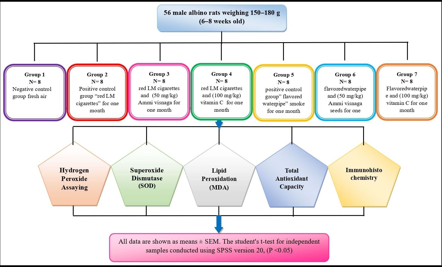
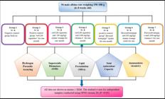
In this study, 56 male albino rats (Rattus ratas), weighing 150–180 g at 6–8 weeks of age, were employed (Alsarhan et al., 2024) were used in this experiment. They were bought from the animal center at the University of Science and Technology and housed in the best possible food and temperature requirements. Seven groups of eight rats each were assigned to the rodents at random. Groups 2-4 were exposed to the most widely available brands of cigarettes in Jordan (red LM cigarettes) at a daily of 1 cigarette per rat for 30 days, while groups 6-7 were exposed side stream to water-pipe smoke made from the whole combustion of 20 g of moassal for 30 days, one session per day. Group 1: The control group was simply exposed to fresh air. An additional month of non-exposure (cessation) was given to the animals in an attempt to facilitate their recovery from the effects of smoking cigarettes and using water pipes. Following each phase, biochemical tests and immunohistochemical and histological studies were conducted. The following was done to divide the groups exposed to cigarette smoke (see Scheme 1):
-
Group 1: The negative control group consisted of 8 rats exposed only to fresh air.
-
Group 2: This positive control group consisted of eight rats that were exposed to red LM cigarettes for a month, followed by a month-long recuperation time.
-
Group 3: This group received treatment with Ammi visnaga seeds (50 mg/kg) for one month concurrently with exposure to red LM cigarettes. A further one-month recuperation period was observed.
-
Group 4: This group was exposed to red LM cigarettes and at the same time treated with (100 mg/kg) vitamin C (ascorbic acid) for one month, followed by a recovery period for one month.
-
Group 5: This positive control group was exposed to water pipe smoke for one month, followed by a one-month recuperation period.
-
Group 6: This group received a one-month treatment of Ammi visnaga seeds at a dose of 50 mg/kg and was exposed to a water pipe. A subsequent one-month recuperation time was observed.
-
Group 7: This group was exposed to a flavored water-pipe and at the same time treated with (100 mg/kg) vitamin C (ascorbic acid) for one month, followed by a one-month recovery period. The procedures used on the experimental animals were observed by the scientific committee of the School of Science/The University of Jordan, which approved the study, which expressed following international standard principles for laboratory animal use and care (NIH Publication No. 8023).
3. Preparation of Antioxidant
3.1. Extraction of Ammi visnaga seeds
The seeds of Ammi visnaga were bought from the local market, and a taxonomist from the Faculty of Science/The University of Jordan approved their classification. After the seeds had been ground into a fine powder, 1500 mL of distilled water and 50 g of the powder were combined and boiled. The mixture was allowed to cool to ambient temperature before filtering through a Buckner funnel. The filtrate was administered orally (gavage) via the rat's vianaso-gastric tube (Khan et al., 2001).
3.2. Vitamin C (Ascorbic Acid)
Rats received a daily dose of Vitamin C (Sigma Chemical Company dissolved in 1 mL of water through a gastric tube. (Sigma Chemical Company) (Ola-Davies et al., 2014).
3.3. The digital smoking machine
Shraideh and Najjar (2011) developed the digital smoking device that was used in the experiment, which is useful for exposing rats to water pipes or cigarette smoke (Shraideh and Najjar, 2011).
4. Measuring Technology
4.1. H2O2 concentration
One metabolic consequence of reactive oxygen, hydrogen peroxide (H2O2), is a critical regulator of several oxidative stress-related states. A very sensitive, straightforward, easy-to-read colorimetric method for determining H2O2 in biological samples is the hydrogen peroxide assay. The OxiRed Probe interacts with H2O2 in the presence of Horse Radish Peroxidase (HRP) to generate a product that is red-fluorescent (Ex/Em=535/587 nm) and has a color (max = 570 nm)(Vicente et al., 2015).
4.2. Superoxide Dismutase (SOD)
The reduction of superoxide anions to hydrogen peroxide, which is thereafter eliminated by catalase enzymes, is catalyzed by superoxide dismutase (SOD). Cells need to protect themselves against the harmful effects of oxygen radicals. Nitric oxide (NO) and superoxide anion (SOD) fight for the superoxide anion, which inactivates NO to create peroxy nitrite. It has been discovered that superoxide dismutase and polyethylene glycol (PEG) covalently conjugate to lengthen the circulatory half-life and offer protection against partly reduced oxygen species. The measurement of superoxide dismutase activity is the suppression of the superoxide radical's rate of cytochrome c reduction, which is seen at 550 nm (Ighodaro and Akinloye, 2018; Beckman et al., 1988).
4.3. Total antioxidant capacity
The assay can detect small molecules when our patented Protein Mask is present or measure small molecules with antioxidants and proteins. Proteins and tiny molecules both convert the Cu2+ ion to Cu+. By blocking protein's ability to reduce Cu2+, the Protein Mask makes it possible to analyze small molecule antioxidants. A colorimetric probe is used to chelate the reduced Cu+ ion, producing a wide absorbance peak at around 570 nm that is proportionate to the overall antioxidant capacity (Alsarhan et al., 2024; Khan et al., 2001).
4.4. Lipid peroxidation (MDA)
Lipid peroxidation, a marker for oxidative stress and a sensitive method for detecting malondialdehyde (MDA) in a variety of samples, is the breakdown of lipids brought on by oxidative damage. MDA is a naturally occurring by-product of lipid peroxidation, and its measurement is commonly employed as a diagnostic for lipid peroxidation together with 4-hydroxynonenal (4-HNE). An MDA-TBA adduct is produced when the sample's MDA and thiobarbituric acid (TBA) combine. Colorimetric measurement of the MDA-TBA adduct is simple and requires an OD of 532 nm (Qiao et al., 2016).
4.5. Immunohistochemistry
Tissue specimens from paraffin wax-embedded tissue blocks were cut at 3-4 µm, mounted on coated slides, air-dried, and baked at 70 °C for 30 minutes. Sections of tissue were deparaffinized by immersing slides in xylene for forty minutes. And then drying in the air. Endogenous peroxidase was inactivated by incubating the slice in a mixture of 0.1-1% hydrogen peroxide for thirty minutes. The slides were washed by running tap water for 10 min. The tissue section was incubated for one hour in 1.5% blocking serum in PBS to block non–specific reactions. The slides were washed in Phosphate Buffer Saline (PBS) solution for 5 min. 3, 3-Diaminobenzidine (DAB) chromagen prepared and was applied to the tissue, and the slides were washed in water for 10 min, followed by counterstaining in Hematoxylin Meyers for 2 min. Slides were dehydrated in xylene and then mounted using DPX for further observation; slides were examined microscopically. By utilizing the outcomes obtained from Adobe Photoshop version 22.0.0, we examined the appearance of P53 variations. Antibodystained section micrographs were analyzed using pixels. The pixels revealed the presence of the biomarker (brown) and residual tissue (blue).
To evaluate the expression ratio, we computed the product of the total pixel count (including both blue and brown pixels) and the pixel count corresponding to the biomarker color. The data analysis was conducted using the Statistical Analysis in Social Science (SPSS) version 23.0 software. An independent t-test was employed to determine the group means. A p-value below 0.05 indicates the statistical significance of the difference. The results were utilized to determine the average ± deviation of p53 isoforms for each group.
4.6. Statistical analysis
Applying SPSS version 22, data management and analysis were carried out. In the physiological section, all data are shown as means ± SEM. The student's t-test for independent samples was used to determine the significance of differences in mean response values between control and experimental tissues. A change with a probability less than 0.05 (P < 0.05) is significant. The analysis was carried out using Graph Pad Prism 8 software. Normal distribution was assessed using the D’Agostino–Pearson omnibus normality test. Parametric data were evaluated using one-way ANOVA with Sidak’s post hoc and Dunnett test. Statistical significance was represented as follows: * for p < 0.05, ** for p < 0.01, *** for p < 0.001, and **** for p < 0.0001.
5. Results
5.1. The effect of Ammi visnaga seeds on oxidative stress biomarkers of rats exposed to sub-chronic smoking
A statistical investigation of specific oxidative biomarker activities (H2O2, TAC, MDA, and SOD) following sub-chronic exposure to waterpipe and cigarette smoking, as well as following cessation with ongoing Ammi visnaga seed treatment, is presented in Tables 1, 2, 3, and 4. The result of treating Ammi visnaga seeds on the H2O2 level is shown in Table 1. Observing both cigarette and water-pipe smoking, the treatment has a considerable impact. The control group had a significant increase in H2O2 levels to 0.05± 0.01 ng/mL despite being subjected to both water-pipe (0.25 ± 0.02, p = 0.0) and cigarette (0.29 ± 0.0, p = 0.0) smoking. Ammi visnaga seed treatment considerably decreased H2O2 values in the water-pipe and cigarette smoking groups (0.20 ± 4.8 ng/mL, p = 0.4 and 0.19± 0.03, respectively). After quitting smoking, all smoke-exposed groups showed a significant decrease in H2O2 levels.
Table 2: shows the effect of treatment of Ammi visnagaon TAC level. The treatment indicates no significant effect on either cigarette or water-pipe smoking.
Table 3 illustrates the impact of Ammi visnaga seed treatment on MDA levels. The treatment has an important effect on waterpipe and cigarette smoking. The control group's MDA level was 0.09 ± 0.1 nmol/µL, and it significantly increased after being exposed to water-pipe and cigarette smoking (0.36± 0.13, p= 0.00 and 0.34 ± 0.15, p= 0.00, respectively). The MDA levels in the water-pipe smoking group (0.15 ± 0.02 nmol/µL, p=0.02) and cigarette group (0.11± 0.01, p=0.02) were considerably reduced by treatment with Ammi visnaga seeds. All smoke-exposed groups had a substantial drop in MDA levels after quitting.
Table 4: indicates the effect of treatment of Ammi visnaga seeds on the %SOD inhibition rate. It appears that the treatment significantly reduces the use of waterpipes and cigarettes. The control group's level of SOD inhibition rate was 98 ± 0.7, and it substantially dropped after being exposed to water-pipe smoking (86 ± 1.7, p= 0.03) and cigarette smoking (74 ± 1.8, p= 0.01). The percentage SOD inhibition rate increased considerably (93 ± 1.3, p= 0.02) in the cigarette group after treatment with Ammi visnaga seeds but not significantly (89 ± 1.5, p= 0.71) in the water pipe smoking group. Following cessation of smoking, the level of % SOD inhibition rate increased significantly in all smoke-exposed groups.
5.2. The effect of Vitamin C (Ascorbic Acid) on oxidative stress biomarkers of rats exposed to sub-chronic smoking
A statistical analysis of certain oxidative biomarker activity (H2O2, TAC, MDA, and SOD levels) following intermittent exposure to water pipe and cigarette smoke, as well as following cessation with ongoing vitamin C (ascorbic acid) treatment, is presented in Tables 5, 6, 7, and 8.
Table 5 shows how ascorbic acid treatment affects H2O2 levels. The treatment appears to have a considerable influence on cigarette and water pipe. The control group's H2O2 level was 0.05 ± 0.01 ng/mL, which considerably increased after exposure to cigarette smoking (0.29 ± 0.0, p = 0.0) and water-pipe smoking (0.25 ± 0.02, p = 0.0). Ascorbic acid treatment has no noticeable impact on H2O2 levels in the water-pipe smoking group (0.20 ± 0.01 ng/mL, p = 0.92) or cigarette smoking group (0.20 ± 0.0, p = 0.84). The levels of H2O2 did not change appreciably in any of the smoke-exposed groups.
Table 6 shows the effect of the treatment of ascorbic acid on TAC level. The treatment shows no significant effect on either cigarette or water-pipe smoking.
Table 7 shows how ascorbic acid treatment affects MDA levels. The treatment has a considerable effect on cigarette and water pipe smoking. MDA levels in the control group were 0.09±0.1 nmol/µL, but significantly increased following exposure to cigarette smoking (0.36±0.13, p=0.00) and water pipe smoking (0.34±0.15, p=0.00). Ascorbic acid treatment significantly reduced MDA levels in cigarette smokers (0.11±0.01, p=0.01) and water pipe smokers (0.11±0.03 nmol/µL, p = 0.04). Subsequently giving up smoking, the level of MDA decreased markedly in all smoke-exposed groups.
Table 8 shows the effect of the treatment of ascorbic acid on %SOD inhibition rate. The treatment shows a significant effect on both cigarette and water-pipe smoking. The level of %SOD inhibition rate in the control group was 98± 0.7 and significantly decreased following exposure to cigarette smoking (74 ± 1.8, p = 0.01), and water-pipe smoking (86 ± 1.7, p = 0.03). Ascorbic acid treatment significantly raised the %SOD inhibition rate in both the cigarette (97 ± 1.3, p = 0.01) and water-pipe smoking groups (91 ± 1.1, p = 0.04). After quitting smoking, the percentage of SOD inhibition rate increased significantly in all smoke-exposed groups.
5.3. Immunohistochemistry results
5.3.1. Expression of p53 of heart ventricle tissues
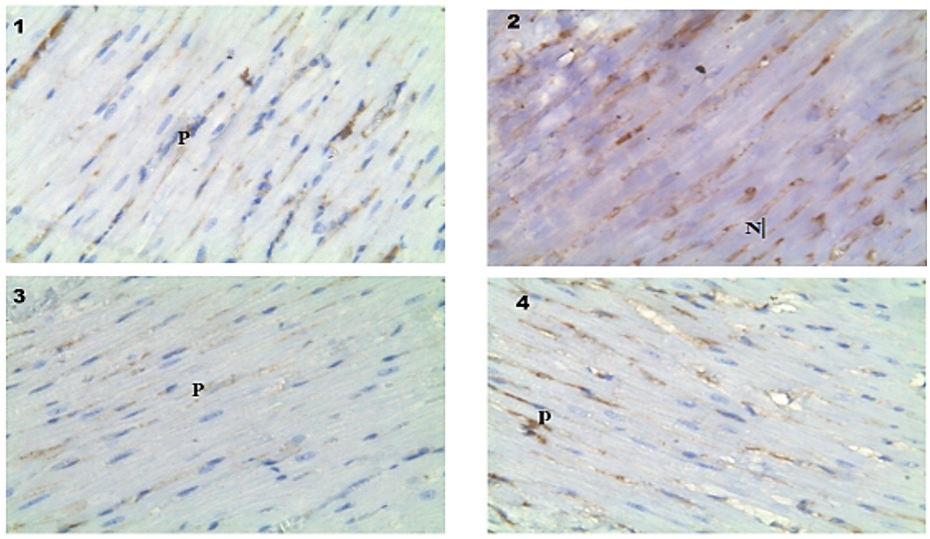
Under physiological conditions, the expression of p53 in the heart ventricle tissues Figure 1-1.
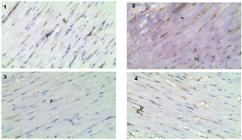
Expression of p53 in the heart tissues among study groups/ cigarette smoking, (1): Expression of p53 in heart ventricle/ Normal group. (2): Expression of p53 in the heart ventricle of cigarette smoking exposed rat. The expression was observed in the nucleus (N). (3): The effect of using Ammi visnaga seeds on the expression of p53 on the heart ventricle of cigarette smoking. (4): The effect of treatment with vitamin C on expression of p53 in the heart ventricle of cigarette smoking exposed rat. The expression of p53 still exists (P). (40× magnification).
Figure 1-2 shows an increasing expression of p53 in heart tissues of underexposed rats to cigarette smoking. Critical Figures 1-3, and 4, show the effect of treating the rats with vitamin C and Ammi seeds extract on the expression of p53 in heart ventricle tissues, which decreases upon treatment with Vitamin C due to its antioxidant activity. Also, Ammi seed extract decreases the re-expression of p53, which was observed in the nucleus. It seems that the treatment with vitamin C was less effective than the treatment with Ammi visnaga seeds in lowering the expression of p53. As demonstrated in Figure 2-2, Exposure to water-pipe smoking induced a noticeable expression of p53 in the heart ventricle. While treating the rats with vitamin C and Ammi seed extract decreased on the expression of p53 in heart ventricle tissues (see Figures 2-3, and 2-4)

Expression of p53 in the heart tissues among study groups / Water-pipe smoking. (1): Expression of p53 in heart ventricle/ Normal group. (2): Expression of p53 in the heart ventricle of water-pipe smoking exposed. (3): The effect of treatment with Ammi visnaga seeds on the expression of p53 in the heart ventricle of water-pipe smoking exposed rat. (4): The effect of treatment with vitamin C on expression of p53 in the heart ventricle of water-pipe smoking exposed rat.
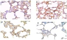
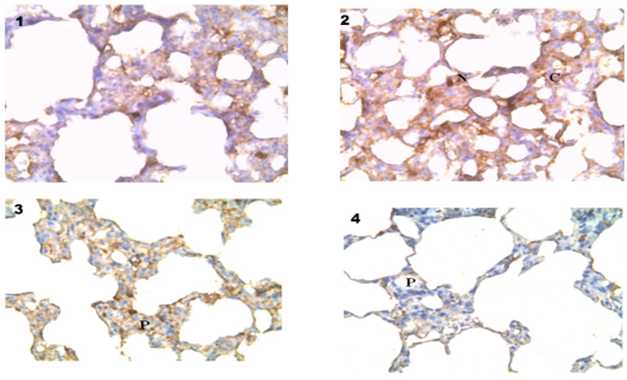
Expression of p53 in the lung tissues among study groups/cigarette smoking. (1): Expression of p53 in lung tissues / Normal group (2): Expression of p53 in the lung of cigarette smoking exposed rat. The expression was observed in the nucleus (N). (3): The effect of treatment with Ammi visnaga seeds on the expression of p53 in the lung of water-pipe exposed rat. The expression of p53 (P) (4): The effect of treatment with vitamin C on expression of p53 in the lung of cigarette-smoking exposed rats. The expression of p53 still exists (P).

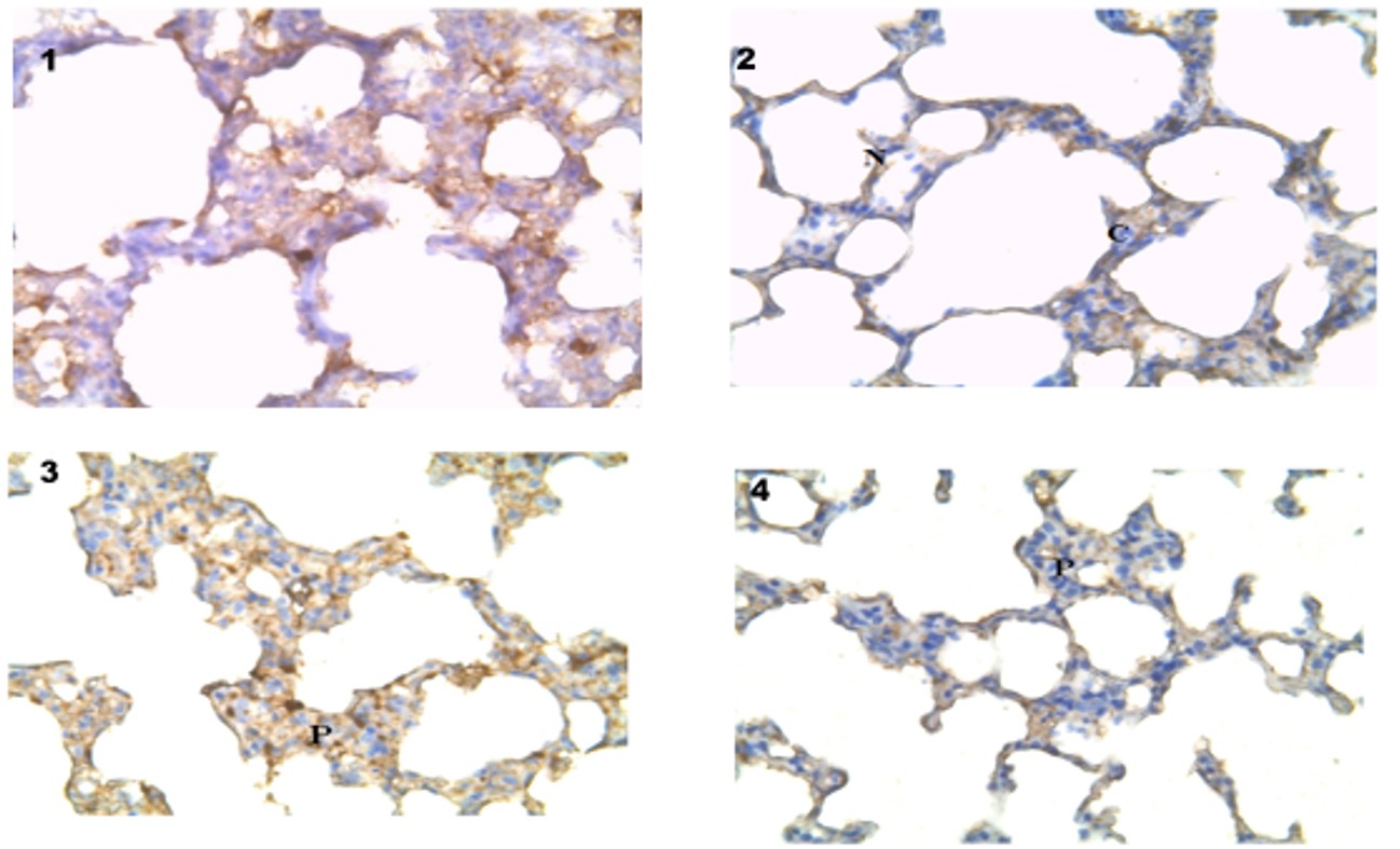
Expression of p53 in the lung tissues among study groups / Water-pipe smoking. (1): Expression of p53 in lung tissues / Normal group (2): Expression of p53 in the lung cells of water-pipe smoking exposed rat. The expression was observed in the nucleus and cytoplasm of lung cells (N& C). 400X. (3): The effect of treatment with Ammi visnaga seeds on the expression of p53 in the lung of water-pipe exposed rat. The expression of p53 (P). (4): The effect of treatment with vitamin C on expression of p53 in the lung of rats exposed to water-pipe smoking. The expression of p53 (P).
5.3.2. Expression of p53 of lung tissues
The expression of p53 in the lungs exhibited some re-expression of p53 under physiological conditions (Figure 3-1). As demonstrated in Figure 3-2, there was an induced expression of p53 due to exposure to cigarette smoking in the hepatic tissues, which was observed in the nucleus. The treatment of vitamin C for cigarette smoking-exposed rats was able to lower the expression of p53 partial smoke exposure in the hepatic-tissues (Figures 3-3, and 3-4). It seems that the treatment with vitamin C was less effective than the treatment with Ammi seed extract in lowering the expression of p53 in the lung tissues of rats exposed to cigarette smoking.
Exposure to water-pipe smoking induced a noticeable expression of p53 in the lungs of rats (Figure 4-2). The treatment with Ammi seed extract exposed to water-pipe smoking showed ameliorating effects of Ammi extract that reduced expression of p53 in the lung, but slight re-expression still presents. Figure 4-4 shows treatment with vitamin C reduced expression in lung tissues as related to water-pipe smoking.
To better understand the impact of vitamin C and Ammi seed extract on lung tissue at the molecular level, we examined the expression of p53 markers in all experimental groups. When the p53 levels were compared to the G1 control group, it was found that smoking waterpipes (G5) and cigarettes (G2) increased the expression of p53 significantly (p < 0.0001) in comparison to the control. After being exposed to cigarette smoking, we noticed a significant decrease in p53 expression in rats treated with vitamin C (G4) (p = 0.048) or Ammi seed extract (G3) (p = 0.044) (see Figure 5A). In lung tissues, we also observed a significant decrease in p53 expression in rats treated with Ammi seed extract (G6) (p = 0.006) or vitamin C (G7) (p = 0.019) after being exposed to water-pipe smoking.

P53 marker expression of experimental groups. A: P53 expression in lung tissue. B: P53 expression in heart tissues. All data are presented as mean ± SEM. one-way ANOVA was analyzed with Sidak’s post hoc multiple comparisons test. * p < 0.05,** p < 0.01, *** p < 0.001, and ****p < 0.0001.
In heart tissues, both cigarette smoking (G2) (p = 0.0001) and water-pipe smoking (G5) (p < 0.0001) significantly increase the expression of p53. Moreover, there is no significant effect of treating rats with either Vitamin C or Ammi extract on the expression of p53 when compared with the control (G1) (figure 5B).
Furthermore, after a month of exposure to cigarette smoking, researchers found a highly significant difference (p < 0.0001) in the expression of p53 in the lung tissue of rats treated with Ammi seed extract (G3) or vitamin C (G4). This was in comparison to the expression of p53 during cigarette smoking (G2). When comparing the p53 expression levels during water-pipe (G5) to the expression of p53 in lung tissue of rats treated with Ammi seed extract (G6) or Vitamin C (G7) after being exposed to water-pipe smoking for one month, researchers observed a significant decrease in p53 expression with Ammi seed extract treatment (p = 0.01) and a very significant decrease with vitamin C treatment (p = 0.0034) (Figure 6).

P53 marker expression of experimental groups in lung tissues. A: P53 expression levels during cigarette smoking compared with treatment with Ammi seed extract or Vitamin C. B: P53 expression with water-pipe smoking compared with treatment with Ammi seed extract or Vitamin C. All data are presented as mean ± SEM. one-way ANOVA was analyzed with Sidak’s post hoc multiple comparisons test. * p < 0.05, ** p < 0.01, *** p < 0.001, and ****p < 0.0001.
In heart tissue, there is a significant reduction in p53 expression when compared to the p53 expression during cigarette smoking (G2) and p53 expression during treatment of Ammi seed extract (G3) and vitamin C (G4) (both p= 0.0001), also there is a significant decrease in p53 expression when compared to the p53 expression during water-pipe smoking (G5) and p53 expression during treatment of Ammi seed extract (G6) and vitamin C (G7)(both p < 0.0001)(Figure 7).

P53 marker expression of experimental groups in heart tissues. A: P53 expression levels during cigarette smoking compared with treatment with Ammi seed extract or Vitamin C. B: P53 expression with water-pipe smoking compared with treatment with Ammi seed extract or Vitamin C. All data are presented as mean ± SEM. one-way ANOVA was analyzed with Sidak’s post hoc multiple comparisons test. * p < 0.05,** p < 0.01, *** p < 0.001, and ****p < 0.0001.
6. Discussion
This work addresses the rising global incidence of smoking and its dangers, which include cardiovascular, pulmonary, and cancer-related diseases. Nicotine intake in rats disturbed the antioxidant defense in rats, increased lipid peroxidation, and reduced the antioxidants in tissues (Abdel-Hamid, 2018). There is a need to investigate the detrimental effects of smoking on biochemical and histopathological levels, as well as the efficacy of employing antioxidant-rich natural products such as vitamin C and Ammi visnaga seeds to alleviate smoking-related side effects.
Medicinal plants are rich in physiologically active substances that exhibit a broad range of molecular diversity. It is recognized that these sources aid in creating novel medications (Alsarhan et al., 2024; Altemimi et al., 2017). It has distinct efficacy, safety, and economic effects on conditions. Moreover, much research has found that natural medicinal plants exhibit anti-cancer and anti-inflammatory activity (Nahar et al., 2023; Kim and Kim, 2015). Polyphenolic chemicals, which include flavonoids and phenols, are among the most efficient in this respect. Despite aiding in capturing free radicals, they exhibit anti-inflammatory and antibacterial characteristics (Raeeszadeh et al., 2022; Kim & Kim, 2015). Scientists have recently become interested in the vast variety of impacts of free radicals. Consequently, the function of free radicals in the pathophysiology of several disorders, especially cancer, has been demonstrated (Ríos-Arrabal et al., 2013). It is also proven that antioxidants play a considerable function in plant compounds that limit the beginning or release of oxidation chain reactions, which can halt or delay the oxidation of other molecules as well as lipids (Tungmunnithum et al., 2018; Ríos-Arrabal et al., 2013; Raeeszadeh and Fallah, 2018).
The current study found that smoking significantly raised the levels of oxidative stress indicators such as H2O2 and MDA, as well as the rate of % SOD inhibition, as likened to the control group. However, the level of TAC did not differ significantly across all smoke-exposed treatment groups. It has been shown that smoke contains radicals per puff and could generate H2O2 and superoxide radicals (Park et al., 2021). Additionally, it has been noted that smoking raises MDA levels (Xi et al., 2021). Kashinakunti et al. (2011) reported significantly higher levels of MDA in smokers contrasted with non-smokers (Kashinakunti et al., 2011). MDA is most commonly used as a biomarker to assess oxidative stress in a variety of health conditions, including cancer and psychiatric disorders, to chronic obstructive pulmonary disease, asthma, and cardiovascular diseases (Park et al., 2021; Kashinakunti et al., 2011). Cigarette smoke contains considerable amounts of reactive oxygen species (ROS) and reactive species such as nitrogen alkoxyl and peroxyl radicals, as well as smokers and secondhand smoke (Park et al., 2021; Kashinakunti et al., 2011). Other studies have found that this is caused by an elevated amount of lipid peroxidation. (Xi et al., 2021; Kashinakunti et al., 2011). Through smoking sessions, free radicals are generated and activate inflammatory cells ending with large amounts of ROS(Park et al., 2021; Kashinakunti et al., 2011). Overexposure to ROS can harm proteins and DNA, which may impact aging, heart disease, cancer, and other diseases (Hong et al., 2024; Essick and Sam, 2010).
Cells use different pathways to get rid of ROS or adjust ROS levels to help with important cell functions like apoptosis. Even though the process of apoptosis triggered by p53 is connected to the p53-dependent increase in ROS, the normal expression of p53 leads to various antioxidant reactions that can reduce oxidative stress (Maddocks and Vousden, 2011). Under normal conditions or slight stress, p53 enhances the antioxidant response and protects cells from potential oxidative injury, as well as that caused by p53-dependent activation of oxidative phosphorylation. However, under severe stress, p53 can use its capability to promote ROS to facilitate apoptosis (Essick and Sam, 2010).
With an imbalance between oxidants and antioxidants, smokers are more likely to experience oxidative stress (Astori et al., 2022). We conducted immunohistochemical analysis to examine p53 expression in heart and lung tissues using different smoking methods, including cigarettes and water pipes. The importance of our data comes from the role that the p53 gene plays as a tumor suppressor gene, which implies that its expression prevents the formation of tumors. Within the cell, the p53 protein binds DNA and acts to stimulate another gene ending with the production of a protein called smoke-exposed p21 which, in turn, inhibits further progression of the real stage of cell divisions (Aubrey et al., 2016; Feroz & Sheikh, 2020). The results indicated a very low-level expression of p53 in the heart ventricle tissue and moderate expression in the lung tissue (control group). In this study, when rats were exposed to cigarette smoking or water-pipe smoking, it led to a significant increase in the p53 expression of both lung and heart ventricle tissues, by the damaging effect of smoking, either cigarette smoking or water-pipe smoking on the cellular environment is obvious. Different mechanisms have been suggested to explain this increase, with one of them being Heitzer et al.(Heitzer et al., 2000) which provides the first evidence of how smoking contributes to increasing ROS formation and dysfunctional uncoupled eNOS. This highlights the key function of ROS in lowering vascular NO bioavailability and promoting endothelial dysfunction.
In the existing work, vitamin C was shown to exacerbate the damaging effects of nicotine. The administration of 100mg vitamin C / kg of rats decreased the expression of p53 comparable to the rat exposure to cigarette or water pipe smoking due to its interaction with the hydroxyl radical, superoxide, and singlet oxygen due to its hydrophilic antioxidant properties and scavenging of reactive oxygen species (Abdel-Hamid, 2018). Furthermore, vitamin C significantly improved the histopathological and ultrastructure changes in lung and heart ventricular tissues due to nicotine exposure.
On the other hand, treatment with Ammi visnaga seeds extract showed good expression against damaging molecular effects of p53. The present findings of histopathological studies were stable with the previous biochemical data, revealing that treated rats showed degenerative lung and heart alterations. Ammi visnaga antioxidant and free radical scavenging properties may assist in relieving these variations in the lung and heart tissues.
Our study found that increased biomarker volume (biomarker effects) is associated with histological abnormalities in the lung and heart, as well as p53 expression in smoker-exposed rats.
7. Conclusion
Our findings verified that smoking water-pipes or cigarettes had negative health impacts. We investigated the effects of tobacco use on several tissues at different levels, including lung and heart histological changes. We showed the detrimental effects of smoking on tissue at all research levels. Treatment with either Ammi visnaga seeds or vitamin C extract showed ameliorative effects against the adverse effects of smoking using all smoke-exposed previous techniques.
References
-
ABDEL-HAMID, G.A., 2018. Ameliorative effect of vitamin C on nicotine-induced histological and ultrastructural changes in zona fasciculata in albino rats. MOJ Anatomy & Physiology, vol. 5, no. 2, pp. 120-125. http://doi.org/10.15406/mojap.2018.05.00175
» http://doi.org/10.15406/mojap.2018.05.00175 -
ADDISSOUKY, T.A., EL SAYED, I.E.T., ALI, M.M.A., WANG, Y., EL BAZ, A., ELARABANY, N. and KHALIL, A.A., 2024. Oxidative stress and inflammation: elucidating mechanisms of smoking-attributable pathology for therapeutic targeting. Bulletin of the National Research Center, vol. 48, no. 1, pp. 16. http://doi.org/10.1186/s42269-024-01174-6
» http://doi.org/10.1186/s42269-024-01174-6 -
ALSARHAN, A.A., KHWALDEH, A.S., AL-SHAWABKEH, J.D., SHOIAB, A.A., AL-SHDEFAT, R., AL-FAWAEIR, S. and YOUSEF, I., 2024. Investigating the hepato-protective properties of chamomile oil and olive leaves extracts against ribociclib-induced hepatotoxicity. Brazilian Journal of Biology = Revista Brasileira de Biologia, vol. 84, pp. e287535. http://doi.org/10.1590/1519-6984.287535 PMid:39442156.
» http://doi.org/10.1590/1519-6984.287535 -
ALSARHAN, A.A., KHASHROUM, A.O., AL-SHAWABKEH, J.D., ABABNHEH, S.K., KHWALDEH, A., LABAN, N.M.F.A. and BANI-KHALED, A., 2024. Levels of expression of Hsp70 and iNOS and effect of Artemisia Sieberi (A. herba-alba) on activity of Hypothalamic-Pituitary-Thyroid (HPT). Axis in Diabetic Rats., vol. 17, no. June, pp. 999-1008. http://doi.org/10.13005/bpj/2917
» http://doi.org/10.13005/bpj/2917 -
ALTEMIMI, A., LAKHSSASSI, N., BAHARLOUEI, A., WATSON, D.G. and LIGHTFOOT, D.A., 2017. Phytochemicals: Extraction, isolation, and identification of bioactive compounds from plant extracts. Plants, vol. 6, no. 4, pp. 42. http://doi.org/10.3390/plants6040042 PMid:28937585.
» http://doi.org/10.3390/plants6040042 -
AOURABI, S., DRIOUCH, M., SFAIRA, M., MAHJOUBI, F., HAMMOUTI, B. and EMRAN, K.M., 2019. Influence of phenolic compounds on antioxidant and anticorrosion activities of Ammi visnaga extracts obtained ultrasonically in three solvent systems. International Journal of Electrochemical Science, vol. 14, no. 7, pp. 6376-6393. http://doi.org/10.20964/2019.07.02
» http://doi.org/10.20964/2019.07.02 -
ASENSI-FABADO, M.A. and MUNNÉ-BOSCH, S., 2010. Vitamins in plants: occurrence, biosynthesis and antioxidant function. Trends in Plant Science, vol. 15, no. 10, pp. 582-592. http://doi.org/10.1016/j.tplants.2010.07.003 PMid:20729129.
» http://doi.org/10.1016/j.tplants.2010.07.003 -
ASTORI, E., GARAVAGLIA, M.L., COLOMBO, G., LANDONI, L., PORTINARO, N.M., MILZANI, A. and DALLE-DONNE, I., 2022. Antioxidants in smokers. Nutrition Research Reviews, vol. 35, no. 1, pp. 70-97. http://doi.org/10.1017/S0954422421000093 PMid:33926594.
» http://doi.org/10.1017/S0954422421000093 -
AUBREY, B.J., STRASSER, A. and KELLY, G.L., 2016. Tumor-suppressor functions of the TP53 pathway. Cold Spring Harbor Perspectives in Medicine, vol. 6, no. 5, pp. a026062. http://doi.org/10.1101/cshperspect.a026062 PMid:27141080.
» http://doi.org/10.1101/cshperspect.a026062 -
BECKMAN, J.S., MINOR JUNIOR, R.L., WHITE, C.W., REPINE, J.E., ROSEN, G.M. and FREEMAN, B.A., 1988. Superoxide dismutase and catalase conjugated to polyethylene glycol increases endothelial enzyme activity and oxidant resistance. The Journal of Biological Chemistry, vol. 263, no. 14, pp. 6884-6892. http://doi.org/10.1016/S0021-9258(18)68727-7 PMid:3129432.
» http://doi.org/10.1016/S0021-9258(18)68727-7 -
BONNIE, R.J., STRATTON, K. and KWAN, L.Y., 2015. The effects of tobacco use on health. In: R.J. BONNIE, K. STRATTON and L.Y. KWAN, eds. Public health implications of raising the minimum age of legal access to tobacco products. Washington, DC: National Academies Press. http://doi.org/10.17226/18997
» http://doi.org/10.17226/18997 -
BUDANOV, A.V., 2014. The role of tumor suppressor p53 in the antioxidant defense and metabolism. Sub-Cellular Biochemistry, vol. 85, pp. 337-358. http://doi.org/10.1007/978-94-017-9211-0_18 PMid:25201203.
» http://doi.org/10.1007/978-94-017-9211-0_18 -
BUTLER, M., D’ANGELO, S., PERRIN, A., RODILLAS, J., MILLER, D., ARADER, L., CHANDERENG, T., CHEUNG, Y.K., SHECHTER, A. and DAVIDSON, K.W., 2023. A series of remote melatonin supplement interventions for poor sleep: protocol for a feasibility pilot study for a series of personalized (N-of-1) trials. JMIR Research Protocols, vol. 12, pp. e45313. http://doi.org/10.2196/45313 PMid:37535419.
» http://doi.org/10.2196/45313 -
BYRNES, Y.M., CIVANTOS, A.M., GO, B.C., MCWILLIAMS, T.L. and RAJASEKARAN, K., 2020. Effect of the COVID-19 pandemic on medical student career perceptions: a national survey study. Medical Education Online, vol. 25, no. 1, pp. 1798088. http://doi.org/10.1080/10872981.2020.1798088 PMid:32706306.
» http://doi.org/10.1080/10872981.2020.1798088 -
CHÍRICO, M.T.T., BEZERRA, F.S., GUEDES, M.R., SOUZA, A.B., SILVA, F.C., CAMPOS, G., DE NORONHA, S.R., MESQUITA, L.B.T., REIS, T.O., CANGUSSÚ, S.D., CHIANCA-JR, D.A. and DE MENEZES, R.C., 2018. Tobacco-Free cigarette smoke exposure induces anxiety and panic-related behaviours in male wistar rats. Scientific Reports, vol. 8, no. 1, pp. 4943. http://doi.org/10.1038/s41598-018-23425-z PMid:29563583.
» http://doi.org/10.1038/s41598-018-23425-z -
DJUARTINA, T., BUDIANTO, I.R., STEVEN, A., STEFANI, M., KAWILARANG, M. and KAWILARANG, M., 2023. The effect of cigarette smoke exposure on rat’s spermatogenesis: A systematic literature review and meta-analysis. European Journal of Inflammation, vol. 21. http://doi.org/10.1177/1721727X231207720
» http://doi.org/10.1177/1721727X231207720 -
ELGAMAL, M.H.A., SHALABY, N.M.M., DUDDECK, H. and HIEGEMANN, M., 1993. Coumarins and coumarin glucosides from the fruits of Ammi majus. Phytochemistry, vol. 34, no. 3, pp. 819-823. http://doi.org/10.1016/0031-9422(93)85365-X
» http://doi.org/10.1016/0031-9422(93)85365-X -
ESAKKY, P., HANSEN, D.A., DRURY, A.M. and MOLEY, K.H., 2015. Cigarette smoke-induced cell cycle arrest in spermatocytes [GC-2spd (ts)] is mediated through crosstalk between Ahr–Nrf2 pathway and MAPK signaling. Journal of Molecular Cell Biology, vol. 7, no. 1, pp. 73-87. http://doi.org/10.1093/jmcb/mju049 PMid:25548370.
» http://doi.org/10.1093/jmcb/mju049 -
ESSICK, E.E. and SAM, F., 2010. Oxidative stress and autophagy in cardiac disease, neurological disorders, aging and cancer. Oxidative Medicine and Cellular Longevity, vol. 3, no. 3, pp. 168-177. http://doi.org/10.4161/oxim.3.3.12106 PMid:20716941.
» http://doi.org/10.4161/oxim.3.3.12106 - FEROZ, W. and SHEIKH, A.M.A., 2020. Exploring the multiple roles of guardian of the genome: P53. The Egyptian Journal of Medical Human Genetics, vol. 21, no. 1, pp. 1-23.
-
FISCHER, B.M., VOYNOW, J.A. and GHIO, A.J., 2015. COPD: balancing oxidants and antioxidants. International Journal of Chronic Obstructive Pulmonary Disease, vol. 10, pp. 261-276. http://doi.org/10.2147/COPD.S42414 PMid:25673984.
» http://doi.org/10.2147/COPD.S42414 - HASHEMI, M., AZARNIA, M., HAJEBRAHIMI, Z. and NEJAD EBRAHIMI, S., 2023. The effect of hydroalcoholic extract of Cichorium intybus leaf on aryl hydrocarbon receptor expression in the testis of Wistar rats exposed to cigarette smoke. Avicenna Journal of Phytomedicine, vol. 13, no. 1, pp. 58-69. PMid:36698732.
-
HEITZER, T., BROCKHOFF, C., MAYER, B., WARNHOLTZ, A., MOLLNAU, H., HENNE, S., MEINERTZ, T. and MÜNZEL, T., 2000. Tetrahydrobiopterin improves endothelium-dependent vasodilation in chronic smokers: evidence for a dysfunctional nitric oxide synthase. Circulation Research, vol. 86, no. 2, pp. E36-E41. http://doi.org/10.1161/01.RES.86.2.e36 PMid:10666424.
» http://doi.org/10.1161/01.RES.86.2.e36 -
HONG, Y., BOITI, A., VALLONE, D. and FOULKES, N.S., 2024. Reactive oxygen species signaling and oxidative stress: transcriptional regulation and evolution. Antioxidants, vol. 13, no. 3. http://doi.org/10.3390/antiox13030312 PMid:38539845.
» http://doi.org/10.3390/antiox13030312 -
IGHODARO, O.M. and AKINLOYE, O.A., 2018. First line defence antioxidants-superoxide dismutase (SOD), catalase (CAT) and glutathione peroxidase (GPX): their fundamental role in the entire antioxidant defence grid. Alexandria Journal of Medicine, vol. 54, no. 4, pp. 287-293. http://doi.org/10.1016/j.ajme.2017.09.001
» http://doi.org/10.1016/j.ajme.2017.09.001 - KAMAL, F.Z., STANCIU, G.D., LEFTER, R., COTEA, V.V., NICULAUA, M., ABABEI, D.C., CIOBICA, A. and ECH-CHAHAD, A., 2022. Chemical composition and antioxidant activity of Ammi visnaga L. essential oil. Antioxidants, vol. 11, no. 2, pp. 347. PMid:35204230.
- KASHINAKUNTI, S.V., KOLLUR, P., KALLAGANADA, G.S., RANGAPPA, M. and INGIN, J.B., 2011. Comparative study of serum MDA and vitamin C levels in non-smokers, chronic smokers and chronic smokers with acute myocardial infarction in men. Journal of Research in Medical Sciences: The Official Journal of Isfahan University of Medical Sciences, vol. 16, no. 8, pp. 993-998. PMid:22279473.
- KHAN, Z.A., ASSIRI, A.M., AL-AFGHANI, H.M.A. and MAGHRABI, T.M.A., 2001. Inhibition of oxalate nephrolithiasis with Ammi visnaga (AI-Khillah). International Urology and Nephrology, vol. 33, no. 4, pp. 605-608. PMid:12452606.
- KIM, J.H. and KIM, J.K., 2015. Protective effect against oxidative stress in medicinal plant extracts. Journal of Radiation Protection, vol. 25, no. 1, pp. 1-37.
-
KIM, J., SONG, H., LEE, J., KIM, Y.J., CHUNG, H.S., YU, J.M., JANG, G., PARK, R., CHUNG, W., OH, C.-M. and MOON, S., 2023. Smoking and passive smoking increases mortality through mediation effect of cadmium exposure in the United States. Scientific Reports, vol. 13, no. 1, pp. 3878. http://doi.org/10.1038/s41598-023-30988-z PMid:36890267.
» http://doi.org/10.1038/s41598-023-30988-z -
LI, J., HUANG, X., LUO, L., SUN, J., GUO, Q., YANG, X., ZHANG, C. and NI, B., 2024. The role of p53 in male infertility. Frontiers in Endocrinology, vol. 15, pp. 1457985. http://doi.org/10.3389/fendo.2024.1457985 PMid:39469578.
» http://doi.org/10.3389/fendo.2024.1457985 -
LIU, X., FAN, L., LU, C., YIN, S. and HU, H., 2020. Functional role of p53 in the regulation of chemical-induced oxidative stress. Oxidative Medicine and Cellular Longevity, vol. 2020, pp. 6039769. http://doi.org/10.1155/2020/6039769 PMid:32190175.
» http://doi.org/10.1155/2020/6039769 -
MADDOCKS, O.D.K. and VOUSDEN, K.H., 2011. Metabolic regulation by p53. Journal of Molecular Medicine, vol. 89, no. 3, pp. 237-245. http://doi.org/10.1007/s00109-011-0735-5 PMid:21340684.
» http://doi.org/10.1007/s00109-011-0735-5 - MENDEL, J.R., BAIG, S.A., HALL, M.G., JEONG, M., BYRON, M.J., MORGAN, J.C., NOAR, S.M., RIBISL, K.M. and BREWER, N.T., 2018. Brand switching and toxic chemicals in cigarette smoke: A national study. PLoS One, vol. 13, no. 1, pp. e0189928. PMid:29324749.
- MOHAMED, Z. and ABDEL-MOTAAL, F.F., 2023. Antioxidant and Antifungal Activities of Ammi visnaga Aqueous Extract Prevent Oxidative Stress Biomarkers and DNA Damage in Experimental Rats Infected with Aspergillus glaucus. The Biological Bulletin, vol. 50, no. 6, pp. 1125-1142.
- MOSER, M.A. and CHUN, O.K., 2016. Vitamin C and heart health: a review based on findings from epidemiologic studies. International Journal of Molecular Sciences, vol. 17, no. 8, pp. 1328. PMid:27529239.
- MUSTAFA, N., BASHIR, A., SOHAIL, R., KUMAR, S., KHATRI, M. and VARRASSI, G., 2023. Knowledge, attitude, and practice of cigarette smoking among medical students of quaid-e-azam medical college, bahawalpur: a web-based cross-sectional study. Cureus, vol. 15, no. 10, pp. e46459. PMid:37927705.
-
NAHAR, J., BOOPATHI, V., RUPA, E.J., AWAIS, M., VALAPPIL, A.K., MORSHED, M.N., MURUGESAN, M., AKTER, R., YANG, D.U., MATHIYALAGAN, R., YANG, D.C. and JUNG, S.K., 2023. Protective effects of Aquilaria agallocha and Aquilaria malaccensis edible plant extracts against lung cancer, inflammation, and oxidative stress: In Silico and In Vitro study. Applied Sciences, vol. 13, no. 10. http://doi.org/10.3390/app13106321
» http://doi.org/10.3390/app13106321 - NIRUMAND, M.C., HAJIALYANI, M., RAHIMI, R., FARZAEI, M.H., ZINGUE, S., NABAVI, S.M. and BISHAYEE, A., 2018. Dietary plants for the prevention and management of kidney stones: preclinical and clinical evidence and molecular mechanisms. International Journal of Molecular Sciences, vol. 19, no. 3, pp. 765. PMid:29518971.
-
OLA-DAVIES, O., AJANI, O.S. and OYEYEMI, M.O., 2014. Spermatozoa morphology and characteristics of Spondias mombin L.(Anacardiaceae) protected male Wistar rats exposed to sodium arsenite. Journal of Veterinary Medicine and Animal Health, vol. 6, no. 2, pp. 63-66. http://doi.org/10.5897/JVMAH2013.0245
» http://doi.org/10.5897/JVMAH2013.0245 - PARK, J.-M., JEONG, H., SEO, Y.-S., DO, V.Q., CHOI, S.-J., LEE, K., CHOI, K.-C., CHOI, W.J. and LEE, M.-Y., 2021. Cigarette smoke extract produces superoxide in aqueous media by reacting with bicarbonate. Toxics, vol. 9, no. 11, pp. 316. PMid:34822707.
-
PENA, K.B., RAMOS, C.O., SOARES, N.P., SILVA, P.F., BANDEIRA, A.C.B., COSTA, G.P., CANGUSSÚ, S.D., TALVANI, A. and BEZERRA, F.S., 2016. The administration of a high refined carbohydrate diet promoted an increase in pulmonary inflammation and oxidative stress in mice exposed to cigarette smoke. International Journal of Chronic Obstructive Pulmonary Disease, vol. 11, no. 1, pp. 3207-3217. http://doi.org/10.2147/COPD.S119485 PMid:28008246.
» http://doi.org/10.2147/COPD.S119485 - PETROVIĆ, A., PLAVŠIĆ-JANJATOVIĆ, I., LISOV, N., ČEBELA, M., ČAKAR, U., STANKOVIĆ, I. and ĐORĐEVIĆ, B., 2023. Antioxidant properties and biological activity of fruit wines. In: Proceedings-1st International Symposium on Biotechnology, 17-18 March 2023, Čačak, Serbia. Čačak: SYMBIOTECH, pp. 405-411.
-
QIAO, Y.-F., GUO, W., LI, L., SHAO, S., QIAO, X., SHAO, J., ZHANG, Q., LI, R. and WANG, L., 2016. Melatonin attenuates hypertension-induced renal injury partially through inhibiting oxidative stress in rats. Molecular Medicine Reports, vol. 13, no. 1, pp. 21-26. http://doi.org/10.3892/mmr.2015.4495 PMid:26531807.
» http://doi.org/10.3892/mmr.2015.4495 - RAEESZADEH, M. and FALLAH, M., 2018. The comparison of the effect of origanum vulgar aqueous extract and vitamin C on the control of cadmium chloride damage in testicular tissue in male rats. Majallah-i Danishgah-i Ulum-i Pizishki-i Babul, vol. 20, no. 8, pp. 44-50.
-
RAEESZADEH, M., BEHESHTIPOUR, J., JAMALI, R. and AKBARI, A., 2022. The antioxidant properties of Alfalfa (Medicago sativa L.) and its biochemical, antioxidant, anti-inflammatory, and pathological effects on nicotine-induced oxidative stress in the rat liver. Oxidative Medicine and Cellular Longevity, vol. 2022, no. 1, pp. 691577. http://doi.org/10.1155/2022/2691577
» http://doi.org/10.1155/2022/2691577 -
RIETJENS, I.M.C.M., LOUISSE, J. and BEEKMANN, K., 2017. The potential health effects of dietary phytoestrogens. British Journal of Pharmacology, vol. 174, no. 11, pp. 1263-1280. http://doi.org/10.1111/bph.13622 PMid:27723080.
» http://doi.org/10.1111/bph.13622 -
RÍOS-ARRABAL, S., ARTACHO-CORDÓN, F., LEÓN, J., ROMÁN-MARINETTO, E., DEL MAR SALINAS-ASENSIO, M., CALVENTE, I. and NÚÑEZ, M.I., 2013. Involvement of free radicals in breast cancer. SpringerPlus, vol. 2, no. 1, pp. 404. http://doi.org/10.1186/2193-1801-2-404 PMid:24024092.
» http://doi.org/10.1186/2193-1801-2-404 - SAMOH, F., ALOO, B. and ISSA, M., 2024. Evaluation of antioxidant activity of tea blends formulated using pineapple peels, ginger and lemon grass. FUW Trends in Science & Technology Journal, vol. 8, no. 3, pp. 100-105.
-
SHARAREH, P., LEILI, T., ABBAS, M., JALAL, P. and ALI, G., 2020. Determining correlates of the average number of cigarette smoking among college students using count regression models. Scientific Reports, vol. 10, no. 1, pp. 8874. http://doi.org/10.1038/s41598-020-65813-4 PMid:32483160.
» http://doi.org/10.1038/s41598-020-65813-4 -
SHARMA, A., 2017. Male infertility; evidences, risk factors, causes, diagnosis and management in human. Annals of Clinical and Laboratory Research, vol. 5, no. 3, pp. 188. http://doi.org/10.21767/2386-5180.1000188
» http://doi.org/10.21767/2386-5180.1000188 - SHRAIDEH, Z.A. and NAJJAR, H.N., 2011. Histological changes in tissues of trachea and lung alveoli of albino rats exposed to the smoke of two types of narghile tobacco products. Jordan Journal of Biological Sciences, vol. 4, no. 4, pp. 219-224.
-
SLAVIN, J.L. and LLOYD, B., 2012. Health benefits of fruits and vegetables. Advances in Nutrition, vol. 3, no. 4, pp. 506-516. http://doi.org/10.3945/an.112.002154 PMid:22797986.
» http://doi.org/10.3945/an.112.002154 -
THIVIYA, P., GAMAGE, A., PIUMALI, D., MERAH, O. and MADHUJITH, T., 2021. Apiaceae as an important source of antioxidants and their applications. Cosmetics, vol. 8, no. 4, pp. 111. http://doi.org/10.3390/cosmetics8040111
» http://doi.org/10.3390/cosmetics8040111 -
TUNGMUNNITHUM, D., THONGBOONYOU, A., PHOLBOON, A. and YANGSABAI, A., 2018. Flavonoids and other phenolic compounds from medicinal plants for pharmaceutical and medical aspects: an overview. Medicines, vol. 5, no. 3, pp. 93. http://doi.org/10.3390/medicines5030093 PMid:30149600.
» http://doi.org/10.3390/medicines5030093 -
VICENTE, C.S.L., IKUYO, Y., SHINYA, R., MOTA, M. and HASEGAWA, K., 2015. Catalases induction in high virulence pinewood nematode Bursaphelenchus xylophilus under hydrogen peroxide-induced stress. PLoS One, vol. 10, no. 4, pp. e0123839. http://doi.org/10.1371/journal.pone.0123839 PMid:25894519.
» http://doi.org/10.1371/journal.pone.0123839 -
WHITE, E., 2016. Autophagy and p53. Cold Spring Harbor Perspectives in Medicine, vol. 6, no. 4, pp. a026120. http://doi.org/10.1101/cshperspect.a026120 PMid:27037419.
» http://doi.org/10.1101/cshperspect.a026120 -
XI, S., ZHAI, X., WANG, Y., GONG, Y., FU, B., GAO, C., GUO, X., LI, Y., WANG, Z., HUANG, S., LU, D., ZHAO, Y., QIAN, L. and WANG, Y., 2021. The Ciji-Hua’ai-Baosheng II formula attenuates chemotherapy-induced anorexia in mice With H22 hepatocellular carcinoma. Frontiers in Pharmacology, vol. 12, pp. 715824. http://doi.org/10.3389/fphar.2021.715824 PMid:34489705.
» http://doi.org/10.3389/fphar.2021.715824 -
ZAKARIA, Z.Z., ALADWI, S.A., BENSLIMANE, F., AL-ABSI, E.S., AL-SHAFAI, M., YALCIN, H.C., KHALIL, A., AL MOUSTAFA, A.E. and AL-ASMAKH, M., 2021. Effect of water-pipe smoking on the normal development of zebrafish. International Journal of Environmental Research and Public Health, vol. 18, no. 21, pp. 11659. http://doi.org/10.3390/ijerph182111659 PMid:34770174.
» http://doi.org/10.3390/ijerph182111659