Essential oil from Otacanthus azureus (Linden) Ronse: preparation of a bioactive nanoemulsion through a low-energy/solvent-free/non-heating method
Abstract
Oil-in-water nanoemulsions are in the spotlight of novelty for viable herbal derivatives, being a valuable strategy for better availability of poor-water soluble compounds. The present study shows the preparation of a nanoemulsion with the essential oil of Otacanthus azureus. The systems were constituted by 90% (w/w) of water, 5% (w/w) of essential oil and 5% (w/w) of surfactant (s). The analysis by gas-chromatograph revealed a predominance of mono- and sesquiterpenes. No major droplet growth was observed for the nanoemulsion prepared with polysorbate 80 during 30 days of storage, remaining around 130 nm. A remarkable improvement of antibacterial activity was observed, especially against S. aureus as follows: MIC = 0.050 and MBC = 0.1 for the essential oil, and MIC – 0.025 and MBC = 0.025 for the nanoemulsion. In conclusion, it must be highlighted that the utilization of a low energy/solvent-free/non-heating method was a suitable ecofriendly approach, suggesting the great potential of this plant based nanoemulsion.
Keywords:
antimicrobial; low cost; Otacanthus azureus; terpenoids
Resumo
Nanoemulsões do tipo óleo em água estão na vanguarda para obtenção de devivados vegetais viáveis, sendo uma estratégia valiosa para melhor disponibilidade de substâncias pouco solúveis em água. O presente trabalho apresenta a preparação de uma nanoemulsão à base do óleo essencial de Otacanthus azureus. Os sistemas foram constituídos por 90% (p/p) de água, 5% (p/p) de óleo essencial e 5% (p/p) de tensoativo (s). A análise por cromatografia em fase gasosa revelou uma predominância de mono- e sesquiterpenos. Não foi observado aumento de gotícula para a nanoemulsão preparada com polisorbato 80 durante 30 dias de armazenamento, permanecendo em torno de 130 nm. Um aumento da atividade antibacteriana foi observado, especialmente frente à S. aureus conforme à seguir: CMI = 0,050 e CMB = 0,1 para o óleo essencial, e CIM – 0,025 e CMB = 0,025 para a nanoemulsão. Salienta-se que a utilização de um método de baixa energia, livre de solvente e sem aquecimento foi uma estratégia amigável ao meio ambiente, sugerindo o potencial biotecnológico dessa espécie vegetal nanoemulsificada.
Palavras-chave:
antimicrobiano; baixo custo; Otacanthus azureus; terpenoides
1. Introduction
The medicinal plants have contributed to the development of new therapeutic strategies due to a wide range of secondary metabolites that they produce. They are recognized by the ability of acting directly or indirectly on living organism, therefore inhibiting or activating molecular and cellular targets (Calixto, 2005). The utilization of phytomedicines and even the folk use of medicinal plants makes necessary continuous research, for better understanding and confirmation of plant action, aiming to reduce undesirable and toxic effects and providing a safe use (Firmo et al., 2011).
Essential oils are one of most common mixtures of natural compounds with this characteristic. They can be extracted from different plant organs and have a wide range of industrial applications (Burt, 2004). The lipophilic nature of essential oils is evidenced by the low water solubility of their compounds. In fact, they are often obtained by steam distillation or hydrodistillation, being separated from the aqueous hydrolate due to the immiscibility of essential oils in water. Therefore, it is worth mentioning the potential of colloidal systems, such oil-in-water nanoemulsions to solve this main problem (McClements and Rao, 2011).
Oil-in-water nanoemulsions are colloidal systems constituted by fine droplets dispersed around an external aqueous phase. The nanoemulsions differ from the microemulsions due to fact that the last are thermodynamically stable (McClements, 2012). However, a main characteristic of this colloidal system is related to its kinetic stability, being also refereed as “approaching thermodynamic stability” due to long-term physical maintenance of characteristics. This high stability is due to small droplets that makes them resistant to gravity forces (Solans and García-Celma, 2017).
Despite the size is specifically important to describe a nanostructure, such nanoemulsions, the exact criteria regarding the threshold to assume that a nanostructure was generated is until unsolved. Opinions and analyses of documents (e.g. European Union) reiterates that merely an upper limit is not appropriate (Bhattacharjee, 2016). Basically, a suitable criterion must consider not only the size, but the intended application, which certainly must differ in any aspect when compared to the bulk material. Despite this issue remain open, specialists consider that an upper limit of 200 nm for diameter (r<100 nm) is appropriate (Solans and Solè, 2012). The low size of internal phase of nanoemulsions makes them a suitable additive for aqueous products. This fact is intrinsically associated to maintenance of desirable appearance of the final product, since the nanoemulsions has a transparent or slightly turbid aspect. Therefore, it would be possible to add lipophilic compounds through the generation of oil-in-water nanoemulsions with no impairment in the aspect, for example, in case of beverages (Rao and McClements, 2011).
A data collection of medicinal plants performed in a riverside community close to Mazagão river (Amapá State, Brazil), an affluent of Amazon River, indicated that several species have been used at this locality as the main strategy for health treatment. People from this locality suffer with absence of basic health assistance, since they are geographically isolated from urban center. Among the species with ethnopharmacology utilization, it can be highlighted the use of Otacanthus azureus. It is popularly known as “copaibinha” and this species was highly representative on the quantitative index of most used species, being cited as an antimicrobial, among other actions (Sarquis et al., 2019).
Otacanthus azureus (Linden) Ronse (syn. Stemodia azurea Linden, Otacacanthus caeruleus Lindley, Tetrapla custauberti Mez) belongs to the family Plantaginaceae. It is a sub-shrub originary from Southeastern region of Brazil and found on North region of this country, where it was naturalized and also used as an ornamental plant (Ronse, 2001). It was introduced on Eastern Amazon by indigenous communities. The most studied derivative from this species is a terpene-rich essential oil. The main constituents previously found were the monoterpenes trans-pinocarveol, myrtenal, pinocarvone and the sesquiterpenes β-copaen-4-α-ol, viridiflorole and α-copaene (Andrade et al., 2006; Houel et al., 2014). This essential oil presented antibacterial, antioxidant (Reddy et al., 2012), antifungal (Houel et al., 2014) and leishmanicidal (Houel et al., 2015) activities.
Bio-prospection of natural products as new biological (e.g. antimicrobials) is a continuous effort (Astolfi Filho et al., 2014). Nowadays, an innovative approach through nanobiotechnology develop a main role on the development of novel formulations (Jaiswal et al., 2015). Nanostructured colloidal systems can modulate the release of compounds (Schaffazick et al., 2003) and even improve the bioactivity. Thus, considering the lack of data in relation to bioactive nanoemulsions prepared with the essential oil from O. azureus, as part of our ongoing bioprospective studies with essential oils and preparation of novel nanoemulsions through ecofriendly methods, the main aims of the present study are developing this type of nanoformulation and evaluate its bioactive potential using gram positive and gram negative bacteria.
2. Material and Methods
2.1. Plant material
The botanical material, constituted by aerial flowering parts of O. azureus, were collected at the municipality of Mazagão, Amapá State, Brazil (00° 06’ 54” S and -51° 17’ 20” W). The identification was performed by comparing the samples with exsiccate located at a regional herbarium. Voucher specimen was deposited at the Herbário IAN of Embrapa of Eastern Amazon under the register number 195972.
2.1.1. Scanning electronic microscopy (SEM)
The leaf and flower from O. azureus were hand free sectionized and directly introduced without any previous treatment on the scanning electronic microscope Tabletop microscope TM3030Plus (Hitachi). The electronic micrographs were obtained using the software TM3030Plus.
2.2. Extraction of the essential oil
Fresh flowering aerial parts (1707.3 g) of O. azureus were crushed with water and placed on a 5 L glass bottom. Then, the plant material was hydrodistillated during 3 h using a Clevenger-type apparatus. After this period, the essential oil was collected and stored protected from light under controlled temperature (4 °C) for further utilization.
2.3. Identification of the essential oil compounds
Gas chromatography-mass spectrometry (GC–MS) analyses was performed on a Shimadzu GC 2010 chromatograph coupled to a mass spectrometry detector (Shimadzu MS2010 Plus) with an electron impact of 70 eV. The capillary column used on the analysis was a of DB-5MS fused silica column (Agilent Advanced J & W, 30 m × 0.25 mm × 0.25 μm). The parameters were as follows: split ratio, 1:20; helium as carrier gas (65 kPa); injection volume, 1.0 μL; injector temperature, 250 °C; detector temperature, 250 °C; initial column temperature, 50 °C for 1 min; increased at a 5 °C min−1 rate to 250 °C. The identification of compounds was performed using the NIST 5.0 equipment library.
2.4. Nanoemulsification
The systems were constituted by 90% (w/w) of water, 5% (w/w) of essential oil and 5% (w/w) of surfactant (s) (Fernandes et al., 2013) at a final mass of 4 g. It was used a low energy, non-heating and solvent-free titration method. Briefly, the oil phase containing the essential oil and single surfactant were homogenized in a magnetic stirrer. Then, deionized water was added dropwise (1 mL/min) under agitation to the oil phase. The nanoemulsions were placed on screw top vials and stored protected from light at room temperature (25 °C).
2.4.1. Parameters of influence on nanoemulsification
Two different systems were prepared by using distinct non-ionic surfactants from the polysorbate series. Polysorbate 20 (HLB = 16.7) or polysorbate 80 (HLB = 15.0) obtained from Praid (SP, Brasil) were used in order to evaluate the influence on nanoemulsification formation and stabilization.
2.4.2. Characterization of the nanoemulsions
The nanoemulsions were characterized by dynamic light scattering (DLS) using a Zetasizer (Nano ZS, Malvern, UK) equipped with a 10 mW “red” laser (λ = 632.8 nm) and the nanodispersions were measured at a 90° scattering detector angle for size measurements. The size of the dispersed droplets is obtained from the Stokes-Einstein equation (Equation 1):
where, Dt is the diffusion coefficient, kB is the Boltzmann’s constant (1.38064852×10⁻²³ J/K), T is the temperature, η is the absolute viscosity and RH is the hydrodynamic radius. Prior to the analysis, the nanoemulsions were diluted in deionized water and results (in triplicate) are expressed as mean ± standard deviation.
2.4.3. Droplet growth (DG)
The droplet growth (DG) was used as a parameter for determining the stability of the nanoemulsions (Mehmood, 2015; Guttoff et al., 2015) through the storage (30 days). The nanoemulsions were analyzed by DLS on the day of preparation and after 1, 21 and 30 days of storage. The DG at different periods was calculated according to the follow equation: DGdx,ty = 100 x [DSdy – DSdx] / DSdx], where DSdy is average droplet size measured in day y, DSdx is average droplet size measured in day x, and dx < dy.
2.5. In vitro antibacterial activity
2.5.1. Microorganisms and sample preparation
The following standard strains were used on the antibacterial assay. Gram-positive bacteria: Staphylococcus aureus (ATCC 6538), Staphylococcus epidermidis. Gram-negative bacteria: Pseudomonas aeruginosa (ATCC 25853), Escherichia coli (ATCC 8739), Klebsiella pneumonie (ATCC 4352) and Salmonella typhi. All strains were obtained from the INCQS/FIOCRUZ (National Institute for Health Quality Control, Rio de Janeiro, Brazil) and maintained in the Laboratory of Quality Control, Bromatology and Microbiology in Amapá Federal University (Brazil). For the tests, all strains were grown in Petri dishes with specific media to each bacterium and they were incubated at 37 °C for 24 h to induce exponential growth. The nanoemulsion was diluted in deionized water and the essential oil was dissolved in a polysorbate 80 aqueous solution (10%, w/v) at the concentration of 1.000 µg mL-1 and serial two-fold dilutions were performed.
2.5.2. Determination of the minimum inhibitory concentration (MIC) and minimum bactericidal concentration (MBC)
The strains were grown in the exponential phase in Mueller–Hinton broth (Merck, Darmstadt, Germany) at 37 °C for 18 h in order to prepare the bacterial inoculum. According the Clinical and Laboratory Standards Institute (CLSI, 2012), the turbidity was adjusted to 0.5 on the McFarland scale by diluting fresh cultures and then diluted to 103 CFU/mL. MIC and MBC assays were performed using the broth microdilution method (CLSI, 2012). MIC is defined as the lowest concentration of the sample that inhibits the growth of microorganism. After 24h of incubation, 30 µL of resazurin was added. This is a blue non-fluorescent oxidation–reduction indicator that is used to evaluate bacterial growth through the development of pink and fluorescent when reduced to resorufin (Sarker et al., 2007). MIC was read as the lowest concentration that caused a change in color. The negative control for the assays consisted of 100 µL of the bacterial inoculum in 100 µL of aqueous solution of surfactant. Chloramphenicol (50 mg mL-1) and gentamicin (10 mg mL-1) were used as positive controls for Gram-positive and Gram-negative bacteria, respectively. The MBC was defined as the lowest concentration of the sample that resulted in either no growth or fewer than three colonies (99% killing) as described in Matos Lopes et al. (2015). To determine the MBC, 10 µL of each well was incubated in Mueller–Hinton agar at 37 °C for 24 h in three replicates.
3. Results and Discussion
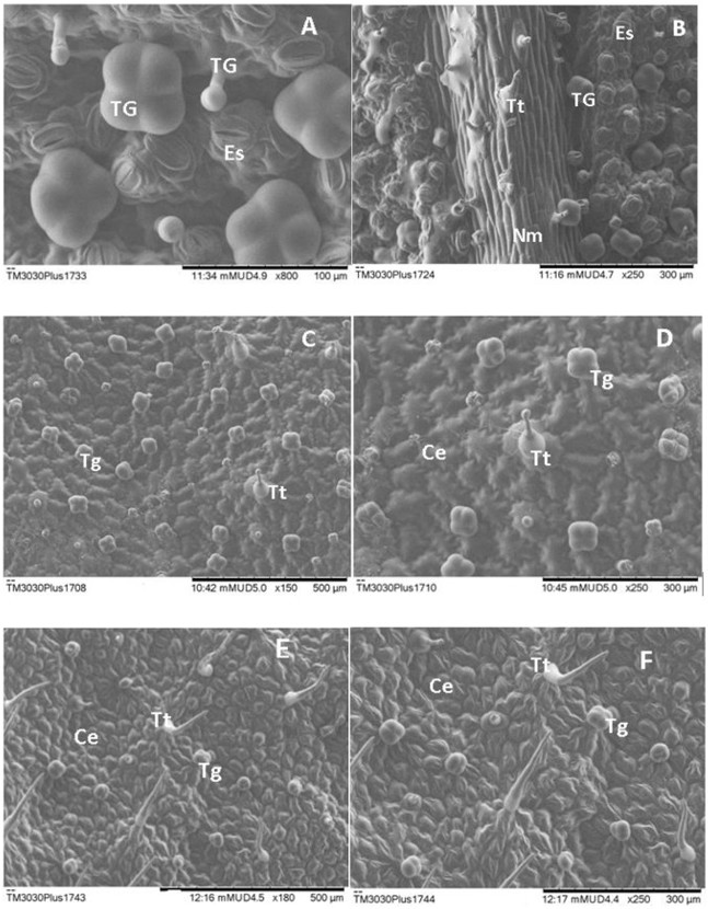
The O. azureus leaf epidermis presents stomata of anomocytic type, occurring only on the abaxial face, therefore being considered a hypostomatic leaf (Figure 1A). Tector and multicellular glandular trichomes (Figures 1A-D) were observed on both sides of the leaf. The tector trichomes are unisseriate (Figure 1B) and long with an oval base (Figures 1C and 1D), more chipboard under the mean vein (Figure 1B) on the abaxial face. The adaxial face presents sparse tector trichomes (Figures 1C and 1D). Multicellular chipboard glandular trichomes were observed on the abaxial and adaxial faces with single-celled stalk and head alveolar round shape (Figures 1A-D). Some of them were wrinkled, probably because their content was released, while others looks broken (Figures 1C and 1D). Petal tector trichomes were observed and glandular trichomes were found only on the abaxial face (had fled) (Figures 1E and 1F). The presence of trichomes may be interpreted as stomata protection of the mesophyll against excess of heat by the reflection of light and by the elimination of allelopathic substances (Taiz and Zeiger, 2008; Potiguara et al., 2013). According to Ronse et al. (1998), the essential oil of O. azureus consists on a complex mixture of monoterpenes and sesquiterpenes, which are secreted in glandular trichomes on the surface of the leaves. In this study, it was observed that the plant presents glandular trichomes throughout whole aerial part.

The epidermis of the leaf and petal from Otacanthus azureus. Photographs obtained after analysis by scanning electron micrography. (A, B) Abaxial face with multicellular glandular trichomes, tector trichomes and stoma; (C, D) Adaxial face with glandular trichomes and tector trichomes; (E, F) Epidermis of the petal with glandular trichomes and tector trichomes. Abbreviations: TG: multicellular glandular trichome; Es: stomata; Tt: tector trichome; Ce: epithelial cell; Nm: medium nervure.
The extraction of aerial flowering parts of O. azureus yielded 0.1% of an orange to yellowish essential oil. The chromatogram obtained by GC/MS revealed a predominance of mono- and sesquiterpenes. The main compounds were β-copaen-4-α-ol (20.0%), trans-pinocarveol (15.0%), myrtenol (13.4%), pinocarvone (6.8%) and α-copaene (5.2%). Similar yield and chemical composition were observed by another authors (Ronse et al., 1997; Houël et al., 2014). All suggested compounds can be found on Table 1.
Table 2 shows the droplet size diameter and polydispersity index of the nanoemulsions prepared with the essential oil of O. azureus. The nanoemulsion prepared with polysorbate 80 presented a bluish reflect which is associated to the generation of the nanostructures, being caused by the Tyndall effect. The droplet size distribution revealed a mean diameter around 130.0 nm and polydispersity index around 0.290. The mean droplet size remained below 150.0 nm during the 30 days of storage, while the pdi remained below 0.300 during this period. Despite the system prepared with polysorbate 20 presented lower pdi (<0.1) on the day of preparation and after 1 day of storage, at these time points it presented higher mean droplet size, slight above up 200 nm. Moreover, further analysis revealed a dramatically increase in droplet size and pdi during storage.
Droplet size distribution of nanoemulsions prepared with essential oil from Otacanthus azureus.
Analysis of the droplet growth during storage revealed a pattern of high and increasing disistabilization for the nanoemulsion prepared with polysorbate 20 along the time, when analyzed this parameter at different intervals from the day 0. After an almost no alteration after 1 day of storage (DG0,1 = 0.02), a high increase (>50%) was observed after 21 and 30 days. After reaching this high level of droplet growth at 21 days, a low increase was observed between 21 and 30 days (D21,30 = 0.9), remaining around 325 nm.
On another hand, considerable lower droplet growth was observed for the nanoemulsion prepared with polysorbate 80 during storage, as evidenced by analyses of this parameter at different intervals from day 0 (DG0,21 = 5.3; DG0,30 = 11.8). In fact, a slightly decrease in droplet size was observed after 1 day of storage (DG0,1 = -2.6), while a low droplet growth was observed using this surfactant, when compared DG0,21 and DG0,30 of the nanoemulsion prepared with polysorbate 80 to the nanoemulsion prepared with polysorbate 20 (DG0,21 = 57.3; DG0,30 = 58.7), being respectively around 10 and 20% for these periods. Higher droplet growth for the nanoemulsion prepared with polysorbate 80, when compared to the nanoemulsion prepared with polysorbate 20, was observed only regarding the analysis day 21 and 30. However, it must be mentioned that the level of droplet growth can be considered low and the mean droplet size remained below 150 nm. Raiser et al. (2024) obtained droplets in this range associated to a natural product-based nanoemulsion, however, using a high energy method instead of low energy method that was used in the present study and which relies in an advantaged utilization of chemistry energy of the system.
Some essential oils with great potential for aqueous products were well-studied regarding obtainment of nanoemulsions. Best performance was achieved with polysorbate 20 for generation of lower droplets of rosemary essential oil-based nanoemulsions, when compared to blends of polysorbate 20/sorbitan monooleate (Fernandes et al., 2013). Lemon essential oil was nanoemulsified using polysorbate 80 (Rao and McClements 2012a, b). In some cases, the utilization of a single surfactant is not sufficient to ensure the stability of the nanoemulsion, through a droplet growth or even a droplet “decrease”. In fact, some aspects of the own essential oil components can develop a main role on the stability of the nanoemulsion (Rao and McClements 2012a), as it will be further discussed.
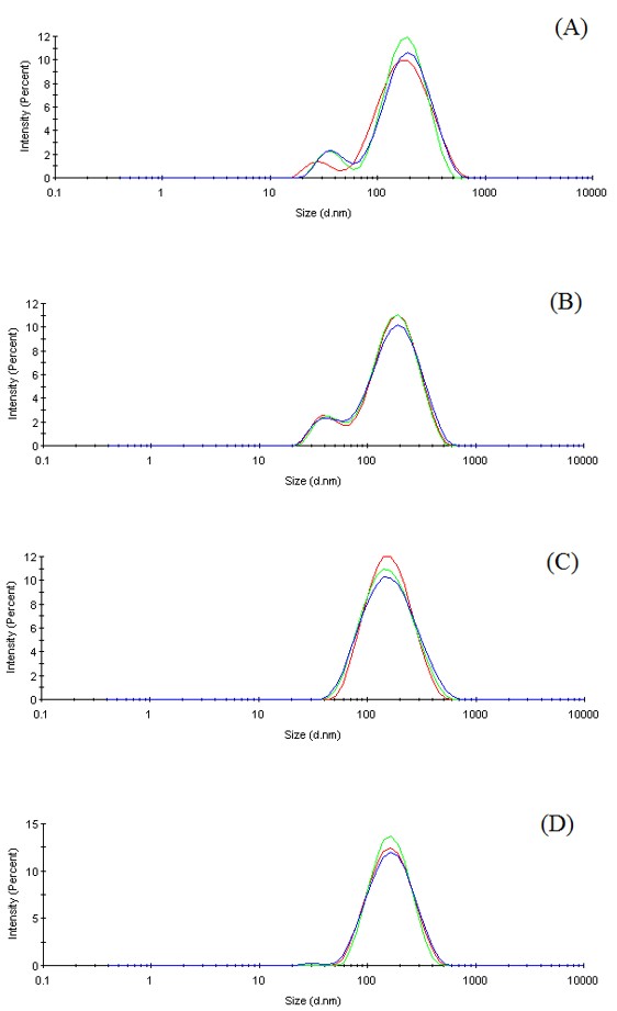
For better understanding of the droplet size distribution changes, the size diameter distribution graphs of the nanoemulsion prepared with polysorbate 80, which was considered the optimal nanoemulsion of the present study, were interpreted along the time of storage. Figures 2A and 2B shows that some considerable droplet population below 100 nm could be observed, respectively on the day of preparation (Day 0) and after 1 day of storage (Day 1). The presence of this population together with a more abundant peak corresponding to a droplet population with higher size is responsible by the pdi around 0.300 initially observed. It is also remarkable the similarity between the two profiles of droplet distribution, responsible by close size and pdi values observed on day 0 and day 1. However, after 21 days (Figure 2C) and 30 days (Figure 2D) of storage, it can be observed the disappearance of this lower-size droplet population and relative maintenance of the principal peak related to the droplets around 130-150 nm. The transition from a more polymodal profile to a more monomodal profile is responsible by the pdi reduction. On another hand, the slightly tendency for a broader base of the curve of principal droplet size population was responsible by the increase on droplet size observed on day 21 and 30. However, the low droplet growth and high degree of similarity on droplet size distribution pattern, suggest a suitable stability along 30 days.

Particle size distribution graphs of nanoemulsions prepared with the essential oil from O. azureus and polysorbate 80 on (A) day 0, (B) day 1, (C) day 21 and (D) day 30.
The physicochemical properties of the major compounds of the O. azureus essential oil were analyzed using an in silico chemical database (Table 3) as follows. Copaene presented the highest logP value (6.17), lowest density (0.9 g cm-3) and lowest water solubility (0.3163 mg/L). The density of remaining compounds were all 1.0 g cm-3. The pinocarvone presented the lowest logP value (1.88) and higher water solubility than copaene, which was (117.2 mg/L). This parameter was not higher than the water solubilities of myrtenol (426.9 mg/L) and trans-pinocarveol (958.1 mg/L). The expected inverse correlation between water solubilities and logP values of myrtenol and trans-pinocarveol were found, being observed the respectively logP values of 3.22 and 2.45. The surface tension (dyne/cm) ranged from 30.7 (copaene) to 33.5 (trans-pinocarveol), the boiling point (°C) ranged from 217.5 (trans-pinocarveol) to 248.5 (copaene) and the index of refraction ranged from 1.496 (pinocarvone) to 1.510 (trans-pinocarveol) Compounds with high water solubility (> 5 mg/L) are associated to low logP values, where high logP values are considered when > 4.3. The last is especially important since it reflects the capacity of compounds to partitioning. The water solubility is associated to high unstable systems, due to Ostwald ripening effect (Rao and McClements, 2012a). Moreover, a better stability against this phenomenon may occur when higher surface tension is observed (Rao and McClements, 2012b). The Ostwald ripening is associated to release of more water-soluble substances from small to large droplets. Therefore, smaller droplets become more concentrated in low water-soluble components and larger droplets become more concentrated in high water-soluble components. The compositional ripening effect may therefore trigger the inhibition of droplet growth that could be expected through an Ostwald ripening effect (Rao and McClements, 2012a).
The boiling points (BP) are considered high for each compound when BP > 214 °C (Rao and McClements, 2012a). This characteristic would lead to a minor loss of compounds from the external aqueous phase to the headspace of the flasks, saturating the continuous phase. Therefore, it could inhibit partitioning of compounds, keeping the droplet size almost constant. A high refractive index of a compound is defined above 1.47 and when a major difference between the value of the compounds from the internal phase and the external phase, a greater turbidity is observed instead of a transparent nanoemulsion (Rao and McClements, 2012a).
Studies regarding antimicrobial activities of essential oils have been widely explored and demonstrate that they are effective against different strains (Carson et al., 2002; Mevy et al., 2007). The antibacterial action of essential oils involve different targets of bacterium cell, interfering on the structure of cell wall, loss of cellular constituents and destruction of genetic material (Kalemba and Kunicka, 2003; Seow et al., 2014). This may be due to relative lipophilicity of their chemical constituents, which facilitates the interaction with the cellular membranes (Santos et al., 2012; Salvia-Trujillo et al., 2015). It has also been established that, in addition to a better dispersability of essential oils when formulated as nanoemulsions, improved bioactivity may also be observed (Donsì et al., 2011; Al-Jabri and Hossain, 2014; Ahmad et al., 2014). Therefore, we used the antibacterial assay as an experimental model to investigate the possible enhancement of the bioactivity, by comparing the essential oil with the optimal nanoemulsion.
No improvement on the bioactivity was observed against E. coli and P. aeruginosa, as evidenced by the same MIC and MBC values of essential oil and nanoemulsion. The MIC value of the nanoemulsion against K. pneumonie was 50% lower, suggesting improved bioactivity. No difference was observed regarding the MBC. All remaining MIC and MBC values were lower for the nanoemulsion, showing a better antibacterial activity against S. aureus, S. epidermidis and S. typhi, suggesting improvement of the bioactivity, as previously hypothesized. Barraqui et al. (2025) reported that the nanoemulsification of an essential oil also improved the bioactivity by evaluating the colloid against bacteria strains. All the values corresponding to the antibacterial assay are presented in Table 4.
Minimum inhibitory concentration (MIC) and minimum bactericidal concentration (MBC) for essential oil and nanoemulsions of Othacantus azureus.
4. Conclusion
The successful achievement of a true ecofriendly nanoemulsion must take into account needs for “green techniques” in accordance to sustainable and ecofriendly concepts. On the present study, it should be highlight that i) a single non-ionic surfactant was used, being not necessary to use a co-surfactant; ii) equal amount of surfactant to essential oil was used; iii) the own essential oil was the oily phase and source of bioactive compounds. Moreover, in some cases it is necessary to use temperature or high-energy methods to obtain the nanoemulsions. In both cases, it elevates the cost of the process due to the needs for energy input. Moreover, heating could lead to volatile loss and may induce difference between the bulk material used for the nanoemulsion preparation and the overall composition of this raw material on the nanoemulsion. Thus, the present study provides a great approach for obtaining O. azureus essential oil nanoemulsions, which proved to exert improved bioactivity against different bacteria strains, therefore being promising as additive for herbal-based products.
Acknowledgements
Authors would like to thank FAPEAP (Prodetec Araguari—Process nº 250.203.035/2013) and PROPESPG/UNIFAP (PAPESQ) for the financial support. We also thank Iann Rodrigues Sarquis for the collection of plant material.
Data Availability Statement
The entire data set that supports the results of this study was published in the article itself.
References
-
AHMAD, N., RAMSCH, R., LLINÀS, M., SOLANS, C., HASHIM, R. and TAJUDDIN, H.A., 2014. Influence of non ionic branched-chain alkyl glycosides on a model nanoemulsion for drug delivery systems. Colloids and Surfaces. B, Biointerfaces, vol. 115, pp. 267-274. http://doi.org/10.1016/j.colsurfb.2013.12.013 PMid:24384142.
» http://doi.org/10.1016/j.colsurfb.2013.12.013 -
AL-JABRI, N.N. and HOSSAIN, M.A., 2014. Comparative chemical composition and antimicrobial activity study of essential oils from two imported lemon fruits samples against pathogenic bacteria. Beni-Suef University Journal of Basic and Applied Sciences, vol. 3, no. 4, pp. 247-253. http://doi.org/10.1016/j.bjbas.2014.10.011
» http://doi.org/10.1016/j.bjbas.2014.10.011 -
ANDRADE, E.H., CARREIRA, L.M., ZOGHBI, M.G. and CONCEIÇÃO, C.C., 2006. Volatiles of the leaves, stems and flowers of Otacanthes azureus (Linden) Ronse. The Journal of Essential Oil Research, vol. 18, no. 5, pp. 481-482. http://doi.org/10.1080/10412905.2006.9699147
» http://doi.org/10.1080/10412905.2006.9699147 - ASTOLFI FILHO, S., SILVA, C.G.N. and BIG, M.F.M.A., 2014. Bioprospecção e biotecnologia. Parcerias Estratégicas, vol. 19, pp. 45-80.
-
BARRAQUI, N.S.G., GARCIA, E.F., LOPES, G.A., FERNANDES, C.P., SILVA, Q.M. and FRANÇA, H.S., 2025. Antibacterial activity of nanoemulsion of essential oil from Brazilian pepper tree (Schinus terebinthifolia Raddi) against sensitive and multidrug-resistant strains. Brazilian Journal of Biology = Revista Brasileira de Biologia, vol. 85, e286942. http://doi.org/10.1590/1519-6984.286942
» http://doi.org/10.1590/1519-6984.286942 -
BHATTACHARJEE, S., 2016. DLS and zeta potential: what they are and what they are not? Journal of Controlled Release, vol. 235, pp. 337-351. http://doi.org/10.1016/j.jconrel.2016.06.017 PMid:27297779.
» http://doi.org/10.1016/j.jconrel.2016.06.017 -
BURT, S., 2004. Essential oils: their antibacterial properties and potential applications in foods: a review. International Journal of Food Microbiology, vol. 94, no. 3, pp. 223-253. http://doi.org/10.1016/j.ijfoodmicro.2004.03.022 PMid:15246235.
» http://doi.org/10.1016/j.ijfoodmicro.2004.03.022 -
CALIXTO, J.B., 2005. Twenty-five years of research on medicinal plants in Latin America: a personal view. Journal of Ethnopharmacology, vol. 100, no. 1-2, pp. 131-134. http://doi.org/10.1016/j.jep.2005.06.004 PMid:16006081.
» http://doi.org/10.1016/j.jep.2005.06.004 -
CARSON, C.F., MEE, B.J. and RILEY, T.V., 2002. Mechanism of action of Melaleuca alternifolia (tea tree) oil on Staphylococcus aureus determined by time-kill, lysis, leakage and salt tolerance assays and electron microscopy. Antimicrobial Agents and Chemotherapy, vol. 46, no. 6, pp. 1914-1920. http://doi.org/10.1128/AAC.46.6.1914-1920.2002 PMid:12019108.
» http://doi.org/10.1128/AAC.46.6.1914-1920.2002 - CLINICAL AND LABORATORY STANDARDS INSTITUTE – CLSI, 2012. Methods for dilution antimicrobial susceptibility tests for bacteria that grow aerobically Wayne, PA: CLSI. CLSI Document M07-A9.
-
DONSÌ, F., ANNUNZIATA, M., SESSA, M. and FERRARI, G., 2011. Nanoencapsulation of essential oils to enhance their antimicrobial activity in foods. Lebensmittel-Wissenschaft + Technologie, vol. 44, no. 9, pp. 1908-1914. http://doi.org/10.1016/j.lwt.2011.03.003
» http://doi.org/10.1016/j.lwt.2011.03.003 -
FERNANDES, C.P., MASCARENHAS, M.P., ZIBETTI, F.M., LIMA, B.G., OLIVEIRA, R.P., ROCHA, L. and FALCÃO, D.Q., 2013. HLB value, an important parameter for the development of essential oil phytopharmaceuticals. Revista Brasileira de Farmacognosia, vol. 23, no. 1, pp. 108-114. http://doi.org/10.1590/S0102-695X2012005000127
» http://doi.org/10.1590/S0102-695X2012005000127 - FIRMO, W.D.C., MENEZES, V.A.M., DE PASSOS, C.E.D.C., DIAS, C.N., ALVES, L.P.L., DIAS, I.C.L., SANTOS NETO, M. and OLEA, R.S.G., 2011. Contexto histórico, uso popular e concepção científica sobre plantas medicinais. Cadernos de Pesquisas, vol. 18, pp. 90-95.
-
GUTTOFF, M., SABERI, A.H. and MCCLEMENTS, D.J., 2015. Formation of vitamin D nanoemulsion-based delivery systems by spontaneous emulsification: factors affecting particle size and stability. Food Chemistry, vol. 171, pp. 117-122. http://doi.org/10.1016/j.foodchem.2014.08.087 PMid:25308650.
» http://doi.org/10.1016/j.foodchem.2014.08.087 -
HOUËL, E., GONZALEZ, G., BESSIÈRE, J.M., ODONNE, G., EPARVIER, V., DEHARO, E. and STIEN, D., 2015. Therapeutics witching: from antidermatophytic essential oils to new leishmanicidal products. Memorias do Instituto Oswaldo Cruz, vol. 110, no. 1, pp. 106-113. http://doi.org/10.1590/0074-02760140332 PMid:25742270.
» http://doi.org/10.1590/0074-02760140332 -
HOUËL, E., RODRIGUES, A.M.S., JAHN-OYAC, A., BESSIÈRE, J.M., EPARVIER, V., DEHARO, E. and STIEN, D., 2014. In vitro antidermatophytic activity of Otacanthus azureus (Linden) Ronse essential oil alone and in combination with azoles. Journal of Applied Microbiology, vol. 116, no. 2, pp. 288-294. http://doi.org/10.1111/jam.12377
» http://doi.org/10.1111/jam.12377 -
JAISWAL, M., DUDHE, R. and SHARMA, P.K., 2015. Nanoemulsion: an advanced mode of drug delivery system. 3 Biotech, vol. 5, no. 2, pp. 123-127. http://doi.org/10.1007/s13205-014-0214-0 PMid:28324579.
» http://doi.org/10.1007/s13205-014-0214-0 -
KALEMBA, D. and KUNICKA, A., 2003. Antibacterial and antifungal properties of essential oils. Current Medicinal Chemistry, vol. 10, no. 10, pp. 813-829. http://doi.org/10.2174/0929867033457719 PMid:12678685.
» http://doi.org/10.2174/0929867033457719 -
MATOS LOPES, T.R., OLIVEIRA, F.R., MALHEIROS, F.F., ANDRADE, M.A., MONTEIRO, M.C. and BAETAS GONÇALVES, A.C., 2015. Antimicrobial bioassay-guided fractionation of a methanol extract of Eupatorium triplinerve. Pharmaceutical Biology, vol. 53, no. 6, pp. 897-903. http://doi.org/10.3109/13880209.2014.948634 PMid:25430540.
» http://doi.org/10.3109/13880209.2014.948634 -
MCCLEMENTS, D.J., 2012. Nanoemulsions versus microemulsions: terminology, differences, and similarities. Soft Matter, vol. 8, no. 6, pp. 1719-1729. http://doi.org/10.1039/C2SM06903B
» http://doi.org/10.1039/C2SM06903B -
MCCLEMENTS, D.J. and RAO, J., 2011. Food-grade nanoemulsions: formulation, fabrication, properties, performance, biological fate, and potential toxicity. Critical Reviews in Food Science and Nutrition, vol. 51, no. 4, pp. 285-330. http://doi.org/10.1080/10408398.2011.559558 PMid:21432697.
» http://doi.org/10.1080/10408398.2011.559558 -
MEHMOOD, T., 2015. Optimization of the canola oil based vitamin E nanoemulsions stabilized by food grade mixed surfactants using response surface methodology. Food Chemistry, vol. 183, pp. 1-7. http://doi.org/10.1016/j.foodchem.2015.03.021 PMid:25863602.
» http://doi.org/10.1016/j.foodchem.2015.03.021 -
MEVY, J.P., BESSIERE, J.M., DHERBOMEZ, M., MILLOGO, J. and VIANO, J., 2007. Chemical composition and some biological activities of the volatile oils of a chemotype of Lippia chevalieri Moldenke. Food Chemistry, vol. 101, no. 2, pp. 682-685. http://doi.org/10.1016/j.foodchem.2006.01.052
» http://doi.org/10.1016/j.foodchem.2006.01.052 - POTIGUARA, R.C., SILVA, R.J.F.S., KIKUCHI, T.Y.S., LUCAS, F.C.A. and MACEDO, E. G., 2013. Estruturas vegetais em microscopia eletrônica de varredura Belém: Museu Paraense Emílio Goeldi, Universidade do Estado do Pará, 116 p.
-
RAISER, A.L., HOSHINO, T.T., TORRES, M.P.R., DEBIASI, B.W., COTRIM, A.C.M., RIBEIRO, E.B. and VALLADÃO, D.M.S., 2024. Potential application of munguba butter in the formulation of nanoemulsified systems. Brazilian Journal of Biology = Revista Brasileira de Biologia, vol. 84, e281236. http://doi.org/10.1590/1519-6984.281236 PMid:38775525.
» http://doi.org/10.1590/1519-6984.281236 -
RAO, J. and MCCLEMENTS, D.J., 2011. Food-grade microemulsions, nanoemulsions and emulsions: fabrication from sucrose monopalmitate and lemon oil. Food Hydrocolloids, vol. 25, no. 6, pp. 1413-1423. http://doi.org/10.1016/j.foodhyd.2011.02.004
» http://doi.org/10.1016/j.foodhyd.2011.02.004 -
RAO, J. and MCCLEMENTS, D.J., 2012a. Impact of lemon oil composition on formation and stability of model food and beverage emulsions. Food Chemistry, vol. 134, no. 2, pp. 749-757. http://doi.org/10.1016/j.foodchem.2012.02.174 PMid:23107687.
» http://doi.org/10.1016/j.foodchem.2012.02.174 -
RAO, J. and MCCLEMENTS, D.J., 2012b. Food-grade microemulsions and nanoemulsions: role of oil phase composition on formation and stability. Food Hydrocolloids, vol. 29, no. 2, pp. 326-334. http://doi.org/10.1016/j.foodhyd.2012.04.008
» http://doi.org/10.1016/j.foodhyd.2012.04.008 - REDDY, L.J., BEENA, J., SPANDANA, G. and RESHMA, D.J., 2012. Evaluation of antibacterial and antioxidant activities of the leaf essential oil and leaf extracts of Otacanthus caerulus(Linden) Ronse. International Journal of Pharmacy and Pharmaceutical Science Research, vol. 2, pp. 20-25.
-
RONSE, A., DE POOTER, H. and DE PROFT, M., 1997. Essential oils of Otacanthus. Phytochemistry, vol. 46, no. 8, pp. 1365-1368. http://doi.org/10.1016/S0031-9422(97)00505-0
» http://doi.org/10.1016/S0031-9422(97)00505-0 - RONSE, A., POOTER, H., VYVER, A.V., 1998. Medicinal and aromatic plants X. Heidelberg: Springer, pp. 306-319. Biotechnology in Agriculture and Forestry, no. 41.
-
RONSE, A., 2001. A revision of OtacanthusLindl. (Scrophulariaceae). Brittonia, vol. 53, no. 1, pp. 137-153. http://doi.org/10.1007/BF02805406
» http://doi.org/10.1007/BF02805406 -
SALVIA-TRUJILLO, L., ROJAS-GRAÜ, A., SOLIVA-FORTUNY, R. and MARTÍN-BELLOSO, O., 2015. Physicochemical characterization and antimicrobial activity of food-grade emulsions and nanoemulsions incorporating essential oils. Food Hydrocolloids, vol. 43, pp. 547-556. http://doi.org/10.1016/j.foodhyd.2014.07.012
» http://doi.org/10.1016/j.foodhyd.2014.07.012 -
SANTOS, R.C.V., ALVES, C.F.D.S., SCHNEIDER, T., LOPES, L.Q.S., AURICH, C., GIONGO, J.L., BRANDELLI, A. and DE ALMEIDA VAUCHER, R., 2012. Antimicrobial activity of Amazonian oils against Paenibacillus species. Journal of Invertebrate Pathology, vol. 109, no. 3, pp. 265-268. http://doi.org/10.1016/j.jip.2011.12.002 PMid:22200645.
» http://doi.org/10.1016/j.jip.2011.12.002 -
SARKER, S.D., NAHAR, L. and KUMARASAMY, Y., 2007. Microtitre plate-based antibacterial assay incorporating resazurin as an indicator of cell growth, and its application in the in vitro antibacterial screening of phytochemicals. Methods, vol. 42, no. 4, pp. 321-324. http://doi.org/10.1016/j.ymeth.2007.01.006 PMid:17560319.
» http://doi.org/10.1016/j.ymeth.2007.01.006 -
SARQUIS, R.D.S.F.R., RODRIGUES SARQUIS, Í., RODRIGUES SARQUIS, I., FERNANDES, C.P., ARAÚJO DA SILVA, G., BORJA LIMA E SILVA, R., GONÇALVES JARDIM, M.A., SÁNCHEZ-ORTÍZ, B.L. and CARVALHO, J.C.T., 2019. The use of medicinal plants in the riverside community of the Mazagão River in the Brazilian Amazon, Amapá, Brazil: ethnobotanical and ethnopharmacological studies. Evidence-Based Complementary and Alternative Medicine, vol. 2019, pp. 6087509. http://doi.org/10.1155/2019/6087509 PMid:31093296.
» http://doi.org/10.1155/2019/6087509 -
SCHAFFAZICK, R.S., GUTERRES, S.S., FREITAS, L.L. and POHLMANN, A.R., 2003. Caracterização e estabilidade físico-química de sistemas poliméricos nanoparticulados para administração de fármacos. Quimica Nova, vol. 26, no. 5, pp. 726-737. http://doi.org/10.1590/S0100-40422003000500017
» http://doi.org/10.1590/S0100-40422003000500017 -
SEOW, Y.X., YEO, C.R., CHUNG, H.L. and YUK, H.G., 2014. Plant essential oils as active antimicrobial agents. Critical Reviews in Food Science and Nutrition, vol. 54, no. 5, pp. 625-644. http://doi.org/10.1080/10408398.2011.599504 PMid:24261536.
» http://doi.org/10.1080/10408398.2011.599504 -
SOLANS, C. and SOLÈ, I., 2012. Nanoemulsions: formation by low energy methods. Current Opinion in Colloid & Interface Science, vol. 17, no. 5, pp. 246-254. http://doi.org/10.1016/j.cocis.2012.07.003
» http://doi.org/10.1016/j.cocis.2012.07.003 - SOLANS, C. and GARCÍA-CELMA, M.J., 2017. Microemulsions and nanoemulsions for cosmetic applications. In: K. SAKAMOTO, R.Y. LOCHHEAD, H.I. MAIBACH and Y. YAMASHITA, eds. Cosmetic science and technology: theoretical principles and applications. Amsterdam: Elsevier, pp. 507-518.
- TAIZ, T. and ZEIGER, E., 2008. Fisiologia vegetal. 4. ed. Porto Alegre: Artmed, 719 p.
Edited by
-
Editor:
Ana Paula Peron