Antibacterial effect of cinnamaldehyde in a microemulsion system against oral colonizing biofilms
Abstract
In the oral biofilm there are the presence of Streptococcus mutans, which is considered the main microorganism related to caries, and Streptococcus mitis and Streptococcus oralis that are considered commensal microorganisms. Mechanical disorganization of the biofilm associated with antimicrobial agents represents an effective method of injury prevention. Thus, the aim of this study was to evaluate the antibacterial effect of cinnamaldehyde-loaded microemulsion formulated in the presence (CEQ) and absence (CE) of chitosan. Single-species biofilms of S. mutans (UA159), S. mitis (ATCC 903) and S. oralis (ATCC 49456) were staggered (108 CFU/mL) and seeded in 96-well plates in BHI medium + 1% sucrose. The samples were incubated for 24 h and 48 h and then remained exposed for 24 h to CE and CEQ microemulsions. Microemulsions were produced by sonicator using cinnamaldehyde (1-10%). The concentration of chitosan included in the microemulsions was 0.1%. 0.12% chlorhexidine and BHI medium + 1% sucrose were used as positive and negative controls, respectively. Subsequently, an analysis of cellular metabolism was performed using the MTT test. Data were analyzed by Kruskal-Wallis test (α=5%). For S. oralis at 24 h, it was assumed that the 2.5% and 1% CE system showed activity similar to chlorhexidine (p>0.05). For the other biofilms, the effect of all concentrations of the CE and CEQ systems showed lower antimicrobial activity than chlorhexidine 0.12% (p<0.05). At 48 h, all microemulsion also showed lower activity than chlorhexidine (p<0.05). The antibacterial activity of cinnamaldehyde-loaded microemulsions was not dependent on concentration but varied according to the tested microorganism. The chitosan-based systems had low antimicrobial activity.
Keywords:
biofilms; natural product; chlorhexidine; chitosan; emulsions
Resumo
O biofilme dentário é composto por espécies bacterianas pertencentes ao grupo dos Streptococcus. Destaca-se a presença de espécies como S. mutans, que é considerado o principal microrganismo relacionado à cárie, e Streptococcus mitis e Streptococcus oralis que são considerados microrganismos comensais. O objetivo deste estudo foi avaliar o efeito antibacteriano do cinamaldeído disperso em microemulsão formulada na presença (CEQ) e ausência (CE) de quitosana. Biofilmes uniespécies de S. mutans (UA159), S. mitis (ATCC 903) e S. oralis (ATCC 49456) foram padronizados (108 UFC/mL) e semeados em placas de 96 compartimentos, em meio BHI com 1% de sacarose. As amostras (n=12/grupo) foram cultivadas por 24 h e 48 h e em seguida, submetidas a exposição as microemulsões CE e CEQ, durante 24 h. As microemulsões foram produzidas por sonicação usando cinemaldeído (1-10%). CEQ contendo 10% de cinemaldeído foi formulada com quitosana a 0,1%. Clorexidina 0,12% e meio BHI com 1% de sacarose foram utilizados como controle positivo e negativo, respectivamente. Posteriormente foi realizada a análise do metabolismo celular por meio do teste de MTT. Os dados foram analisados pelo teste de Kruskal-Wallis (α=5%). As formulações otimizadas CE e CEQ apresentaram tamanho de gotículas de 125,1±1,7nm e 556,8±13,4nm com potencial zeta de -18,0±0,2mV e +14,7±0,2mV, respectivamente. Para o S. oralis no tempo de 24 h, verificou-se que as microemulsões CE 2,5% e 1% apresentaram atividade semelhante à clorexidina (p>0,05). Para os demais biofilmes, o efeito de todas as concentrações dos sistemas CE e CEQ apresentaram atividade antimicrobiana inferior a clorexidina 0,12% (p<0,05). No tempo de 48 h, todas as formulações também apresentaram atividade inferior à clorexidina (p<0.05). As microemulsões de cinamaldeído apresentaram atividade antibacteriana não dependente de concentração, mas dependente de microrganismo avaliado. As formulações contendo quitosana apresentaram baixa atividade antimicrobiana.
Palavras-chave:
biofilmes; produto natural; clorexidina; quitosana; emulsões
1. Introduction
The cariogenic biofilm is composed of microorganisms inserted into an extracellular matrix adhered to the tooth, in the presence of adsorbed salivary proteins. The composition of the biofilm is varied, considering its stage of maturation, with the vast majority of streptococci present. Streptococcus mutans is involved in the etiology of dental caries and is considered the main microorganism related to this disease. It has distinct characteristics, such as aciduricity and acidogenicity, which promote the demineralization of hard dental tissues (Jailani et al., 2022; Wassel et al., 2023).
S. mitis and S. oralis are among the primary colonizers of the salivary film and are considered commensal microorganisms, as they form the basis of the dental biofilm by providing adherence sites for secondary colonizers (Ramadugu et al., 2021; Engen et al., 2017; Wan et al., 2021). S. mutans is characterized by promoting the formation of the climax community in the presence of demineralization, as indicated by the diagnosis of active white spot lesions (Jailani et al., 2022; Wassel et al., 2023).
The development of dental biofilm consists of a survival strategy adopted by microbial cells, allowing greater tolerance to antimicrobials, present in the saliva itself, compared to their planktonic counterparts (Costa et al., 2020). Mature biofilms pose challenges for removal as they secrete a polymeric extracellular matrix that makes it difficult for antimicrobial substances to penetrate (Ali et al., 2021; Dudek-Wicher et al., 2022). Therefore, interfering with biofilm development during its early stages and attenuating bacterial virulence are considered promising, innovative and high-value strategies to avoid therapeutic challenges associated with infections (Ali et al., 2021).
Disorganization or mechanical removal of dental biofilm, through brushing, is the main preventive method to contain the appearance of caries lesions. However, antimicrobial agents are used as an adjunct to biofilm control, with chlorhexidine as the gold standard of topical antimicrobial substance (Pitts et al., 2021; Poppolo and Ouanounou, 2022). Its mechanism of action makes it effective against planktonic cells, reducing adhesion and inhibiting biofilm formation in the early stages of development. (Dudek-Wicher et al., 2022). However, prolonged use of chlorhexidine may lead to microbial resistance and the occurrence of side effects, such as mucosal dryness, tooth staining, and taste alterations (Wang et al., 2018; Ferreira et al., 2018).
Thus, there is a need to develop antimicrobial agents that effectively inhibit biofilm formation, presenting different molecular targets and fewer side effects. Phytocompounds and their derivatives, such as cinnamaldehyde, geraniol, and terpineol, enhance the ability to reduce bacterial pathogenicity and are considered alternatives to conventional antibacterial agents (Guimarães et al., 2019; Yanakiev, 2020). Its advantages include abundant sources of plant species, high chemical diversity and biochemical specificity (He et al., 2019).
Cinnamaldehyde, predominantly present in cinnamon essential oils, is used as a flavoring agent and condiment (Shreaz et al., 2016). In the health sector, it has been demonstrated that this molecule and its derivatives have antimicrobial and antibiofilm activity against a wide range of pathogenic bacteria and fungi, such as S. mutans, E. faecalis and C. albicans, and its inhibitory effects occur through inhibition of ATPase activity, inhibition of cell wall biosynthesis and alteration of membrane structure and integrity (Jailani et al., 2022; Ali et al., 2021; Shreaz et al., 2016).
However, the main challenge in using cinnamaldehyde as an antimicrobial agent is its hydrophobicity, which limits its availability in biofilms and necessitates high concentrations to achieve inhibitory effects (Jailani et al., 2022). Therefore, new delivery systems must be developed to enhance the penetration and inhibitory efficacy of cinnamaldehyde in biofilms.
In this context, the use of microemulsions represents an efficient strategy to increase the availability of bioactive molecules that are insoluble or poorly soluble in water. Such systems can be directly bactericidal or designed to increase the aqueous solubility of drugs, and can be adjusted to allow controlled release of the drug and induce a target-specific action (Jailani et al., 2022; Benoit et al., 2019; Souto et al., 2022).
Another compound reported in the literature to exhibit antimicrobial activity against bacteria, fungi, and yeast is chitosan. This biopolymer has amino groups distributed along its chain, which confer a polycationic nature in an acidic environment due to the ionization of these groups (Matica et al., 2019). Its mechanisms of action include the neutralization of negative charges on the microbial surface (Yan et al., 2021). Additionally, Shreaz et al. (2016) highlight that the combination of chitosan with cinnamaldehyde holds significant potential for the development of new antimicrobial agents.
Therefore, the objective of this study was to evaluate, in vitro, the antimicrobial activity of cinnamaldehyde, in a microemulsion system in the presence and absence of chitosan, in different concentrations against unispecies of S. mutans, S. oralis and S. mitis biofilms.
2. Materials and Methods
2.1. Pre-formulation study
For the production of microemulsions, a pre-formulation study of samples was carried out combining different concentrations of the components of the oily phase, namely cinnamaldehyde (1-10%), Kolliphor® ELP (4%) and Lipoid® S100 (1-2%), and the aqueous phase, with Pluronic® F-68 (1-4%) and Tween 80 (1%) solubilized in ultrapure water, as shown in Table 1.
2.2. Microemulsions preparation
Microemulsions were prepared using the sonication technique. The final microemulsion was composed of cinnamaldehyde (10%), Kolliphor® ELP (4%) and Pluronic® F-68 solution (4%) in ultrapure water. For the bioadhesive formulation, a chitosan solution (0.1%) was used, replacing Pluronic® F-68 in the aqueous phase. Two systems were produced, in the presence (CEQ) and absence (CE) of chitosan. The microemulsions were produced by sonication for 1.5min at 60% (Eco-sonique QR350, São Paulo, Brazil).
2.3. Microemulsions characterization
2.3.1. Particle size analysis and polydispersity index
The mean hydrodynamic diameter and polydispersity index (pdi) of the particles were determined by the dynamic light scattering technique using the Zetasizer Lab (Malvern Instruments, Malvern, United Kingdom). The samples were diluted in Milli® Q water in a 1:100 ratio and tested at 25 °C with a detection angle set at 90º.
2.3.2. Zeta potential analysis
The zeta potential of the microemulsions was determined by the electrophoretic light scattering technique using the Zetasizer Lab device (Malvern Instruments, Malvern, United Kingdom). The samples were diluted in a 1 mmol/L NaCl solution in a ratio of 1:100 and analyzed at 25 °C.
2.4. Biofilm formation
Reference strains of Streptococcus mutans (UA159), Streptococcus mitis (ATCC 903) and Streptococcus oralis (ATCC 49456) were used, reactivated in BHI (Brain Heart Infusion) broth (KASVI®, Brazil) supplemented with 1% sucrose, in microaerophilia for 24 h at 37 °C. After this period, the set was centrifuged, the supernatant removed and the cells resuspended in 0.9% saline solution to standardize the inoculum in a spectrophotometer (LGL Scientific 0741/16, Brazil) at a wavelength of 600nm, with cell density adjusted to 1.0 x 10⁸ CFU/mL (Wan et al., 2021; Sampaio et al., 2019). For the formation of unispecies biofilms, 100 μL of inocula were added to 96-well plates. The biofilms were cultivated for 24 h and 48 h in microaerophilia at 37 °C. The culture medium was changed every 24 hours.
2.5. Exposure to substances
The concentrations of cinnamaldehyde were 10%, 5%, 2.5% and 1%, diluted in BHI medium with 1% sucrose. As a positive control, 0.12% chlorhexidine solution (Colgate-Palmolive, São Paulo, Brazil) was used. The biofilm grown in culture medium (BHI + 1% sucrose) was considered as a growth control. After the incubation period (24 and 48h), the culture medium was removed and 100μL of the test substances, from the positive control and negative control were added to 96-well plates (n=12/group). Again, the plates were incubated in microaerophilia for 24 h at 37 °C.
2.6. Biofilm metabolic analysis
After the incubation period, the culture medium was removed and 100 μL of BHI medium containing 10% methyltetrazolium salt (MTT) was inserted. Thus, the biofilms were incubated for 3 hours at 37 °C in the presence of MTT salt and the system was protected from light. Then, the medium was removed and 200 μL of acidic isopropanol were inserted, with subsequent homogenization of the samples. The reading was then carried out on a spectrophotometer at 550 nm. The entire experiment was performed in duplicate.
2.7. Data analysis
The tabulation and analysis of the collected data were carried out using the Statistical Package for the Social Sciences (SPSS®) software, version 21. The normality and homoscedasticity of the data were determined by the Shapiro Wilk and Levene tests, respectively. After checking the aforementioned determinations, statistical analysis was performed using the Kruskal-Wallis test, with the significance level adopted for this study being 5% (α=5%).
3. Results
The physicochemical parameters of the cinnamaldehyde microemulsions in the pre-formulation study are summarized in Table 2. The average particle size of the samples ranged from 125.1 to 556.8 nm. The Polydispersity Index (PdI) values varied from 0.083 to 0.256. The zeta potential of the formulations varied between -15.8 and +14.7 mV.
The S. oralis biofilm cultivated for 24h was sensitive to the use of the 2.5% and 1% CE systems in the same way as 0.12% chlorhexidine (p>0.05). The other concentrations showed intermediate antimicrobial activity, compared to the growth control (p<0.05) and between the test substances. The S. oralis biofilm exposed to 10% CEQ showed greater metabolism, with this concentration having a lower antimicrobial effect (Figure 1).

Metabolic activity represented by the absorbance of the S. oralis biofilm, after 24 h of cultivation (n=12/group). Box representing the median and interquartile distance. Equal letters represent absence of statistical differences (p>0.05, Kruskal-Wallis). CHX: Chlorhexidine.
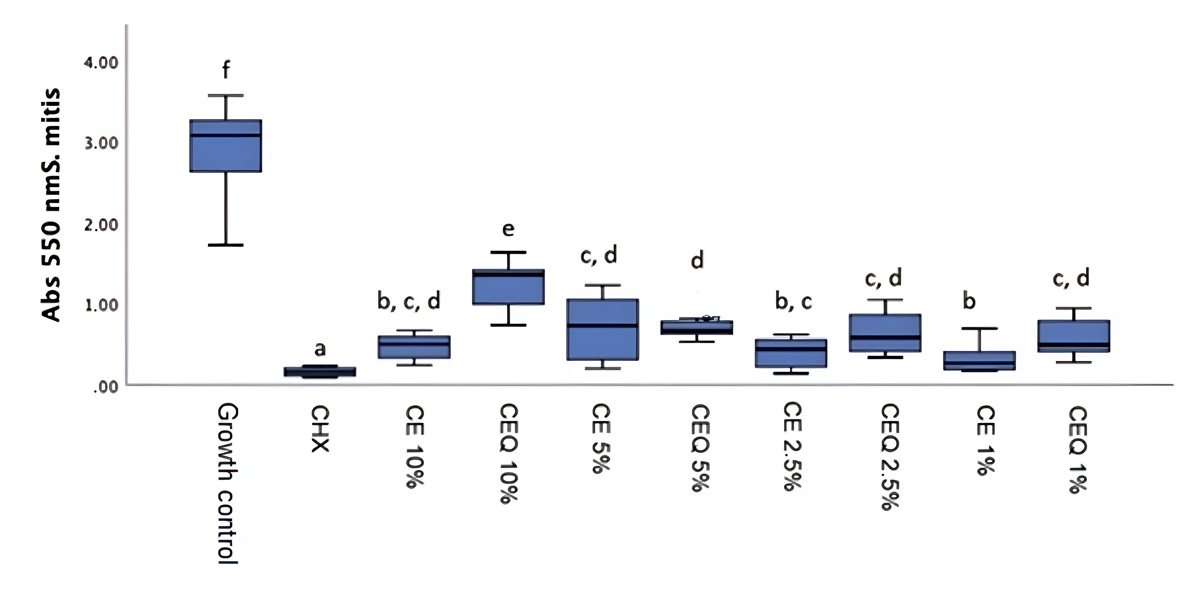
For S. mitis, all concentrations of the CE and CEQ systems showed lower activity than 0.12% chlorhexidine (p<0.05). However, among the systems, CE 1% presented the best performance, while CEQ 10% presented the lowest antibacterial potential, represented by the greater metabolic activity of the biofilm (Figure 2).

Metabolic activity represented by the absorbance of the S. mitis biofilm, after 24 h of cultivation (n=12/group). Box representing the median and interquartile distance. Equal letters represent absence of statistical differences (p>0.05, Kruskal-Wallis). CHX: Chlorhexidine.
For S. mutans, CE concentrations of 10%, 2.5% and 1% were the most effective compared to the other concentrations (p<0.05), however they did not present activity similar to chlorhexidine (p<0.05). The CEQ 10% system showed the smallest reduction in bacterial metabolism among the test substances (Figure 3).

Metabolic activity represented by the absorbance of the S. mutans biofilm, after 24 h of cultivation (n=12/group). Box representing the median and interquartile distance. Equal letters represent absence of statistical differences (p>0.05, Kruskal-Wallis). CHX: Chlorhexidine.
Considering the biofilms cultivated for 48 hours, it was found that for S. oralis, the CE 10%, 2.5% and 1% systems did not show statistical difference between them (p>0.05), but were more effective when compared to other cinnamaldehyde concentrations (p<0.05). Chlorhexidine showed the lowest rate of bacterial metabolism (p<0.05), while the growth control showed the highest (p<0.05) (Figure 4).

Metabolic activity represented by the absorbance of the S. oralis biofilm, after 48 h of cultivation (n=12/group). Box representing the median and interquartile distance. Equal letters represent absence of statistical differences (p>0.05, Kruskal-Wallis). CHX: Chlorhexidine.
For S. mitis, the 1% CE concentration showed the best antimicrobial activity compared to other cinnamaldehyde concentrations (p<0.05). The CEQ 10% system had the lowest effectiveness among the test substances (Figure 5).
Metabolic activity represented by the absorbance of the S. mitis biofilm, after 48 h of cultivation (n=12/group). Box representing the median and interquartile distance. Equal letters represent absence of statistical differences (p>0.05, Kruskal-Wallis). CHX: Chlorhexidine.
Against the S. mutans biofilm, the concentrations of CE 10%, 2.5% and 1% were also those that showed the greatest reduction in bacterial metabolism in relation to the other concentrations (p<0.05), while CEQ 10% and 2.5% presented the highest absorbance values, with the exception of the growth control (p<0.05) (Figure 6).

Metabolic activity represented by the absorbance of the S. mutans biofilm, after 48 h of cultivation (n=12/group). Box representing the median and interquartile distance. Equal letters represent absence of statistical differences (p>0.05, Kruskal-Wallis). CHX: Chlorhexidine.
In all biofilms analyzed within 48 hours, chlorhexidine was the substance with the greatest antibacterial potential when compared to the other systems (p<0.05). At both times and for all biofilms, all tested substances differed statistically from the growth control (p<0.05).
4. Discussion
This study evaluated the antibacterial effect of a phytocompound against biofilms present in the oral cavity, which are related to the development of caries lesions. Despite advances in public policies, dental caries remains the most prevalent and costly oral disease worldwide, representing a global public health problem to be managed by authorities and dental professionals (Freires et al., 2015).
Effective caries prevention methods have been developed and changed in recent decades, based on scientific evidence that mechanical control of biofilm is one of the most efficient strategies. This method may be associated with the use of antimicrobials, with the main chemical agents currently available being fluorine, chlorhexidine, triclosan, cetylpyridinium chloride and products of natural origin (Freires et al., 2015; Maguire, 2014; Liao et al., 2017).
In recent years, essential oils and their components have received significant attention as potential antimicrobial agents due to their relative safety and low long-term genotoxicity (Freires et al., 2015; Miranda-Cadena et al., 2021). However, there is a predominance in the literature of in vitro studies that evaluate essential oils as a whole, rather than isolated components (Miranda-Cadena et al., 2021).
Thus, this study evaluated the in vitro antibacterial effect of cinnamaldehyde, a molecule classified as a phenylpropanoid, present in essential oils from trees of the Cinnamomum genus of the Lauraceae family (Albano et al., 2019). The molecule was inserted into a microemulsion system and evaluated for its antibacterial potential against biofilms, as they are communities of adherent sessile cells that have properties distinct from planktonic cells (Nobile and Johnson, 2015).
Cinnamaldehyde was evaluated in a microemulsion system, characterized as single-phase dispersions on the nanometer scale containing water, oil and amphiphiles (co-surfactants and surfactants) that have a high scattering coefficient and low surface tension (Nagai and Otake, 2022). Therefore, the study of this formulation was carried out to evaluate the influence of different concentrations of oil (cinnamaldehyde) and lipophilic (Kolliphor® ELP and Lipoid® S100) and hydrophilic (Pluronic® F-68 and Tween 80) surfactants on the formation of microemulsions, considering size, PdI and zeta potential.
The chitosan solution was added to the CE microemulsion, replacing the aqueous medium containing the surfactant. Thus, the addition of chitosan to the formulation contributed to the increase in particle size. This was due to the fact that electrostatic interactions of the NH3+ ions present in the chitosan chain with the surface of the negatively charged oily droplet cause the increase in size and, consequently, the inversion of the surface charge, presenting a positive zeta potential (Makeen et al., 2021).
It was found that the microemulsions were within the nanometric scale standard, containing water, oil and amphiphiles (co-surfactants and surfactants). The results indicate that increasing the concentration of cinnamaldehyde, without a proportional increase in surfactants (CE2 and CE3), directly impacts the size of the micelles, which confirms the direct interaction between the concentration of surfactants associated with the concentration of oil in the system.
Regarding the analysis of bacterial metabolism, it was found in all biofilms analyzed that cinnamaldehyde, in the absence of chitosan, at a concentration of 1% (CE 1%) was effective as an antimicrobial agent. It was found that the antibacterial effect was not dependent on the concentration used. This result has not been frequently reported in the literature, as studies have generally demonstrated that higher concentrations of cinnamaldehyde exhibit greater antimicrobial effects (Jailani et al., 2022; Bakhtiari et al., 2019; Souza et al., 2021; Xu et al., 2022). However, the use of lower concentrations of cinnamaldehyde is reported when compared to this study, with values ranging from 0.125 mg/mL (0.0125%) (Bakhtiari et al., 2019) to 10 mg/mL (1%) (Souza et al., 2021).
Souza et al. (2021) used concentrations closer to this study, reporting good antimicrobial efficacy of 1% cinnamaldehyde, corroborating the efficient action of this substance at this concentration. Furthermore, cinnamaldehyde is used in the literature in different formulations, such as in its pure form (Bakhtiari et al., 2019; Souza et al., 2021; Xu et al., 2022), in the form of microparticles associated with porous silicon (pSi) (Jailani et al., 2022) and associated with chitosan nanoparticles (Xu et al., 2022), differing from this experiment, which may also have contributed to the divergent results.
On the other hand, the microemulsion functionalized with chitosan particles at a concentration of 10% (CEQ 10%) in this study presented, at all times and biofilms analyzed, the lowest rate of reduction in microbial metabolism. Studies have analyzed the association of cinnamaldehyde with chitosan and in most of them, the antibacterial effect is improved compared to the use of the substances alone (Negi and Kesari, 2022; Cheng et al., 2022). However, Xu et al. (2022) reported that within the same concentration range, the inhibitory activity of cinnamaldehyde associated with chitosan nanoparticles was slightly lower than that of free cinnamaldehyde.
Furthermore, Cheng et al. (2022) reported that in a system containing cinnamaldehyde and chitosan, higher concentrations of cinnamaldehyde resulted in lower antimicrobial activity. A possible explanation for this finding is a slower absorption rate by the vehicle used (hydrogel) at higher cinnamaldehyde concentrations.
Chitosan is a polymer with mucoadhesive properties and most of its natural proportion is found in the form of the exoskeletal covering of arthropods (Negi and Kesari, 2022; Nimbeni et al., 2021). Chitosan nanoparticles are used as carriers for controlled release of active substances and can also be used as an emulsion stabilizer (Nimbeni et al., 2021; Rattanaburi et al., 2019; Niu et al., 2022).
The use of these nanoparticles can improve the solubility and stability of antimicrobials, thereby increasing their antibacterial effects and reducing their side effects (Xu et al., 2022). The results presented in this study, which showed the superiority of isolated cinnamaldehyde (CE) compared to its association with chitosan (CEQ), has a possible explanation for the slower release of the drug when chitosan is present. Therefore, the 24-hour exposure time was an important variable, considering the slower release of cinnamaldehyde.
The characterization of the systems determined that although the particles were within the standards, the microemulsion in the presence of chitosan in the formulation contributed to the increase in particle size, verified by the zeta potential. The average size of CE particles is within the range described for microemulsions (Nagai and Otake, 2022) and the highest zeta potential among other samples, which can contribute to the stabilization of the system, based on the electrostatic repulsion between the particles (Makeen et al., 2021; Nagai and Otake, 2022).
Based on the results, the increase in cinnamaldehyde, a bioactive compound of interest, was tested in association with a higher surfactant concentration. This resulted in a formulation (CE) with an average particle size within the range described for microemulsions and the highest zeta potential among all samples, which may contribute to system stabilization through electrostatic repulsion between particles (Nagai and Otake, 2022).
The mechanism of action of cinnamaldehyde against biofilms may involve the loss of cell wall integrity due to ergosterol depletion and an apoptotic effect on microbial cells (Miranda-Cadena et al., 2021; Khan et al., 2017). Additionally, it can diffuse through the biofilm's polysaccharide matrix and destabilize it, highlighting its potential as a promising lead structure in the development of new antibacterial agents.
Furthermore, differences in biofilm behavior were observed when exposed to antimicrobial substances. The three microorganisms are classified as viridans group streptococci, which are associated with bacteremia in immunocompromised patients and infectious endocarditis (Adams et al., 2017).
The literature highlights that S. mutans possesses key characteristics that contribute to its cariogenicity and resistance to antibiotics and host defenses. These include its ability to synthesize large amounts of extracellular polysaccharides, which facilitate biofilm adhesion and co-aggregation, its capacity to transport and metabolize a wide range of carbohydrates into organic acids (acidogenicity), and its ability to thrive under environmental stress conditions, particularly low pH (aciduricity) (Lemos et al., 2019; Atta et al., 2022).
On the other hand, little is known about the physiology and genetics of S. mitis and S. oralis, however, it is observed in the literature that these microorganisms are associated with rapid resistance to antibiotics, such as penicillin (Lopardo et al., 2022) and daptomycin (Adams et al., 2017).
However, in all biofilms analyzed at 24 and 48 hours, chlorhexidine exhibited the lowest bacterial metabolism rates compared to all other tested substances, confirming its effectiveness as an antimicrobial agent. Its substantivity, a key characteristic that contributes to its status as the gold standard in antimicrobial effectiveness (Chandki et al., 2020), must be considered. Consequently, biofilms exposed to chlorhexidine may have exhibited lower metabolic activity, which could explain the differences observed among the evaluated formulations.
Due to the high lipophilicity of cinnamaldehyde, this study incorporated the compound into a microemulsion system. Emulsions are oil-based drug delivery systems composed of two immiscible phases, such as water and oil, stabilized by surfactants and co-surfactants. This stabilization increases the particle surface area and free energy, enhancing the system's efficiency in transporting substances across the cell membrane (Batisti et al., 2022). The classification of emulsions as macro, nano, or microemulsions is based on particle size; however, there is no consensus in the literature regarding this distinction (Valizadeh et al., 2018; Gupta et al., 2014; Komaiko and McClements, 2016).
Valizadeh et al. (2018) classify nanoemulsions as particles that have a diameter of less than 100 nm, while particles with a size above 100 nm up to a few microns are called microemulsions, and in their study it is reported that cinnamon oil microemulsions had superior antimicrobial activity when compared with nanoemulsions or with the oil in pure form against biofilms of Escherichia coli and Staphylococcus aureus.
In most studies, however, this system has been used in drugs for dermatological diseases (Souto et al., 2022; Banh and Cave, 2020; Patel et al., 2015; Alkholifi et al., 2023), with limited research on emulsions for topical application in the oral cavity.
Therefore, cinnamaldehyde may be effective in preventing biofilm development and the subsequent onset of carious lesions. However, no studies have demonstrated its efficacy against mature biofilms. Additionally, further research is needed to evaluate the effects of cinnamaldehyde at different concentrations on oral biofilms, as well as its combination with chitosan in microemulsion systems.
5. Conclusion
The cinnamaldehyde-based systems exhibited antibacterial activity that was not concentration-dependent but varied according to the microorganism evaluated. In contrast, the chitosan-based systems demonstrated low antimicrobial activity.
Acknowledgements
The authors would like to thank the Federal University of Paraíba (UFPB, João Pessoa, Brazil) for their support, including financial assistance and infrastructure, which were essential for conducting this study. This study was supported by the National Council for Scientific and Technological Development (CNPq).
-
Data Availability Statement
Research data is only available upon request
References
-
ADAMS, H.M., JOYCE, L.R., GUAN, Z., AKINS, R.L. and PALMER, K.L., 2017. Streptococcus mitis and S. oralis lack a requirement for CdsA, the enzyme required for synthesis of major membrane phospholipids in bacteria. Antimicrobial Agents and Chemotherapy, vol. 61, no. 5, e02552-16. http://doi.org/10.1128/AAC.02552-16 PMid:28223392.
» http://doi.org/10.1128/AAC.02552-16 -
ALBANO, M., CRULHAS, B.P., ALVES, F.C.B., PEREIRA, A.F.M., ANDRADE, B.F.M.T., BARBOSA, L.N., FURLANETTO, A., LYRA, L.P.D.S., RALL, V.L.M. and JÚNIOR, A.F., 2019. Antibacterial and anti-biofilm activities of cinnamaldehyde against S. epidermidis Microbial Pathogenesis, vol. 126, pp. 231-238. http://doi.org/10.1016/j.micpath.2018.11.009 PMid:30439400.
» http://doi.org/10.1016/j.micpath.2018.11.009 -
ALI, I.A.A., MATINLINNA, J.P., LÉVESQUE, C.M. and NEELAKANTAN, P., 2021. Trans-cinnamaldehyde attenuates Enterococcus faecalis virulence and inhibits biofilm formation. Antibiotics, vol. 10, no. 6, pp. 702. http://doi.org/10.3390/antibiotics10060702 PMid:34208134.
» http://doi.org/10.3390/antibiotics10060702 -
ALKHOLIFI, F.K., ALAM, A., FOUDAH, A.I. and YUSUFOGLU, H.S., 2023. Phospholipid-based topical nano-hydrogel of mangiferin: enhanced topical delivery and improved dermatokinetics. Gels, vol. 9, no. 3, pp. 178. http://doi.org/10.3390/gels9030178 PMid:36975627.
» http://doi.org/10.3390/gels9030178 -
ATTA, L., KHALIL, R., KHAN, K.M., ZEHRA, M., SALEEM, F., NUR-E-ALAM, M. and UL-HAQ, Z., 2022. Virtual screening, synthesis and biological evaluation of Streptococcus mutans mediated biofilm inhibitors. Molecules, vol. 27, no. 4, pp. 1455. http://doi.org/10.3390/molecules27041455 PMid:35209243.
» http://doi.org/10.3390/molecules27041455 -
BAKHTIARI, S., JAFARI, S., TAHERI, J.B., KASHI, T.S.J., NAMAZI, Z., IMAN, M. and POORBERAFEYI, M., 2019. The effects of cinnamaldehyde (cinnamon derivatives) and nystatin on Candida albicans and Candida glabrata Open Access Macedonian Journal of Medical Sciences, vol. 7, no. 7, pp. 1067-1070. http://doi.org/10.3889/oamjms.2019.245 PMid:31049082.
» http://doi.org/10.3889/oamjms.2019.245 -
BANH, H.L. and CAVE, A., 2020. Determination of efficacy and toxicity of diclofenac microemulsion formulation for musculoskeletal pain: an observational study. BMC Research Notes, vol. 13, no. 1, pp. 285. http://doi.org/10.1186/s13104-020-05120-3 PMid:32532323.
» http://doi.org/10.1186/s13104-020-05120-3 -
BATISTI, D.L.S., CAMARGO, R.B., DE SOUZA, V.K.G., PATTARO JÚNIOR, J.R., GONÇALVES, J.E. and MORITZ, C.M.F., 2022. Evaluation of the antiseptic effect of nanoemulsion with cinnamon essential oil on Streptococcus mutans Research, Society and Development, vol. 11, e33211426314. http://doi.org/10.33448/rsd-v11i4.26314
» http://doi.org/10.33448/rsd-v11i4.26314 -
BENOIT, D.S.W., SIMS JUNIOR, K.R. and FRASER, D., 2019. Nanoparticles for oral biofilm treatments. ACS Nano, vol. 13, no. 5, pp. 4869-4875. http://doi.org/10.1021/acsnano.9b02816 PMid:31033283.
» http://doi.org/10.1021/acsnano.9b02816 -
CHANDKI, R., NIKHIL, V. and KALYAN, S.S., 2020. Comparative evaluation of substantivity of two biguanides: 0.2% polyhexanide and 2% chlorhexidine on human dentin. Journal of Conservative Dentistry, vol. 23, no. 1, pp. 46-50. http://doi.org/10.4103/JCD.JCD_256_19 PMid:33223641.
» http://doi.org/10.4103/JCD.JCD_256_19 -
CHENG, C.H., TU, Y.Y. and LIN, J.C., 2022. Studies of mercaptosuccinic acid-crosslinked chitosan hydrogel with grafted cinnamaldehyde and silver nanoparticles for antibacterial biomedical application. International Journal of Molecular Sciences, vol. 23, no. 23, pp. 14806. http://doi.org/10.3390/ijms232314806 PMid:36499133.
» http://doi.org/10.3390/ijms232314806 -
COSTA, R.C., SOUZA, J.G.S., BERTOLINI, M., RETAMAL-VALDES, B., FERES, M. and BARÃO, V.A.R., 2020. Extracellular biofilm matrix leads to microbial dysbiosis and reduces biofilm susceptibility to antimicrobials on titanium biomaterial: an in vitro and in situ study. Clinical Oral Implants Research, vol. 31, no. 12, pp. 1173-1186. http://doi.org/10.1111/clr.13663 PMid:32894779.
» http://doi.org/10.1111/clr.13663 -
DUDEK-WICHER, R., JUNKA, A.F., MIGDAŁ, P., KORZENIOWSKA-KOWAL, A., WZOREK, A. and BARTOSZEWICZ, M., 2022. The antibiofilm activity of selected substances used in oral health prophylaxis. BMC Oral Health, vol. 22, no. 1, pp. 509. http://doi.org/10.1186/s12903-022-02532-4 PMid:36397044.
» http://doi.org/10.1186/s12903-022-02532-4 -
ENGEN, S.A., RØRVIK, G.H., SCHREURS, O., BLIX, I.J. and SCHENCK, K., 2017. The oral commensal Streptococcus mitis activates the aryl hydrocarbon receptor in human oral epithelial cells. International Journal of Oral Science, vol. 9, no. 3, pp. 145-150. http://doi.org/10.1038/ijos.2017.17 PMid:28621325.
» http://doi.org/10.1038/ijos.2017.17 -
FERREIRA, G.L.S., BEZERRA, L.M.D., RIBEIRO, I.L.A., MORAIS JÚNIOR, R.C.D. and CASTRO, R.D., 2018. Susceptibility of cariogenic microorganisms to phytoconstituents. Brazilian Journal of Biology = Revista Brasileira de Biologia, vol. 78, no. 4, pp. 691-696. http://doi.org/10.1590/1519-6984.174147 PMid:29412250.
» http://doi.org/10.1590/1519-6984.174147 -
FREIRES, I.A., DENNY, C., BENSO, B., ALENCAR, S.M. and ROSALEN, P.L., 2015. Antibacterial activity of essential oils and their isolated constituents against cariogenic bacteria: a systematic review. Molecules, vol. 20, no. 4, pp. 7329-7358. http://doi.org/10.3390/molecules20047329 PMid:25911964.
» http://doi.org/10.3390/molecules20047329 -
GUIMARÃES, A.C., MEIRELES, L.M., LEMOS, M.F., GUIMARÃES, M.C.C., ENDRINGER, D.C., FRONZA, M. and SCHERER, R., 2019. Antibacterial activity of terpenes and terpenoids present in essential oils. Molecules, vol. 24, no. 13, pp. 2471. http://doi.org/10.3390/molecules24132471 PMid:31284397.
» http://doi.org/10.3390/molecules24132471 -
GUPTA, G., MITRA, D., ASHOK, K.P., GUPTA, A., SONI, S., AHMED, S. and ARYA, A., 2014. Efficacy of preprocedural mouth rinsing in reducing aerosol contamination produced by ultrasonic scaler: a pilot study. Journal of Periodontology, vol. 85, no. 4, pp. 562-568. http://doi.org/10.1902/jop.2013.120616 PMid:23855840.
» http://doi.org/10.1902/jop.2013.120616 -
HE, Z., HUANG, Z., JIANG, W. and ZHOU, W., 2019. Antimicrobial activity of cinnamaldehyde on Streptococcus mutans biofilms. Frontiers in Microbiology, vol. 10, pp. 2241. http://doi.org/10.3389/fmicb.2019.02241 PMid:31608045.
» http://doi.org/10.3389/fmicb.2019.02241 -
JAILANI, A., KALIMUTHU, S., RAJASEKAR, V., GHOSH, S., COLLART-DUTILLEUL, P.Y., FATIMA, N., KOO, H., SOLOMON, A.P., CUISINIER, F. and NEELAKANTAN, P., 2022. Trans-cinnamaldehyde eluting porous silicon microparticles mitigate cariogenic biofilms. Pharmaceutics, vol. 14, no. 7, pp. 1428. http://doi.org/10.3390/pharmaceutics14071428 PMid:35890323.
» http://doi.org/10.3390/pharmaceutics14071428 -
KHAN, S.N., KHAN, S., IQBAL, J., KHAN, R. and KHAN, A.U., 2017. Enhanced killing and antibiofilm activity of encapsulated cinnamaldehyde against Candida albicans Frontiers in Microbiology, vol. 8, pp. 1641. http://doi.org/10.3389/fmicb.2017.01641 PMid:28900419.
» http://doi.org/10.3389/fmicb.2017.01641 -
KOMAIKO, J.S. and MCCLEMENTS, D.J., 2016. Formation of food-grade nanoemulsions using low-energy preparation methods: a review of available methods. Comprehensive Reviews in Food Science and Food Safety, vol. 15, no. 2, pp. 331-352. http://doi.org/10.1111/1541-4337.12189 PMid:33371595.
» http://doi.org/10.1111/1541-4337.12189 -
LEMOS, J.A., PALMER, S.R., ZENG, L., WEN, Z.T., KAJFASZ, J.K., FREIRES, I.A., ABRANCHES, J. and BRADY, L.J., 2019. The Biology of Streptococcus mutans Microbiology Spectrum, vol. 7, no. 1, pp. 1-18. http://doi.org/10.1128/microbiolspec.GPP3-0051-2018 PMid:30657107.
» http://doi.org/10.1128/microbiolspec.GPP3-0051-2018 -
LIAO, Y., BRANDT, B.W., LI, J., CRIELAARD, W., VAN LOVEREN, C. and DENG, D.M., 2017. Fluoride resistance in Streptococcus mutans: a mini review. Journal of Oral Microbiology, vol. 9, no. 1, pp. 1344509. http://doi.org/10.1080/20002297.2017.1344509 PMid:28748043.
» http://doi.org/10.1080/20002297.2017.1344509 -
LOPARDO, H.A., VIGLIAROLO, L., BONOFIGLIO, L., GAGETTI, P., GARCÍA GABARROT, G., KAUFMAN, S., MOLLERACH, M., TORESANI, I. and VON SPECHT, M., 2022. Beta-lactam antibiotics and viridans group streptococci. Revista Argentina de Microbiologia, vol. 54, no. 4, pp. 335-343. http://doi.org/10.1016/j.ram.2022.06.004 PMid:36266147.
» http://doi.org/10.1016/j.ram.2022.06.004 -
MAGUIRE, A., 2014. ADA clinical recommendations on topical fluoride for caries prevention. Evidence-Based Dentistry, vol. 15, no. 2, pp. 38-39. http://doi.org/10.1038/sj.ebd.6401019 PMid:24971851.
» http://doi.org/10.1038/sj.ebd.6401019 -
MAKEEN, H.A., MOHAN, S., AL-KASIM, M.A., SULTAN, M.H., ALBARRAQ, A.A., AHMED, R.A., ALHAZMI, H.A. and ALAM, M.I., 2021. Preparation, characterization, and anti-cancer activity of nanostructured lipid carriers containing imatinib. Pharmaceutics, vol. 13, no. 7, pp. 1086. http://doi.org/10.3390/pharmaceutics13071086 PMid:34371776.
» http://doi.org/10.3390/pharmaceutics13071086 -
MATICA, M.A., AACHMANN, F.L., TØNDERVIK, A., SLETTA, H. and OSTAFE, V., 2019. Chitosan as a wound dressing starting material: antimicrobial properties and mode of action. International Journal of Molecular Sciences, vol. 20, no. 23, pp. 5889. http://doi.org/10.3390/ijms20235889 PMid:31771245.
» http://doi.org/10.3390/ijms20235889 -
MIRANDA-CADENA, K., MARCOS-ARIAS, C., MATEO, E., AGUIRRE-URIZAR, J.M., QUINDÓS, G. and ERASO, E., 2021. In vitro activities of carvacrol, cinnamaldehyde and thymol against Candida biofilms. Biomedicine and Pharmacotherapy, vol. 143, pp. 112218. http://doi.org/10.1016/j.biopha.2021.112218 PMid:34649348.
» http://doi.org/10.1016/j.biopha.2021.112218 -
NAGAI, N. and OTAKE, H., 2022. Novel drug delivery systems for the management of dry eye. Advanced Drug Delivery Reviews, vol. 191, pp. 114582. http://doi.org/10.1016/j.addr.2022.114582 PMid:36283491.
» http://doi.org/10.1016/j.addr.2022.114582 -
NEGI, A. and KESARI, K.K., 2022. Chitosan nanoparticle encapsulation of antibacterial essential oils. Micromachines, vol. 13, no. 8, pp. 1265. http://doi.org/10.3390/mi13081265 PMid:36014186.
» http://doi.org/10.3390/mi13081265 -
NIMBENI, S.B., NIMBENI, B.S. and DIVAKAR, D.D., 2021. Role of chitosan in remineralization of enamel and dentin: a systematic review. International Journal of Clinical Pediatric Dentistry, vol. 14, no. 4, pp. 562-568. http://doi.org/10.5005/jp-journals-10005-1971 PMid:34824515.
» http://doi.org/10.5005/jp-journals-10005-1971 -
NIU, B., CHEN, H., WU, W., FANG, X., MU, H., HAN, Y. and GAO, H., 2022. Co-encapsulation of chlorogenic acid and cinnamaldehyde essential oil in Pickering emulsion stablized by chitosan nanoparticles. Food Chemistry: X, vol. 14, pp. 100312. http://doi.org/10.1016/j.fochx.2022.100312 PMid:35492257.
» http://doi.org/10.1016/j.fochx.2022.100312 -
NOBILE, C.J. and JOHNSON, A.D., 2015. Candida albicans biofilms and human disease. Annual Review of Microbiology, vol. 69, no. 1, pp. 71-92. http://doi.org/10.1146/annurev-micro-091014-104330 PMid:26488273.
» http://doi.org/10.1146/annurev-micro-091014-104330 -
PATEL, M.R., PATEL, R., PARIKH, J.R. and PATEL, B.G., 2015. Novel isotretinoin microemulsion-based gel for targeted topical therapy of acne: formulation consideration, skin retention and skin irritation studies. Applied Nanoscience, vol. 6, no. 4, pp. 539-553. http://doi.org/10.1007/s13204-015-0457-z
» http://doi.org/10.1007/s13204-015-0457-z -
PITTS, N.B., TWETMAN, S., FISHER, J. and MARSH, P.D., 2021. Understanding dental caries as a non-communicable disease. Brazilian Dental Journal, vol. 231, no. 12, pp. 749-753. http://doi.org/10.1038/s41415-021-3775-4 PMid:34921271.
» http://doi.org/10.1038/s41415-021-3775-4 -
POPPOLO, D.F. and OUANOUNOU, A., 2022. Chlorhexidine in dentistry: pharmacology, uses, and adverse effects. International Dental Journal, vol. 72, no. 3, pp. 269-277. http://doi.org/10.1016/j.identj.2022.01.005 PMid:35287956.
» http://doi.org/10.1016/j.identj.2022.01.005 -
RAMADUGU, K., BHAUMIK, D., LUO, T., GICQUELAIS, R.E., LEE, K.H., STAFFORD, E.B., MARRS, C.F., NEISWANGER, K., MCNEIL, D.W., MARAZITA, M.L. and FOXMAN, B., 2021. Maternal oral health influences infant salivary microbiome. Journal of Dental Research, vol. 100, no. 1, pp. 58-65. http://doi.org/10.1177/0022034520947665 PMid:32859139.
» http://doi.org/10.1177/0022034520947665 -
RATTANABURI, P., CHAROENRAT, N., PONGTHARANGKUL, T., SUPHANTHARIKA, M. and WONGKONGKATEP, J., 2019. Hydroxypropyl methylcellulose enhances the stability of o/w Pickering emulsions stabilized with chitosan and the whole cells of Lactococcus lactis IO-1. Food Research International, vol. 116, pp. 559-565. http://doi.org/10.1016/j.foodres.2018.08.074 PMid:30716980.
» http://doi.org/10.1016/j.foodres.2018.08.074 -
SAMPAIO, A.A., SOUZA, S.E., RICOMINI-FILHO, A.P., DEL BEL CURY, A.A., CAVALCANTI, Y.W. and CURY, J.A., 2019. Candida albicans increases dentine demineralization provoked by Streptococcus mutans biofilm. Caries Research, vol. 53, no. 3, pp. 322-331. http://doi.org/10.1159/000494033 PMid:30448846.
» http://doi.org/10.1159/000494033 -
SHREAZ, S., WANI, W.A., BEHBEHANI, J.M., RAJA, V., IRSHAD, M., KARCHED, M., ALI, I., SIDDIQI, W.A. and HUN, L.T., 2016. Cinnamaldehyde and its derivatives, a novel class of antifungal agents. Fitoterapia, vol. 112, pp. 116-131. http://doi.org/10.1016/j.fitote.2016.05.016 PMid:27259370.
» http://doi.org/10.1016/j.fitote.2016.05.016 -
SOUTO, E.B., CANO, A., MARTINS-GOMES, C., COUTINHO, T.E., ZIELIŃSKA, A. and SILVA, A.M., 2022. Microemulsions and nanoemulsions in skin drug delivery. Bioengineering, vol. 9, no. 4, pp. 158. http://doi.org/10.3390/bioengineering9040158 PMid:35447718.
» http://doi.org/10.3390/bioengineering9040158 -
SOUZA, M.H.B., RODRIGUES, N.C., BRITO, A.C.M., BEZERRA, I.M. and ALMEIDA, L.F.D., 2021 [viewed 13 February 2025]. Cinamaldehído y terpineol como inhibidores de biopelículas de Candida albicans y Enterococcus faecalis Revista Cubana de Estomatologia [online], vol. 58, no. 2, e3026. Available from: http://scielo.sld.cu/scielo.php?script=sci_arttext&pid=S0034-75072021000200001&lng=es
» http://scielo.sld.cu/scielo.php?script=sci_arttext&pid=S0034-75072021000200001&lng=es -
VALIZADEH, A., SHIRZAD, M., ESMAEILI, F. and AMANI, A., 2018. Increased antibacterial activity of cinnamon oil microemulsionin comparison with cinnamon oil bulk and nanoemulsion. Nanomedicine Research Journal, vol. 3, pp. 37-43. http://doi.org/10.22034/nmrj.2018.01.006
» http://doi.org/10.22034/nmrj.2018.01.006 -
WAN, S.X., TIAN, J., LIU, Y., DHALL, A., KOO, H. and HWANG, G., 2021. Cross-kingdom cell-to-cell interactions in cariogenic biofilm initiation. Journal of Dental Research, vol. 100, no. 1, pp. 74-81. http://doi.org/10.1177/0022034520950286 PMid:32853527.
» http://doi.org/10.1177/0022034520950286 -
WANG, Y., ZHANG, Y., SHI, Y.Q., PAN, X.H., LU, Y.H. and CAO, P., 2018. Antibacterial effects of cinnamon (Cinnamomum zeylanicum) bark essential oil on Porphyromonas gingivalis Microbial Pathogenesis, vol. 116, pp. 26-32. http://doi.org/10.1016/j.micpath.2018.01.009 PMid:29325862.
» http://doi.org/10.1016/j.micpath.2018.01.009 -
WASSEL, M.O., SALMAN, N.S. and METWALLI, N.E., 2023. A preliminarily investigation on oral colonization and counts of Streptococcus mutans and Streptococcus mitis in a group of predentate infants in relation to some maternal and infant factors (a longitudinal observational study). International Journal of Clinical Pediatric Dentistry, vol. 16, no. 1, pp. 79-86. http://doi.org/10.5005/jp-journals-10005-2486 PMid:37020784.
» http://doi.org/10.5005/jp-journals-10005-2486 -
XU, J., LIN, Q., SHENG, M., DING, T., LI, B., GAO, Y. and TAN, Y., 2022. Antibiofilm effect of cinnamaldehyde-chitosan nanoparticles against the biofilm of Staphylococcus aureus Antibiotics, vol. 11, no. 10, pp. 1403. http://doi.org/10.3390/antibiotics11101403 PMid:36290061.
» http://doi.org/10.3390/antibiotics11101403 -
YAN, D., LI, Y., LIU, Y., LI, N., ZHANG, X. and YAN, C., 2021. Antimicrobial properties of chitosan and chitosan derivatives in the treatment of enteric infections. Molecules, vol. 26, no. 23, pp. 7136. http://doi.org/10.3390/molecules26237136 PMid:34885715.
» http://doi.org/10.3390/molecules26237136 -
YANAKIEV, S., 2020. Effects of cinnamon (Cinnamomum spp). dentistry: a review. Molecules, vol. 25, no. 18, pp. 4184. http://doi.org/10.3390/molecules25184184 PMid:32932678.
» http://doi.org/10.3390/molecules25184184
Edited by
-
Editor:
Ana Paula Peron
Research data is only available upon request