Carboxymethyl cellulose and chitosan-based tampon for effective epistaxis management
Abstract
Epistaxis, commonly known as nosebleeds, is a frequent occurrence that places significant strain on public health systems worldwide. Traditional methods for managing epistaxis, such as nasal packing, can be uncomfortable and may result in complications, such as adhesions and mucosal trauma. In response to this, the use of carboxymethylcellulose (CMC), chitosan (CS), and citric acid (CA) as green crosslinkers for nasal packing in epistaxis management was studied. These biomaterials were chosen for their hemostatic properties and biocompatibility. This study aimed to assess the effectiveness of these natural materials in improving patient comfort and treatment outcomes compared with conventional packing materials when used as nasal packs for epistaxis. This study compared CA-crosslinked and freeze-dried sponges with two nasal packs commonly used in clinical practice: cotton gauze and a commercial hemostatic sponge. The goal was to optimize the crosslinking process using CA to refine the formulation parameters of the nasal packing materials and enhance their hemostatic efficacy, biocompatibility, antibacterial properties, and mechanical properties. The CS-CMC2 sponge exhibited exceptional morphological, hydrophilic, and mechanical properties, including the ability to partially degrade in an incubated aqueous medium. It has hemocompatible properties in vitro, confirming its safety in the blood contact material. In comparison to cotton gauze and the commercial sponge, it exhibited increased blood clotting capacity and the ability to adhere to red blood cells and platelets owing to its CS content. CS-CMC2 demonstrated potent antibacterial properties against Staphylococcus aureus, which could be prescribed to the presence of CS. These antibacterial properties may facilitate nasal mucosal repair. In summary, CS-CMC2 exhibits outstanding blood clotting, balanced swelling, and mechanical properties, which can support nasal hemostasis and mucosal repair, making it a promising therapy for epistaxis and valuable instrument for monitoring bleeding.
Keywords:
biomaterial; nasal tampon; epistaxis; hemostasis; hemostatic sponge
Resumo
A epistaxe, comumente conhecida como sangramento nasal, é uma ocorrência frequente que impõe uma pressão significativa aos sistemas de saúde pública em todo o mundo. Os métodos tradicionais para controle da epistaxe, como o tamponamento nasal, podem ser desconfortáveis e resultar em complicações, como aderências e trauma da mucosa. Em resposta a isso, foi estudado o uso de carboximetilcelulose (CMC), quitosana (QS) e ácido cítrico (AC) como reticulantes verdes para tamponamento nasal no contole da epistaxe. Esses biomateriais foram escolhidos por suas propriedades hemostáticas e biocompatibilidade. Este estudo teve como objetivo avaliar a eficácia desses materiais naturais na melhoria do conforto do paciente e dos resultados do tratamento, em comparação com materiais de tamponamento convencionais, quando usados como tampões nasais para epistaxe. Este estudo comparou esponjas reticuladas com AC e liofilizadas com dois tampões nasais comumente utilizados na prática clínica: gaze de algodão e uma esponja hemostática comercial. O objetivo foi otimizar o processo de reticulação utilizando AC para refinar os parâmetros de formulação dos materiais de tamponamento nasal e aprimorar sua eficácia hemostática, biocompatibilidade, propriedades antibacterianas e propriedades mecânicas. A esponja QS-CMC2 apresentou propriedades morfológicas, hidrofílicas e mecânicas excepcionais, incluindo a capacidade de se degradar parcialmente em meio aquoso incubado. Ela possui propriedades hemocompatíveis in vitro, confirmando sua segurança no material de contato com sangue. Em comparação com gaze de algodão e a esponja comercial, ela apresentou maior capacidade de coagulação sanguínea e capacidade de aderir a hemácias e plaquetas devido ao seu teor de QS. A CS-CMC2 demonstrou potentes propriedades antibacterianas contra Staphylococcus aureus, o que pode ser atribuído à presença de QS. Essas propriedades antibacterianas podem facilitar o reparo da mucosa nasal. Em resumo, a QS-CMC2 apresenta excelentes propriedades de coagulação sanguínea, equilíbrio do edema e propriedades mecânicas, que podem auxiliar na hemostasia nasal e no reparo da mucosa, tornando-a uma terapia promissora para epistaxe e um instrumento valioso para o monitoramento de sangramentos.
Palavras-chave:
biomaterial; tampão nasal; epistaxe; hemostasia; esponja hemostática
1. Introduction
Epistaxis, commonly referred to as nosebleeds, is a prevalent phenomenon encountered in emergency departments worldwide and exerts a substantial burden on public health systems (Durmuş et al., 2020; Mylonas et al., 2023). Annually, a significant portion of the population, ranging from 6% to 10%, seeks medical attention for active nasal bleeding, underscoring the pervasive nature of this condition, and severe epistaxis represents approximately 30% of all emergency otolaryngology cases (Beck et al., 2018; Durmuş et al., 2020; Tunkel et al., 2020). However, recurrent or severe cases require referral to specialists for further assessment and treatment (Khan et al., 2017). Postoperative care following functional endoscopic sinus surgery (FESS) is essential for maintaining adequate drainage and preventing complications (Abdur Razzak et al., 2017; Ishii et al., 2017; Kanodia et al., 2021). Various treatment modalities are employed in the management of epistaxis, with cauterization and nasal packing representing cornerstone interventions. Cauterization using heat or chemicals promotes blood vessel coagulation to achieve hemostasis. Nasal packing applies pressure to the bleeding vessels in the nasal cavity to facilitate clot formation and offers an alternative approach for cauterization failures or in cases of posterior epistaxis (Beck et al., 2018; Bayat Tork et al., 2016). Nasal packing materials play a crucial role in preventing bleeding, infection, and other postoperative issues, with absorbable packs offering advantages in terms of comfort and facilitating day-case surgery (Khafagy and Maarouf, 2021).
The management of epistaxis or nasal hemorrhage poses significant challenges in clinical practice because of discomfort and complications associated with traditional nasal packing materials (Lorusso et al., 2021). Patients commonly experience discomfort during the removal of nasal packs, primarily due to adhesions formed between the packing material and the nasal mucosa (Bresnihan et al., 2007; Jimenez-Martin et al., 2022). Moreover, some individuals experience transient fish odors during follow-up, originating from the chitosan composition of the tampon material (Khafagy and Maarouf, 2021). Furthermore, the mechanical properties of conventional packing materials can cause mucosal trauma during insertion and removal, thereby complicating the healing process (Acioğlu et al., 2012; Deniz et al., 2014; Franklin and Wright, 2007; Shaw et al., 2000).
These challenges underscore the pressing need for alternative nasal packing materials with improved patient comfort and efficacy. Thus, there is a clear need for the development of novel approaches to enhance the effectiveness and tolerability of nasal packing.
Based on the literature, the rationale for exploring cellulose derivatives and chitosan for nasal packing in the context of epistaxis management is multifaceted. Cellulose derivatives, including oxidized cellulose, oxidized regenerated cellulose, and carboxymethyl cellulose (CMC), have demonstrated remarkable hemostatic capabilities and biocompatibility in various biomedical applications (Cheng et al., 2016; Mahmoodzadeh et al., 2021; Ohta et al., 2015; Zhang et al., 2020). Despite their efficacy, conventional cellulose-based hemostats face challenges such as limited hemostatic efficiency and water solubility, particularly concerning CMC-based materials. However, recent research suggests that the chemical crosslinking of cellulose derivatives could enhance their structural integrity and hemostatic properties, potentially overcoming these limitations (Kim et al., 2018; Pan et al., 2014; Wang et al., 2021, 2023)
Chitosan (CS) is widely used in wound dressings owing to its good hemostatic efficacy, antibacterial properties, biocompatibility, and biodegradability. However, CS wound dressings prepared under acidic conditions are limited by poor stability (Bal-Ozturk et al., 2019; Chaturvedi et al., 2017; Gheorghiță et al., 2023; Liu et al., 2023). CA-mediated crosslinking, which can be achieved through catalyst-free reactions and is known for its biocompatibility, low cost, nontoxicity, and antimicrobial properties, has emerged as an effective crosslinker for chitosan-based films, hydrogels, sponges, and membranes (Cao et al., 2023; Franklin and Guhanathan, 2015; Spinella et al., 2016; Zhuang et al., 2020). Crosslinking agents, such as citric acid (CA), have shown promise in enhancing the mechanical properties of CS and CMC-based materials, expanding their potential applications in biomedical settings (Pan et al., 2023; Zhuang et al., 2020). This green method mitigates the need for harsh chemical catalysts and contributes to the overall biocompatibility and sustainability of the resulting products (Nowakowska et al., 2008).
Therefore, in this study, CS and CMC were prepared by cross-linking CA at low temperatures. When combined with CMC/CS/CA composites, they provide effective hemostatic effects, antibacterial properties, and mechanical strength. The primary objectives of our study were to refine the crosslinking process by CA of natural polymers (CMC-CS) and adjust formulation parameters to tailor nasal packing materials to meet specific clinical requirements, including enhanced hemostatic efficacy, biocompatibility, antibacterial properties, and mechanical properties that are anticipated to prolong residence time within the nasal cavity and minimize patient discomfort during epistaxis management. Furthermore, our research aimed to advance the principles of sustainability by utilizing biodegradable materials. Characterization methods such as mechanical testing, Fourier-transform infrared spectroscopy (FTIR), and scanning electron microscopy (SEM) were employed to evaluate the structural, chemical, and mechanical properties of the materials. In vitro testing protocols will include clotting assays to assess hemostatic efficacy and cell culture studies to evaluate biocompatibility.
2. Materials and Methods
2.1. Materials
Carboxymethyl Cellulose Sodium (CMC, 400 ~ 800 mPaS of viscosity), (Sigma Aldrich, Germany), and Chitosan low molecular weight (CS, Mv=143.2 kDa, degree of deacetylation=97.21%) were purchased from Cahaya Kimia Lab. Chem. (Bandung, Indonesia), Citric Acid (Sigma Aldrich, Germany), trypsin-EDTA were obtained from Lonza, Switzerland, Dimethyl sulfoxide (DMSO), Dulbecco’s minimal Eagle medium (DMEM), was obtained from Sigma Aldrich, fetal bovine serum and penicillin-streptomycin (FBS, Gibco, Thermo Fisher Scientific, USA), BHK-21 [C-13] Cell Line (Elabscience Biotechnology Inc, USA) 3-(4,5-Dimethylthiazol-2-yl)-2,5-diphenyltetrazolium (MTT) Germany, Deionized water(DI), Glycerin with a purity 99.5%, the analytical-grade chemical reagents, such as sodium hydroxide and phosphate buffer saline, were obtained from Sumber Ilmiah Persada(Surabaya, Indonesia) for subsequent examination, and Staphylococcus aureus and Escherichia coli were supplied by the Institute of Tropical Disease Airlangga University. Medical absorbent gauze and Commercial hemostatic Sponge were purchased from Kimia Pharma (Surabaya, Indonesia).
2.2. Preparation of CMC and CS composites
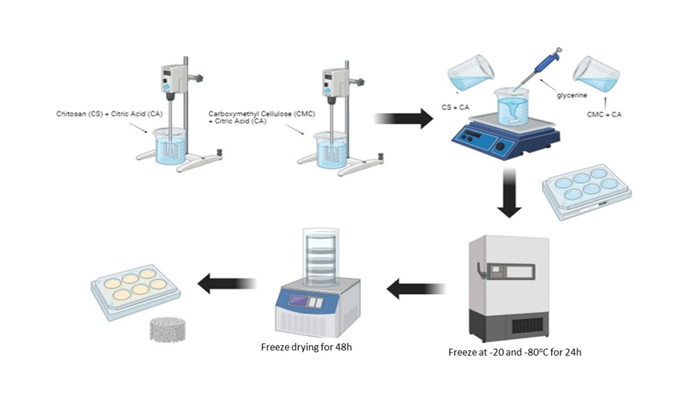
CS Solutions were prepared in concentrations of 1%, 2%, and 3% blended with 160 mg of CA dissolved in 1 mL of DI (Pan et al., 2023). CMC Solution is formulated at a 2% concentration crosslinked with CA, and enriched with Glycerol at 1.5%. Once the individual solutions are prepared, they are meticulously mixed for 3 hours. Following this, the combined solution is transferred into molds and allowed to set for 12 hours. To eliminate any entrapped air bubbles, the samples undergo either a 12-hour degassing process or ultrasonic treatment. The next stage involves a controlled freezing process is initial freezing at -20 °C for 3 hours. Subsequent deep-freezing at -80 °C for approximately 12 hours. Finally, the samples undergo lyophilization at -50 °C for an extended duration of 48 hours. The sponge samples are then stored in a cool, vacuum-sealed environment to maintain their integrity and stability.
2.3. Structural characterization
The evaluation of sponge morphology involves the evaluation of various macroscopic characteristics, such as color, surface features, consistency, and flexibility. High-resolution photography was used to capture detailed images of the sponge specimens in their initial, folded, and recovered states. Scanning electron microscopy with energy-dispersive X-ray microanalysis (SEM-EDS) was employed for microstructural analysis and chemical composition examination, and a 15 kV beam accelerator voltage was used to comprehensively examine the surface and cross-sectional micrographs and to precisely cut 3 mm × 3 mm samples for detailed analysis. The samples were coated with a thin layer of gold before observation to enhance the imaging quality and preserve the sample structure. ImageJ software was used to measure the pore sizes of 20-250 randomly selected pores for each material, with the average pore sizes calculated for each sample. This quantitative assessment provides valuable insights into the physical characteristics of materials. The FT-IR spectra of the materials were acquired using an Agilent Cary 630 FTIR spectrometer, capturing spectra within the wavenumber range of 650-4000 cm−1 with a scanning resolution of ≤ 2 cm−1. Robust data collection was ensured using 15 consecutive scans for each sample.
2.4. Mechanical properties
The mechanical properties of the samples were assessed through various tests, such as visual compressive stress, elongation break, and tensile strength, both in dry and wet conditions. A Universal Testing Machine (UTM) was used to measure the mechanical properties of the sponges. The sponges were cut into rectangular strips and clamped into the steel grip jaws of the UTM instrument for measurement. The tensile strengths of the wet and dry materials were measured at room temperature using a 5 kN load cell at a crosshead speed of 25 mm/min. The results are expressed as the maximum force at rupture and elongation ratio, and a minimum of three samples were used for each condition (Zhao et al., 2019). The elongation calculation involves the use of two parameters: the final length of the sample, denoted as L after it has been broken, and the initial length of the sample, referred to as L0, before testing (Monfared-Hajishirkiaee et al., 2023; Equation 1)
To determine the Young's modulus of each biomaterial, the relevant equation was applied to each point on the stress-strain curve (Jimenez-Martin et al., 2022; Equation 2).
2.5. Blood and water absorbance capacity
Blood absorbance and maximum blood swelling capacity were tested by immersing within 5 seconds reweighed (W0) samples (3 samples per group) of the materials in 500 μL of blood. After 5 seconds and 5 min, to allow complete blood absorption and maximum blood swelling capacity, samples were weighed again (m). The mass ratios of DI water or blood to materials (%) were calculated using the following Equation 3:
m0 and m are the weights of the initial samples, and the sample is immersed in media.
Subsequently, the samples underwent immersion in DI maintained at 37 °C for 10 seconds to attain saturated water absorption. Upon completion of the water absorption phase, each sample was delicately removed and placed on filter paper to eliminate any excess water from the surface. Immediately following this, the wet weight of the sample (m) was measured (Jimenez-Martin et al., 2022; Pan et al., 2023).
2.6. Swelling ratio
The swelling ratio was determined using a previously reported method (Zhou et al., 2022). The sponges were weighed (W0) and immersed in PBS (pH 7.4) at 37 0C for different time points (10, 20, 40, and 60 minutes). After each time point, excess water was removed using filter paper, and the weight of the sponges was denoted as Wi. The swelling ratio was calculated using the following Equation 4:
2.7. Antimicrobial assay
The disk diffusion method was used to assess the antimicrobial properties of sponges against Escherichia coli and Staphylococcus aureus. McFarland standard (0.5), comprising 108 CFU/ml, was prepared from a diluted suspension in Mueller-Hinton agar and placed on plates. The antibiotic discs containing tetracycline served as the control group. Each sponge sample was placed at the center of a culture dish and incubated at 37 °C for 24 h. The diameter of the inhibitory zones was measured to determine antimicrobial activity. The experiment was conducted three times in a sterile environment using sterile equipment.
2.8. In vitro biodegradation test
The assessment of the in vitro sample degradation involved the initial measurement of the weight of the material (Wo) while immersed in 10 mL of PBS (pH 7.4) at 37 °C. The materials were then placed in an incubator at 37 °C. After intervals of 4-5 and 7 d, the samples were freeze-dried for 24 h, and their final weight (Wf) was measured (Wang et al., 2021; Equation 5).
2.9. Hemostatic properties
2.9.1. In vitro whole blood clotting time
The whole-blood clotting time was determined following the method described by Zhou et al, 2022. Briefly, a mixture of CaCl2 solution (375 μL, 25 mM) and whole blood (750 μL) obtained from volunteers was rapidly added to a tube containing the sponge (10 mg). The tube was gently incubated at 37 °C. After precisely 30 seconds, the tube was tilted at an angle of approximately 30°, and clotting observations were made. The coagulation time was recorded when complete clotting of the blood was observed. If the clotting time exceeded 30 minutes, it was noted as noncoagulated. Each sample underwent this procedure three times (n = 3) to ensure the reliability and consistency. Medical gauze and commercial hemostatic sponges were employed as controls (Zhou et al., 2022).
2.10. Biocompatibility tests
2.10.1. Hemolysis test
The assessment of hemolysis ratios for the samples was carried out by immersing them in normal saline and incubating them at 37 °C. Subsequently, 20 μL of whole blood stock dispersion was added to 1 mL of the sample suspension and incubated for an hour at 37 °C. The mixtures were then centrifuged for 10 min at 2000 rpm. The absorbance of the supernatant was measured at 545 nm using a UV-visible spectrophotometer. To ensure the accuracy of the results, normal saline mixed with the samples and distilled water mixed with the samples were used as the negative and positive controls, respectively. The hemolysis rate (HR, %) was calculated using the following Equation 6:
where Dn, Dp, and Ds represent the absorbance of normal saline mixed with samples, distilled water mixed with samples, and the sample, respectively. The experiment was conducted three times to ensure precision (Shakiba-Marani and Ehtesabi, 2023).
2.10.2. Cytotoxicity assay
2.10.2.1. Cell culture
Cells from BHK-21[C-13] baby hamsters were grown in MEM supplemented with FBS (10%) and penicillin-streptomycin (1%). These cell cultures were kept in a humidified environment at 37 °C and 5% CO2 for 24 h until the maximum density was achieved. Following the 24-hour incubation period, the spent medium was discarded and the cells were dissociated using trypsin-EDTA. The resulting cell suspension was centrifuged at 2000(rpm) for 5 minutes. The supernatant was removed and the resulting cell pellet was resuspended in a fresh growth medium. For the preparation of 96-well microplates, 100 µL of the prepared viable cell suspension, was dispensed into each well. Subsequently, the microplates were maintained in the incubator for an additional 24-hour period until they reached a confluence of at least 90%.
2.10.2.2. Cell viability
Sterilized sponge samples were placed into 96-well plates, and BHK-21 (C-13) cells were manually counted using a hemocytometer and subsequently seeded onto their surfaces at a density of 3 × 10³ cells/cm2. The wells were divided into experimental groups, including non-treated (NT), positive control, and sponge-treated groups, each prepared in triplicate. After 24 hours of incubation, 20 μL of MTT solution (5 mg/mL) was added to each well, followed by incubation at 37 °C for 4 hours. The resulting formazan crystals were dissolved in 100 μL of dimethyl sulfoxide (DMSO), and the optical density (OD) was measured using an ELISA reader at a wavelength of 540 nm. A control group, consisting of cells not exposed to the sponge materials and assumed to have 100% viability, was maintained. Cell viability was calculated using the following Equation 7:
This analysis provided a quantitative assessment of the cytotoxic potential of sponge extracts on fibroblast cell lines over various time intervals. The approach supports the evaluation of material biocompatibility in vitro.
2.11. Statistical analysis
Statistical analyses were carried out using Student’s t-tests (non-parametric) and Dunnett tests, with the aid of GraphPad Prism version 9.5.1 software and an ANOVA test to determine statistical significance. The numbers have not been altered, and the data is presented as the mean value ± standard deviation (SD). P< 0.05 was used as the threshold for indicating a statistically significant difference.
3. Results and Discussion
3.1. Morphology of the sponges
The pore architecture of biomaterials designated for application as hemostatic agents or wound dressings plays a pivotal role in their efficacy. In this study, we proposed a novel approach with soft, odorless, white-colored multiporous sponges to reduce and stop bleeding in the nasal cavity, aiming to enhance patient comfort and optimize wound healing conditions. Our innovation builds upon the previous study's emphasis on soft surfaces to minimize mucosal adhesion, providing a gentle yet effective solution for nasal packing interventions (Bayat Tork et al., 2016). Glycerol is commonly employed as a softening agent in polymer production due to its ability to impart flexibility and elasticity to these materials. Their use has a significant impact on the structure of the resulting sponges (Monfared-Hajishirkiaee et al., 2023). A schematic representation of the fabrication process for the composite hemostatic device is shown in Figure 1. Scanning electron microscopy (SEM) analysis of the porous material CS-CMC2 revealed a distinctly interconnected porous framework. The sponges exhibit a surface characterized by varying quantities of interconnected pores, spanning diameters from 25.3 to 230 μm (Figure 2A). Moreover, the cross-sectional morphology of the CS-CMC2 sponge exhibits a porous and stratified arrangement (Figure 2B). These macropores are intricately linked by convoluted mesopores, thereby engendering a hierarchical porous configuration. It is noteworthy that the dimensions of these pores bear a profound relationship with the hemostatic prowess of the material (Zheng et al., 2020). Striking an optimal balance between larger pore dimensions conducive to the internalization of red blood cells and platelets, and smaller pores that facilitate an adequate surface area crucial for effective cell adhesion, represents a paramount consideration (Murphy and O’Brien, 2010).

Schematic illustration of the fabrication process for the composite hemostatic material. The figure was created using BioRender.com.

Scanning electron microscope micrographs of CS-CMC2 groups with surface view (A) and cross-sectional (B) images. The original magnification is 630x and the scale bar is equal to 200 μm. (C) Result of SEM-EDS spectrum analysis of CS-CMC2 sponge composite containing. Elemental mapping of EDS analysis clarified the elemental constituents, namely carbon (C), oxygen (O), sodium (Na), and chlorine (Cl), present within the composite material.
Although the calculated average pore size ranges from 25 to 230 μm, all scaffolds exhibited high porosity levels suitable for hemostasis applications. Our study's average pore size aligns comparably with previous research on freeze-dried scaffolds derived from CS/CMC (50-300 μm) (Dobaj Štiglic et al., 2021) and CMC-NHS crosslinked with CA, which reported diameters spanning 50 to 200 μm (Pan et al., 2023). Additionally, investigations into CS/CA scaffolds have revealed a network structure featuring connected macropores sized between 31 and 78.4 μm, and with the introduction of Ag nanoparticles, an average pore size of 67.2 μm was observed (Cao et al., 2023). In another study involving CS/CMC with Ag nanoparticles, pore sizes ranged from 30 to 400 μm (Hasan et al., 2018). Furthermore, previous research has demonstrated varying pore size distributions across different materials, with gauze exhibiting the highest pore size at 479 μm, while basic gauze, Merocel®, and SP-CH showed pore size distributions 141-144 μm, (Jimenez-Martin et al., 2022) respectively.
The CS-CMC2 sponges were examined using SEM-EDS to identify the types of elements present (Figure 1). The elemental composition of the sample revealed peaks corresponding to carbon, oxygen, and sodium present in the sample. The carbon, nitrogen and oxygen elements were present in EDS images (Akin Sahbaz, 2023; Ali et al., 2018) for CS-CMC2 due to CS and CMC functional groups. After the CS-CMC cross-linking process involving CA, glycerol, and polymer chains, there was an increase in the content of carbon and oxygen elements compared to the untreated samples, based on the results of previous studies (Kumar et al., 2020). This increase indicates the incorporation of additional functional groups containing carbon and oxygen into the material structure owing to cross-linking reactions. The increase in the peak intensity of carbon and oxygen indicates successful cross-linking between the polymer chains, which is facilitated by CA and glycerol. A significant decrease in the sodium (Na) peak intensity was also observed in the EDS spectrum after cross-linking. This reduction indicated a decrease in the presence of sodium-containing species, possibly caused by the formation of new chemical bonds with other elements (Kumar et al., 2020; Maslamani et al., 2022).
3.2. Fourier transform infrared spectroscopy (FTIR)
The formation of mechanically reinforced sponges was accomplished by incorporating CA into the CS-CMC mixture to create sponges. Fourier-transform infrared spectroscopy (FTIR) was used to characterize the synthesized sponges' chemical composition and structural properties. The FT-IR spectra of CMC, CS, CA, glycerol, and 3 groups of CS-CMC sponges are presented in Figure 3A.

FTIR spectrum of the crosslinked CS-CMC sponges and CS, CMC, CA, and Glycerol spectrum before synthesis (A). Results well plate and petri dish molding lightweight sponges (B-F). Shape recovery photographs of compressed CS-CMC2 sponges absorbing water and blood (E-G).
The analysis revealed distinctive peaks corresponding to the specific functional groups present in the materials under investigation. The CS-CMC sponge exhibited characteristic peaks at approximately 3330 and 3362 cm-1, attributed to the stretching vibrations of the -OH and -NH groups, including the amide II and CO-NH stretching bands. Additionally, weak peaks observed at 2111, 2083, and 2100 cm-1 were assigned to the –OH and C-H stretching of CMC, respectively. Notably, the peak at 1636-1638 cm-1 indicates the presence of –NH angular vibrational and carboxyl vibrational bands, with the C–O stretching vibrational band of amide I overlapping with this feature. The appearance of C–O stretching vibration peaks at 1636-1638 cm−1 suggests the successful integration of CA into CS, as supported by previous studies (Cao et al., 2023; Narasagoudr et al., 2020; Suo et al., 2018). Furthermore, the FTIR spectrum of CA displayed three distinct absorption peaks associated with the C–O stretching vibration originating from the vibration coupling of the carboxyl groups. These findings corroborate previous research, highlighting the spectroscopic characteristics of CA (Zhou et al., 2022). Analysis of CS revealed peaks at 3311 cm-1, corresponding to –OH and –NH2 stretching vibrations, and at 1585 cm-1 and 1321 cm-1, attributed to amide I and N–H deformation vibrations of amide, respectively (Fan et al., 2021; Wang et al., 2019). Similarly, CMC samples exhibited three characteristic absorption peaks at approximately 3334, 2877, and 1017 cm-1, representing the stretching vibrations of the –OH, –NH, –CH2, and C–O groups, respectively (Wang et al., 2021). Moreover, the presence of peaks around 897 cm-1 and 1160 cm-1 indicated the saccharide structure, whereas ether C–O–C functional groups were identified around 1017.35 cm-1 for CMC, as reported in recent studies (Xiong et al., 2010; Zhou et al., 2022). In the spectrum of the composite sponges, the –NH stretching vibration band was observed from approximately 3448 cm-1 to 3385 cm-1, indicating good compatibility and miscibility among the constituent materials (Cai et al., 2018; Cai and Kim, 2008). FTIR analysis confirmed the successful integration of carboxymethyl cellulose, chitosan, and citric acid all of which exhibited characteristic absorption peaks in the infrared spectrum.
3.3. Mechanical characterizations
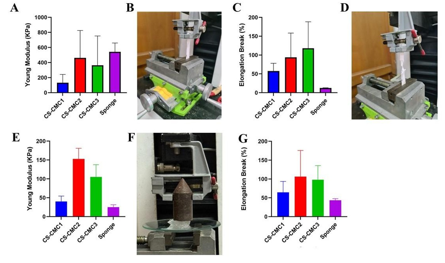
Improving the mechanical properties of nasal tampons is crucial for reducing the injuries associated with their use. These nasal tampons must have favorable mechanical characteristics and deformation resistance. Appropriate handling, clinical efficacy, and patient comfort depend on their development (Jimenez-Martin et al., 2022). Tensile stress and elongation testing revealed significant differences in the mechanical properties of the dry and wet sponges. The Young's modulus and elongation break results for the CS-CMC materials under dry and wet conditions are shown in Figure 4A-G. The tests of the mechanical properties of sponges as nasal packages or tampons for epistaxis under dry and wet conditions allow for comparisons across various environmental conditions and ensure a thorough understanding of their mechanical properties.
Mechanical characterization CS-CMC1, CS-CMC2, CS-CMC3, and commercial hemostatic sponges. (A) Young's modulus of the dry materials. (C)Elongation break of the dry materials. (B, D) Characterization photographs of the procedure carried out for tensile and (F) capacity to absorb fluid under pressure. (E) Young's modulus of wet materials. (G) Elongation break of the wet materials. The mean and standard deviation of the data are portrayed, with a sample size of n = 3.
The results revealed that Young's modulus of dry CS-CMC1, measured at 131±111 kPa, increased to 363 ±389 kPa in CS-CMC3 and further increased to 462±362 kPa. Commercial sponges exhibited a Young's modulus of 548±117 kPa. Upon evaluating the experiment, it became apparent that both CS-CMC2 and commercial sponges exhibited an increase in tensile strength compared to CS-CMC1 sponges. Dry CS-CMC1, CS-CMC2, and CS-CMC3 also showed elongations at break of 57 ± 20%, 93 ± 64%, and 118 ±70%, respectively. In comparison, the commercial hemostatic sponges exhibited an elongation break of only 12±0.6%. In addition, both CS-CMC2 and CS-CMC3 exhibited high elastic deformation before breaking with fluctuations.
The results indicated that Young's modulus of wet CS-CMC1, which was measured at 40±14 kPa, increased to 152±28 kPa in CS-CMC3 and further increased to 105±32 kPa. In contrast, wet commercial sponges displayed a Young's modulus of 25±6 kPa. Upon assessing the experiment, it became evident that both CS-CMC2 sponges exhibited significantly higher tensile strengths than CS-CMC1 and commercial sponges. Wet CS-CMC1, CS-CMC2, and CS-CMC3 also showed elongations at break of 64 ± 29%, 106 ± 69%, and 98 ± 37%, respectively. In comparison, wet commercial hemostatic sponges exhibited an elongation at break of only 43±4%. Furthermore, similar to the dry conditions, CS-CMC2 and CS-CMC3 exhibited high elastic deformations.
However, it is noteworthy that the overall mechanical behavior seems to remain consistent across various sponge-forming conditions, encompassing polymer concentration, ionic cross-linking concentration, and composition. CS-CMC preparation involved cross-linking CA at low temperatures, resulting in the crystallization of water during freeze-drying. The electrostatic attraction between NH3+ in CS and COO- in CA facilitates ionic cross-linking and sponge formation. The specific arrangement of CS chains during vacuum freeze-drying also contributed to sponge formation. In addition, glycerol content increases the flexibility and elasticity of sponges (Cao et al., 2023; Pan et al., 2023; Zhuang et al., 2020). Nevertheless, nasal tampons exert pressure that may induce discomfort during placement and removal, potentially leading to complications, such as rebleeding, mucosal abrasions, demucosalization, and scar detachment (Jimenez-Martin et al., 2022). Figure 3E and 3D shows that the CS-CMC2 sponge compressed and rapidly expanded to return to its original size within 5 s after absorbing water and blood, suggesting its potential utility in nasal tampons. However, the loss of absorbed fluid under pressure poses a challenge for achieving effective hemostasis. Compared to commercial gauze and sponges, CS-CMC2 demonstrated a superior water retention capacity under pressure, which is attributed to its unique microstructure, as shown in Figure 4. Additionally, the use of advanced materials with good mechanical strength under wet and dry conditions, similar to CS-CMC2, may offer a promising solution to improve patient comfort and outcomes. This reduces discomfort during placement and removal, and techniques to minimize the risk of rebleeding and mucosal damage should be explored. These advancements could have a significant positive impact on the effectiveness and safety of nasal tampons in clinical settings.
3.4. Water and blood absorbance
Wound dressings with superior absorption properties can efficiently draw fluid from the bloodstream, thereby aiding the clotting process. In addition, they can maintain a moist environment on the wound surface, thereby promoting healing (Lv et al., 2022; Zheng et al., 2022). The investigation of hemostatic materials revealed significant differences in their capacity to absorb blood and DI, which is crucial for managing profuse epistaxis. Cotton gauze, commercial hemostatic sponges, and CS-CMC sponges were evaluated for comparison (Figure 5). Figure 6 illustrates that the commercial sponges and gauze initially exhibited the highest blood-absorption ratios, with mean values of 1172 ± 157% and 1073 ± 131%, respectively, surpassing the absorption rates of CS-CMC2 (744 ± 116%), CS -CMC3 (585 ± 123%), and CS-CMC1 (442 ± 115%) sponges. Following a 5-minute interval, a notable increase in the blood absorption capacity of the sponges was observed. CS-CMC2 (1193 ± 435%), CS-CMC3 (1278± 241%), and CS-CMC1 (740 ± 86%) surpassed both the commercial sponge (1105 ± 178%) and the gauze (979 ± 202%). This enhancement was attributed to the highly interconnected porous structure and hydrophilic internal interface characteristics of the CS-CMC sponge. Furthermore, this study examined the water absorption capabilities of these materials within 5 s. Cotton displayed a water absorption ratio of 1080 ± 163%, mirroring its blood absorption rate. In contrast, the commercial sponge exhibited a higher ratio (1204 ± 136%). Conversely, CS-CMC1, CS-CMC2, and CS-CMC3 sponges displayed increased water-absorbing ratios of 711 ± 58%, 714 ± 25%, and 675 ± 68%, respectively, compared to their blood-absorbing capacities.

(A) Mass ratios of the absorbed blood at 5 s and 5 min and DI water (B) to CS-CMC sponges, cotton gauze, and commercial Hemostatic sponge (n = 3). Error bars, mean ± SD. The photograph of the proposed mechanism for compressed CS-CMC2 sponges and recovery shape by quick absorption PBS and swelling (C).

(A) The biodegradation properties of CS-CMC sponges, gauze, and commercial sponges were monitored in phosphate-buffered saline (PBS). Quantitative graph of weight loss (%) of samples against immersion time in 4, 7, and 12 days, respectively. n = 3 replicates for each group. (B) The swelling ratio of the CS-CMC groups, commercial sponge, and gauze samples(n=3).
The progressive increase in the blood-swelling capacity of the CS-CMC sponge provided several benefits. Primarily, a greater quantity of absorbed blood enables the creation of a more robust tamponade effect at the injury site, which is crucial for halting bleeding. The maximum blood-swelling capacity achieved by the sponge after 5 min makes it a viable option for addressing epistaxis, without causing continuous swelling. Notably, the CS-CMC sponge offers effective control and cessation of nasal bleeding without causing discomfort or irritation to patients during the treatment process. Intrinsic to the sponge's structure is CS, which has been demonstrated to be effective in facilitating blood clotting and reducing bleeding in the nasal cavity. Simultaneously, the increased blood volume absorbed by the sponge results in a higher concentration of red blood cells and platelets within the biomaterial, leading to enhanced hemostasis and thrombus formation (Jimenez-Martin et al., 2022). Additionally, the abundance of carboxyl groups results in a negatively charged interface that triggers blood coagulation through the intrinsic coagulation pathway potentially exacerbating micropore congestion in the sponge structure (Guo et al., 2021). In conclusion, this method is a promising alternative to nasal packaging for the treatment of epistaxis, providing both efficacy and patient comfort.
3.5. Swelling ratio
The ability of a substance to absorb fluid is a crucial factor in determining its potential to control bleeding in the nasal cavity. This is particularly important for hemostasis. The effects of various component ratios on the degree of swelling and weight loss were evaluated, and the results are shown in Figure 6A. The samples, including commercial gauze and sponge, displayed different swelling profiles, with the maximum swelling capacity ranging between 1614 ± 397% (CS-CMC2) and 1861 ± 997% (CS-CMC1), and the lowest at 1545 ± 533%, (CS-CMC3) of their initial weight after 20 min. During this period, the gauze and sponge absorbed 953 ± 158%, and 1283 ± 453% of their initial weight, respectively. All sponges swelled rapidly at the beginning, and the curves for the gauze, commercial sponge, and CS-CMC2 started to stabilize after 20 min. In contrast, the CS-CMC1 and CS-CMC3 groups continued to absorb water for 40 min. The swelling capacity observed for CA-cross-linked CS-CMC scaffolds(<1000%) was in good agreement with the values obtained for similar polysaccharide scaffolds, including CS/CMC dehydrothermal (DHT) treatment in the dry state after PEC (Dobaj Štiglic et al., 2021), CH (chitin), and soybean nasal scaffolds (Jimenez-Martin et al., 2022), as well as CA-crosslinked and N-hydroxysuccinimide (NHS) ester-activated carboxymethyl cellulose (CMC-NHS)(Pan et al., 2023), as well as CA cross-linked CS with reinforcing agents such as silver nanoparticles (Ag NPs) that absorb a greater amount of fluid than those without Ag NPs(Cao et al., 2023).
Water absorption characteristics are of paramount importance in epistaxis management, as nasal compression achieves hemostasis by applying pressure to the damaged blood vessels. These compresses absorb fluids from the wound and expand within the nostril, maintaining pressure without causing harm to the nasal cavity. However, excessive pressure can lead to complications, such as compression displacement, respiratory issues, reduced sense of smell, and potential tissue damage (Beck et al., 2018; García Callejo et al., 2010; Viehweg et al., 2006). These CS-CMC sponges indicate a shift towards tailored solutions that not only effectively address hemostasis but also prioritize patient comfort and overall nasal health.
3.6. Antimicrobial assay
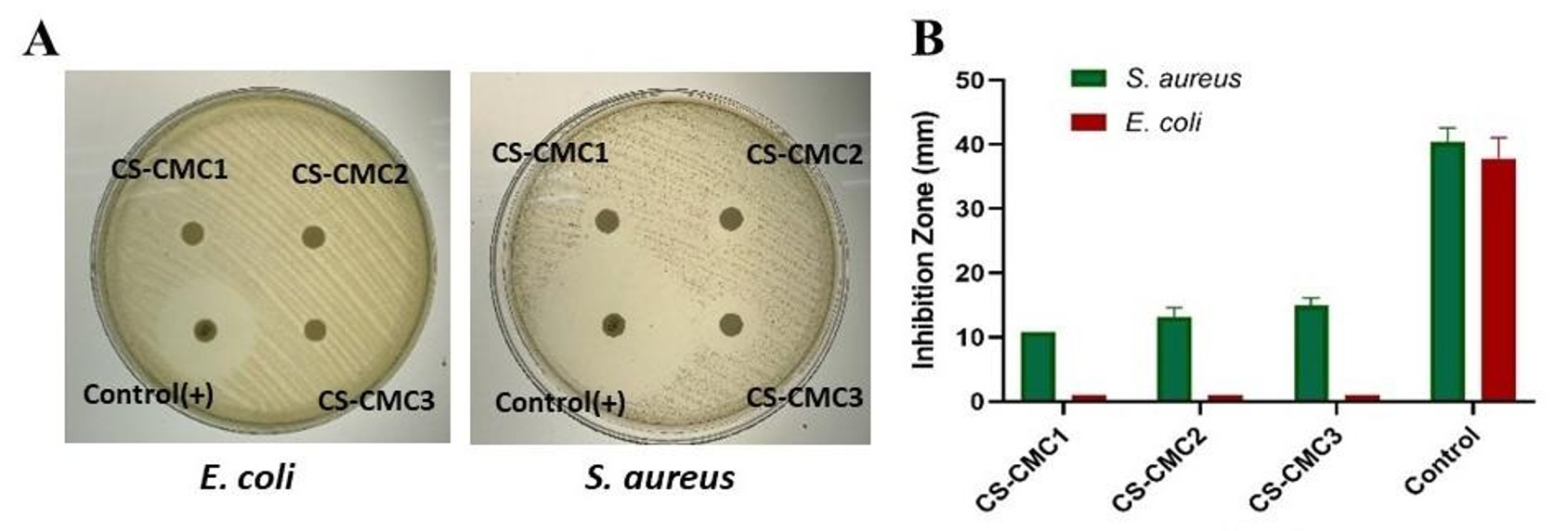
Bacterial infections frequently complicate bleeding wounds (Zhao et al., 2017), underscoring the importance of antibacterial properties in hemostatic agents (Cao et al., 2023; Chen et al., 2021; Zhang et al., 2021). Consequently, nasal tampons should exhibit robust antibacterial characteristics. In evaluating the antibacterial efficacy of CS-CMC sponges, bacterial solutions were co-cultured with the sponges. CS-CMC sponges, composed of CS, CMC, and CA, are distinguished by their abundant carboxyl groups, suggesting potential intrinsic antibacterial efficacy. However, CS-CMC sponges did not manifest an inhibition zone against gram-negative E. coli bacteria. As depicted in Figures 7A and 7B, all CS-CMC sponge groups exhibited inhibition zones compared to the control group, with a gradual reduction observed with decreasing CS concentrations. The bacterial inhibition zones displayed a proportional increase with rising CS concentrations. Furthermore, the Inhibition zone of CS-CMC1, CS-CMC2, and CS-CMC3 sponges against S. aureus was determined to be 9.24 ± 13.4, 11.36 ± 14.98 and 12.37 ± 17.98, respectively. Similarly, the antibacterial activity of CS/CA with a CA concentration of 2.8% on S. aureus and E. coli were 26.03 ± 11.27% and 17.74 ± 4.48% respectively (Cao et al., 2023). Gentamicin-loaded CMC/CS composites inhibit E. coli, and the diameters of the antibacterial circles are 2.35-4.96 mm (Dobaj Štiglic et al., 2021). These findings suggest that within CS-CMC sponges, even at higher concentrations of CS, significant inhibition of E. coli growth was not observed, while the antibacterial efficacy against S. aureus notably increased with CS concentrations. The observed antibacterial efficacy in the samples predominantly originated from CS. This phenomenon can be attributed to CS's inherent antibacterial activity, facilitated by its ability to neutralize negative charges within bacteria through the presence of radical amino groups (-NH2) carrying positive charges. Additionally, since the samples were composed of CMC and CA, both of which are abundant in carboxyl groups, this composition suggested that the samples also possessed antibacterial capabilities (Cai et al., 2018; Pan et al., 2023; Yuan et al., 2020)

Antibacterial assay of CS-CMC sponges (A) A visual representative display picture of the Inhibition zone of E. coli and S. aureus; (B) Depiction of the inhibition zones observed. Error bars, mean ± SD.
3.7. In vitro biodegradation test
The use of aqueous media for weight loss in nasal packaging biomaterials is gaining popularity because of its ability to minimize patient discomfort and rebleeding upon removal (Berlucchi et al., 2009). In this study, the degradation characteristics of various nasal packing materials were assessed, focusing on their weight loss over 12 days in an aqueous environment. Gauze showed no weight loss, highlighting its non-degradable nature, while commercial sponges degraded significantly, losing 65% of their initial weight (Figure 6B). CA-crosslinked sponges, including CS-CMC2 and CS-CMC3, displayed similar degradation patterns, with weight losses ranging from 54% to 65%, indicating a controlled degradation process. CS-CMC1 exhibited a 50% weight loss initially but showed more significant fragmentation after 12 days. The rapid degradation observed in CS-CMC1 and CS-CMC2 sponges, particularly after four days, underscores the hydrophilic nature of CMC, which facilitates dissolution in aqueous environments.
The total weight loss or degradation of the CS-CMC2 and CS-CMC3 samples over 12 days in PBS was comparable to or even better than that of CS/CMC-based materials reported in the literature, where 20–40% degradation within 28 days was observed using biofluids (Dobaj Štiglic et al., 2021). A previous study reported a significant weight loss of approximately 25% of the original weight within 48 h for an SP-CH scaffold for a nasal packaging. This weight loss was attributed to the presence of glycerol, which comprised 30% of the total weight of the scaffold and was subsequently dissolved in water (Jimenez-Martin et al., 2022). The fact that CS-CMC1 and CS-CMC2 degraded 50% of their weight after four days compared to gauze and commercial sponges demonstrates the hydrophilic nature of CMC, which facilitates its dissolution in aqueous environments. Additionally, the interactions between chitosan and CMC chains owing to the presence of amino and carboxyl groups further validated the successful cross-linking achieved through the use of CA. This enhanced wet resilience not only prevents uncontrolled and rapid degradation but also contributes to the balanced stability of the sponges in biofluid environments. The incorporation of additional chemical cross-linking agents, such as CA and glycerol, as plasticizers has provided valuable insights into the broader implications of chemical modifications for enhancing the performance of biomaterials. Furthermore, the suitability of this material as a partially absorptive nasal pack has been established, with advantages that include eliminating concerns such as discomfort during removal and interference with wound healing as well as reducing the likelihood of nasal adhesions (García Callejo et al., 2010; Wormald et al., 2006).
3.8. In vitro whole blood clotting time
The ability to promote blood clotting is critical for biomaterials utilized in treating bleeding wounds, such as nasal hemorrhage (Jimenez-Martin et al., 2022). Therefore, we conducted an in vitro whole blood coagulation assay to evaluate the coagulation ability of CS-CMC sponges compared to a commercial hemostatic sponge and cotton gauze. According to the graph in Figure 8B, the efficacy of CS-CMC1, CS-CMC2, and CS-CMC3 in reducing the blood coagulation time was markedly superior to that of gauze and commercial sponges. The commercial sponge exhibited the fastest blood absorption and, consequently, the slowest blood-clotting time, with an index of 201 ± 91%. In contrast, the gauze demonstrated significantly slower performance at 364 ± 8% compared to CS-CMC1, CS-CMC2, and CS-CMC3, which showed indices of 177 ± 34%, 131 ± 10%, and 115 ± 6%, respectively. The effect of coagulation on CS-CMC sponges exhibited noticeable variation, even when the difference in CS content was minimal. This variation can be prescribed to the interaction between the carboxyl groups of CMC and the amino groups in CS, which resulted in a decrease in the cationic amino groups on the surface of the CS-CMC sponges (Khan and Mujahid, 2019).

In vitro hemocompatibility and hemostatic property materials. (A, C) Hemolysis assay of the CS-CMC sponges with negative control (normal saline water) and positive control (DI water). (B) Hemostatic time of CS-CMC sponges, gauze, and commercial hemostatic sponge in vitro. Tests for each group were replicated 3 times (n = 3). The mean ± standard deviation was used to present the data.
We aimed to ascertain the red blood cell (RBC) adhesion or clotting capacity of the sample in question. The adhesion of RBCs to the surface of the biomaterial plays a critical role in promoting hemostasis by forming stable and strong blood clots (Biranje et al., 2020). This assay reinforced the results obtained in Figure 9B, confirming the higher RBC adhesion to the surfaces of CS-CMC2 and CS-CMC3. The blood absorbed by the sponges formed a strong clot due to its interaction, the CS-CMC2 and CS-CMC3 samples appeared darker due to higher RBC adhesion, and the liquid collected afterward presented a lighter color due to the lower hemoglobin concentration. In contrast to the gauze and commercial sponge, which were unable to form clots that maintained their integrity, the addition of DI to the liquid in each petri dish resulted in a red color, confirming the decreased hemostatic effects of the washed materials compared to the CS-CMC sponges. The interaction of strong blood absorption, porous structure, and cationic amino groups on the surface of CS-CMC2 and CS-CMC3 led to the fortified coagulation efficiency of the sponges. The internal structure of sponges allows for the rapid absorption and clotting of red blood cells when exposed to blood. Studies have shown that the positive charge of the amino groups in chitosan facilitates the aggregation of red blood cells and platelets through electrostatic adsorption, which is believed to enhance clotting and further support the hemostatic properties of the CS-CMC sponges (Zhou et al., 2022).

(A) In vitro, cytocompatibility tests for CS-CMC hemostatic sponges and the control group were cells treated without a sample(n=3). Error bars, mean ± SD. The line indicates 70% cell viability. (B) Photographs depict the visual appearance of the materials at various stages in the process of determining the presence of blood within the hemostatic materials. Whole blood was dropped on each sample until fully swelled, and DW was added and shaken to diffuse the erythrocytes that were not clotted in the sponge. Liquid collected after removing hemostatic materials.
3.9. Hemolysis test
A hemolysis assay was carried out (Figures 8A and 8C) to evaluate the hemocompatibility of the materials, which is crucial to exhibit minimal hemolysis, or the destruction of red blood cells (RBCs) brought about by shear stress or changes in osmotic pressure (Weber et al., 2018). The results of the hemolysis assay offer valuable information regarding the hemocompatibility of the assessed biomaterials. CS-CMC1 showed a minimal hemolysis rate of 0.17 ± 0.3%, indicating its exceptional hemocompatibility. Similarly, CS-CMC2 exhibited a significantly reduced hemolysis rate of 0.7 ± 0.1% compared to both CS-CMC1 and CS-CMC3, further confirming its suitability for blood-contact applications. Although CS-CMC3 showed a slightly higher hemolysis rate of 1.3 ± 0.3%, the absence of a significant difference in hemocompatibility compared to CS-CMC1 suggests its potential for certain blood-contact applications. These findings align with the ISO 10993-4:2002 standard, which specifies a maximum acceptable hemolysis rate of 5% for blood-contact biomaterials (Meng et al., 2022). The results of the hemolysis assay indicate that all tested hemostatic sponges fall well below this threshold, thus establishing their non-hemolytic nature and confirming their hemocompatibility.
3.10. Cytotoxicity assay
Hemostatic agents and wound dressings ideally display good cytocompatibility (Zhang et al., 2022). The direct cytotoxicity was assessed to validate the biocompatibility of the materials under investigation. To evaluate the biocompatibility of the scaffolds, we seeded BHK-21 cells. The experimental procedures followed the guidelines outlined in ISO 10993-5:2009 for biological assessment of medical devices. In cytotoxicity tests, CS-CMC1 and CS-CMC2 exhibited cell viabilities exceeding 70%, thus confirming their biocompatibility. As shown in Figure 9A, the viability of the cells treated with the CS-CMC1(85%) and CS-CMC2(84%) sponges was not significantly different from the control group those treated with cells not exposed to the sponges (86% viability) were used as the control group. Notably, no substantial differences in biocompatibility were observed between CS-CMC2 and CS-CMC3 in either assay. However, specimens CS-CMC3 with CS3% concentration displayed significantly reduced biocompatibility (64%) compared to the CS-CMS2, CS-CMC1, and control groups, and in the direct cytotoxicity evaluation. Although the study detected a reduction in biocompatibility with samples containing a concentration of CS3%, it was attributed to the high degree of deacetylation (DDA) of chitosan utilized in this research. The DDA of chitosan affects the number of free primary amino groups on its main structure when the DDA value is high (>90%). Chitosan with a high DDA level exhibits slight toxicity (Caprifico et al., 2021; Xia et al., 2022).
4. Conclusion
The current study explores the efficacy of CA crosslinked CMC and CS-based sponges for nasal tampons in the management of epistaxis. The sponges developed in this study exhibit desirable characteristics such as softness, lack of odor, and excellent mechanical properties under both wet and dry conditions. These features suggest that sponges have the potential to effectively manage hemostasis. The interconnected pore surfaces of the sponges facilitate the hemostatic process, and their water absorption properties enable them to maintain pressure in the nasal cavity without causing discomfort to the patient. This study emphasizes the potential antibacterial role of CS and CA against S. aureus, which prevents infections and accelerates wound healing in the nasal cavity. CS-CMC2 demonstrates significant weight loss in aqueous media, which indicates that it is a candidate for the biodegradable nasal package. Moreover, the biocompatibility and hemocompatibility of the CS-CMC2 sponges were confirmed through cytotoxicity tests, which highlighted their safety and blood-clotting abilities as an effective treatment for bleeding wounds, such as nasal hemorrhages. However, further comprehensive investigations are necessary to fully assess the effectiveness of these buffers for clinical use in in vivo blood-contacting applications.
Acknowledgements
The authors acknowledge and would like to thank the financial support for this study received from the Indonesian Government: Penelitian Disertasi Doktor year 2023: 011/E5/PG.02.00.PL/2023, and 749/UN3.LPPM/PT.01.03/2023TN/LLDIKTI.
Data Availability Statement
The data is available from the corresponding author on reasonable request.
References
-
ABDUR RAZZAK, M., KHORSHED ALAM, M., WAKILUR RAHMAN, M., SANAUL ISLAM, S., RAKIB HOSSAIN, M. and AL AMIN, Z., 2017. Functional endoscopic sinus surgery (FESS) for the management of chronic rhinosinusitis. Original Article Bangladesh J Otorhinolaryngol, vol. 23, no. 2, pp. 122-126. http://doi.org/10.3329/bjo.v23i2.45139
» http://doi.org/10.3329/bjo.v23i2.45139 -
ACIOĞLU, E., EDIZER, D.T., YIĞIT, Ö., ONUR, F. and ALKAN, Z., 2012. Nasal septal packing: which one? European Archives of Oto-Rhino-Laryngology, vol. 269, no. 7, pp. 1777-1781. http://doi.org/10.1007/s00405-011-1842-1 PMid:22160143.
» http://doi.org/10.1007/s00405-011-1842-1 -
AKIN SAHBAZ, D., 2023. Enhanced adsorption performance cross-linked chitosan/citrus reticulata peel waste composites as low-cost and green bio-adsorbents: kinetic, equilibrium isotherm, and thermodynamic studies. Polymers, vol. 15, no. 15, pp. 3246. http://doi.org/10.3390/polym15153246 PMid:37571140.
» http://doi.org/10.3390/polym15153246 -
ALI, N., AWAIS., KAMAL, T., UL-ISLAM, M., KHAN, A., SHAH, S.J. and ZADA, A., 2018. Chitosan-coated cotton cloth supported copper nanoparticles for toxic dye reduction. International Journal of Biological Macromolecules, vol. 111, pp. 832-838. http://doi.org/10.1016/j.ijbiomac.2018.01.092 PMid:29355628.
» http://doi.org/10.1016/j.ijbiomac.2018.01.092 -
BAL-OZTURK, A., KARAL-YILMAZ, O., AKGUNER, Z.P., AKSU, S., TAS, A. and OLMEZ, H., 2019. Sponge-like chitosan-based nanostructured antibacterial material as a topical hemostat. Journal of Applied Polymer Science, vol. 136, no. 19, pp. 47522. http://doi.org/10.1002/app.47522
» http://doi.org/10.1002/app.47522 -
BAYAT TORK, M., HEMMATI NEJAD, N., GHALEHBAGH, S., BASHARI, A., SHAKERI-ZADEH, A. and KAMRAVA, S.K., 2016. In situ green synthesis of silver nanoparticles/chitosan/poly vinyl alcohol/poly ethylene glycol hydrogel nanocomposite for novel finishing of nasal tampons. Journal of Industrial Textiles, vol. 45, no. 6, pp. 1399-1416. http://doi.org/10.1177/1528083714560255
» http://doi.org/10.1177/1528083714560255 -
BECK, R., SORGE, M., SCHNEIDER, A. and DIETZ, A., 2018. Current approaches to epistaxis treatment in primary and secondary care. Deutsches Ärzteblatt International, vol. 115, no. 1-02, pp. 12-22. http://doi.org/10.3238/arztebl.2018.0012 PMid:29345234.
» http://doi.org/10.3238/arztebl.2018.0012 -
BERLUCCHI, M., CASTELNUOVO, P., VINCENZI, A., MORRA, B. and PASQUINI, E., 2009. Endoscopic outcomes of resorbable nasal packing after functional endoscopic sinus surgery: a multicenter prospective randomized controlled study. European Archives of Oto-Rhino-Laryngology, vol. 266, no. 6, pp. 839-845. http://doi.org/10.1007/s00405-008-0841-3 PMid:18946677.
» http://doi.org/10.1007/s00405-008-0841-3 -
BIRANJE, S.S., MADIWALE, P.V., PATANKAR, K.C., CHHABRA, R., BANGDE, P., DANDEKAR, P. and ADIVAREKAR, R.V., 2020. Cytotoxicity and hemostatic activity of chitosan/carrageenan composite wound healing dressing for traumatic hemorrhage. Carbohydrate Polymers, vol. 239, pp. 116106. http://doi.org/10.1016/j.carbpol.2020.116106 PMid:32414437.
» http://doi.org/10.1016/j.carbpol.2020.116106 -
BRESNIHAN, M., MEHIGAN, B. and CURRAN, A., 2007. An evaluation of Merocel and Series 5000 nasal packs in patients following nasal surgery: a prospective randomised trial. Clinical Otolaryngology, vol. 32, no. 5, pp. 352-355. http://doi.org/10.1111/j.1749-4486.2007.01517.x PMid:17883554.
» http://doi.org/10.1111/j.1749-4486.2007.01517.x -
CAI, Z. and KIM, J., 2008. Characterization and electromechanical performance of cellulose–chitosan blend electro-active paper. Smart Materials and Structures, vol. 17, no. 3, pp. 035028. http://doi.org/10.1088/0964-1726/17/3/035028
» http://doi.org/10.1088/0964-1726/17/3/035028 -
CAI, B., ZHONG, T., CHEN, P., FU, J., JIN, Y., LIU, Y., HUANG, R. and TAN, L., 2018. Preparation, characterization and in vitro release study of drug-loaded sodium carboxy-methylcellulose/chitosan composite sponge. PLoS One, vol. 13, no. 10, e0206275. http://doi.org/10.1371/journal.pone.0206275 PMid:30346976.
» http://doi.org/10.1371/journal.pone.0206275 -
CAO, S., BI, Z., LI, Q., ZHANG, S., SINGH, M. and CHEN, J., 2023. Shape memory and antibacterial chitosan-based cryogel with hemostasis and skin wound repair. Carbohydrate Polymers, vol. 305, pp. 120545. http://doi.org/10.1016/j.carbpol.2023.120545 PMid:36737195.
» http://doi.org/10.1016/j.carbpol.2023.120545 -
CAPRIFICO, A.E., FOOT, P.J.S., POLYCARPOU, E. and CALABRESE, G., 2021. Overcoming the protein corona in chitosan-based nanoparticles. Drug Discovery Today, vol. 26, no. 8, pp. 1825-1840. http://doi.org/10.1016/j.drudis.2021.04.014 PMid:33892141.
» http://doi.org/10.1016/j.drudis.2021.04.014 -
CHATURVEDI, A., DOWLING, M.B., GUSTIN, J.P., SCALEA, T.M., RAGHAVAN, S.R., PASLEY, J.D. and NARAYAN, M., 2017. Hydrophobically modified chitosan gauze: a novel topical hemostat. The Journal of Surgical Research, vol. 207, pp. 45-52. http://doi.org/10.1016/j.jss.2016.04.052 PMid:27979487.
» http://doi.org/10.1016/j.jss.2016.04.052 -
CHEN, C., ZHOU, P., HUANG, C., ZENG, R., YANG, L., HAN, Z., QU, Y. and ZHANG, C., 2021. Photothermal-promoted multi-functional dual network polysaccharide hydrogel adhesive for infected and susceptible wound healing. Carbohydrate Polymers, vol. 273, pp. 118557. http://doi.org/10.1016/j.carbpol.2021.118557 PMid:34560968.
» http://doi.org/10.1016/j.carbpol.2021.118557 -
CHENG, F., HE, J., YAN, T., LIU, C., WEI, X., LI, J. and HUANG, Y., 2016. Antibacterial and hemostatic composite gauze of N,O-carboxymethyl chitosan/oxidized regenerated cellulose. RSC Advances, vol. 6, no. 97, pp. 94429-94436. http://doi.org/10.1039/C6RA15983D
» http://doi.org/10.1039/C6RA15983D -
DENIZ, M., ÇIFTÇI, Z., IŞIK, A., DEMIREL, O.B. and GÜLTEKIN, E., 2014. The impact of different nasal packings on postoperative complications. American Journal of Otolaryngology, vol. 35, no. 5, pp. 554-557. http://doi.org/10.1016/j.amjoto.2014.04.001 PMid:24943408.
» http://doi.org/10.1016/j.amjoto.2014.04.001 -
DOBAJ ŠTIGLIC, A., KARGL, R., BEAUMONT, M., STRAUSS, C., MAKUC, D., EGGER, D., PLAVEC, J., ROJAS, O.J., STANA KLEINSCHEK, K. and MOHAN, T., 2021. Influence of charge and heat on the mechanical properties of scaffolds from ionic complexation of chitosan and carboxymethyl cellulose. ACS Biomaterials Science & Engineering, vol. 7, no. 8, pp. 3618-3632. http://doi.org/10.1021/acsbiomaterials.1c00534 PMid:34264634.
» http://doi.org/10.1021/acsbiomaterials.1c00534 -
DURMUŞ, K., ALTUNTAŞ, E.E. and MILKOV, M., 2020. Management of Epistaxis. In: C. CINGI and N.B. MULUK, eds. All around the nose: basic science, diseases and surgical management. Cham: Springer, pp. 415-429. http://doi.org/10.1007/978-3-030-21217-9_48
» http://doi.org/10.1007/978-3-030-21217-9_48 -
FAN, Y., LU, Q., LIANG, W., WANG, Y., ZHOU, Y. and LANG, M., 2021. Preparation and characterization of antibacterial polyvinyl alcohol/ chitosan sponge and potential applied for wound dressing. European Polymer Journal, vol. 157, pp. 110619. http://doi.org/10.1016/j.eurpolymj.2021.110619
» http://doi.org/10.1016/j.eurpolymj.2021.110619 -
FRANKLIN, D.S. and GUHANATHAN, S., 2015. Investigation of citric acid-glycerol based pH-sensitive biopolymeric hydrogels for dye removal applications: a green approach. Ecotoxicology and Environmental Safety, vol. 121, pp. 80-86. http://doi.org/10.1016/j.ecoenv.2015.05.003 PMid:25982408.
» http://doi.org/10.1016/j.ecoenv.2015.05.003 -
FRANKLIN, J.H. and WRIGHT, E.D., 2007. Randomized, controlled, study of absorbable nasal packing on outcomes of surgical treatment of rhinosinusitis with polyposis. American Journal of Rhinology, vol. 21, no. 2, pp. 214-217. http://doi.org/10.2500/ajr.2007.21.3011 PMid:17424883.
» http://doi.org/10.2500/ajr.2007.21.3011 -
GARCÍA CALLEJO, F.J., MUÑOZ FERNÁNDEZ, N., ACHIQUES MARTÍNEZ, M.A., MOYA-ANGELER, S.F., MONTORO ELENA, M.J. and MARCO ALGARRA, J., 2010. Nasal packing in posterior epistaxis: comparison of two methods. Acta Otorrinolaringologica, vol. 61, no. 3, pp. 196-201. http://doi.org/10.1016/S2173-5735(10)70034-X PMid:20137770.
» http://doi.org/10.1016/S2173-5735(10)70034-X -
GHEORGHIȚĂ, D., MOLDOVAN, H., ROBU, A., BIȚA, A.I., GROSU, E., ANTONIAC, A., CORNESCHI, I., ANTONIAC, I., BODOG, A.D. and BĂCILĂ, C.I., 2023. Chitosan-based biomaterials for hemostatic applications: a review of recent advances. International Journal of Molecular Sciences, vol. 24, no. 13, pp. 10540. http://doi.org/10.3390/ijms241310540 PMid:37445718.
» http://doi.org/10.3390/ijms241310540 -
GUO, B., DONG, R., LIANG, Y. and LI, M., 2021. Haemostatic materials for wound healing applications. Nature Reviews. Chemistry, vol. 5, no. 11, pp. 773-791. http://doi.org/10.1038/s41570-021-00323-z PMid:37117664.
» http://doi.org/10.1038/s41570-021-00323-z -
HASAN, A., WAIBHAW, G., SAXENA, V. and PANDEY, L.M., 2018. Nano-biocomposite scaffolds of chitosan, carboxymethyl cellulose and silver nanoparticle modified cellulose nanowhiskers for bone tissue engineering applications. International Journal of Biological Macromolecules, vol. 111, pp. 923-934. http://doi.org/10.1016/j.ijbiomac.2018.01.089 PMid:29415416.
» http://doi.org/10.1016/j.ijbiomac.2018.01.089 -
ISHII, L.E., TOLLEFSON, T.T., BASURA, G.J., ROSENFELD, R.M., ABRAMSON, P.J., CHAIET, S.R., DAVIS, K.S., DOGHRAMJI, K., FARRIOR, E.H., FINESTONE, S.A., ISHMAN, S.L., MURPHY JUNIOR, R.X., PARK, J.G., SETZEN, M., STRIKE, D.J., WALSH, S.A., WARNER, J.P. and NNACHETA, L.C., 2017. Clinical practice guideline: improving nasal form and function after rhinoplasty. Otolaryngology - Head and Neck Surgery, vol. 156, suppl. 2, pp. S1-S30. http://doi.org/10.1177/0194599816683153 PMid:28145823.
» http://doi.org/10.1177/0194599816683153 -
JIMENEZ-MARTIN, J., LAS HERAS, K., ETXABIDE, A., URANGA, J., DE LA CABA, K., GUERRERO, P., IGARTUA, M., SANTOS-VIZCAINO, E. and HERNANDEZ, R.M., 2022. Green hemostatic sponge-like scaffold composed of soy protein and chitin for the treatment of epistaxis. Materials Today. Bio, vol. 15, pp. 100273. http://doi.org/10.1016/j.mtbio.2022.100273 PMid:35572855.
» http://doi.org/10.1016/j.mtbio.2022.100273 -
KANODIA, A., VERMA, H., JAIN, A., KALSOTRA, G., KUMARI, S., AGRAWAL, S.K., GAUTAM, H., KAUSHAL, D., GUGLIANI, A. and LODHA, J. 2021. Prevention and management of complications. In: H. VERMA and A. THAKAR, eds. Essentials of Rhinology. Singapore: Springer, pp. 277–307. http://doi.org/10.1007/978-981-33-6284-0_8
» http://doi.org/10.1007/978-981-33-6284-0_8 -
KHAFAGY, A.G. and MAAROUF, A.M., 2021. Polyurethane versus chitosan-based polymers nasal packs after functional endoscopic sinus surgery: a prospective randomized double-blinded study. American Journal of Rhinology & Allergy, vol. 35, no. 5, pp. 624-630. http://doi.org/10.1177/1945892420983645 PMid:33430613.
» http://doi.org/10.1177/1945892420983645 -
KHAN, M., CONROY, K., UBAYASIRI, K., CONSTABLE, J., SMITH, M.E., WILLIAMS, R.J., KUHN, I., SMITH, M. and PHILPOTT, C., 2017. Initial assessment in the management of adult epistaxis: systematic review. The Journal of Laryngology and Otology, vol. 131, no. 12, pp. 1035-1055. http://doi.org/10.1017/S0022215117002031 PMid:29280694.
» http://doi.org/10.1017/S0022215117002031 -
KHAN, M.A. and MUJAHID, M., 2019. A review on recent advances in chitosan-based composite for hemostatic dressings. International Journal of Biological Macromolecules, vol. 124, pp. 138-147. http://doi.org/10.1016/j.ijbiomac.2018.11.045 PMid:30447365.
» http://doi.org/10.1016/j.ijbiomac.2018.11.045 -
KIM, G.H., IM, J.N., KIM, T.H., LEE, G.D., YOUK, J.H. and DOH, S.J., 2018. Preparation and characterization of calcium carboxymethyl cellulose/chitosan blend nonwovens for hemostatic agents. Textile Research Journal, vol. 88, no. 16, pp. 1902-1911. http://doi.org/10.1177/0040517517712101
» http://doi.org/10.1177/0040517517712101 -
KUMAR, B., PRIYADARSHI, R., SAURAJ., DEEBA, F., KULSHRESHTHA, A., GAIKWAD, K.K., KIM, J., KUMAR, A. and NEGI, Y.S., 2020. Nanoporous sodium carboxymethyl cellulose-g-poly (Sodium acrylate)/fecl3 hydrogel beads: synthesis and characterization. Gels, vol. 6, no. 4, pp. 1-11. http://doi.org/10.3390/gels6040049 PMid:33322561.
» http://doi.org/10.3390/gels6040049 -
LIU, Z., XU, Y., SU, H., JING, X., WANG, D., LI, S., CHEN, Y., GUAN, H. and MENG, L., 2023. Chitosan-based hemostatic sponges as new generation hemostatic materials for uncontrolled bleeding emergency: Modification, composition, and applications. Carbohydrate Polymers, vol. 311, pp. 120780. http://doi.org/10.1016/j.carbpol.2023.120780 PMid:37028883.
» http://doi.org/10.1016/j.carbpol.2023.120780 - LORUSSO, F., DISPENZA, F., SIRECI, F., MODICA, D.M. and GALLINA, S., 2021. A comparative double blind study of nasal dressing Sponge® versus Merocel® as nasal pack after nasal surgery. Iranian Journal of Otorhinolaryngology, vol. 33, no. 119, pp. 369-373. PMid:35223654.
-
LV, C., ZHOU, X., WANG, P., LI, J., WU, Z., JIAO, Z., GUO, M., WANG, Z., WANG, Y., WANG, L. and ZHANG, P., 2022. Biodegradable alginate-based sponge with antibacterial and shape memory properties for penetrating wound hemostasis. Composites. Part B, Engineering, vol. 247, pp. 110263. http://doi.org/10.1016/j.compositesb.2022.110263
» http://doi.org/10.1016/j.compositesb.2022.110263 -
MAHMOODZADEH, A., MOGHADDAS, J., JAROLMASJED, S., EBRAHIMI KALAN, A., EDALATI, M. and SALEHI, R., 2021. Biodegradable cellulose-based superabsorbent as potent hemostatic agent. Chemical Engineering Journal, vol. 418, pp. 129252. http://doi.org/10.1016/j.cej.2021.129252
» http://doi.org/10.1016/j.cej.2021.129252 -
MASLAMANI, N., BAKHSH, E.M., KHAN, S.B., DANISH, E.Y., AKHTAR, K., FAGIEH, T.M., SU, X. and ASIRI, A.M., 2022. Chitosan@Carboxymethylcellulose/CuO-Co2O3 Nanoadsorbent as a super catalyst for the removal of water pollutants. Gels, vol. 8, no. 2, pp. 91. http://doi.org/10.3390/gels8020091 PMid:35200472.
» http://doi.org/10.3390/gels8020091 -
MENG, L., LIU, X., LIU, L., HONG, Q., CHENG, Y., GAO, F., CHEN, J., ZHANG, Q. and PAN, C., 2022. Comparative investigation of the corrosion behavior and biocompatibility of the different chemical conversion coatings on the magnesium alloy surfaces. Metals, vol. 12, no. 10, pp. 1644. http://doi.org/10.3390/met12101644
» http://doi.org/10.3390/met12101644 -
MONFARED-HAJISHIRKIAEE, R., EHTESABI, H., REZAEI, A. and NAJAFINOBAR, S., 2023. Development of carboxymethyl cellulose/chitosan double-layer hydrogel combining myrtle essential oil and thyme honey to enhance antibacterial and mechanical properties. Journal of Industrial and Engineering Chemistry, vol. 126, pp. 382-397. http://doi.org/10.1016/j.jiec.2023.06.027
» http://doi.org/10.1016/j.jiec.2023.06.027 -
MURPHY, C.M. and O’BRIEN, F.J., 2010. Understanding the effect of mean pore size on cell activity in collagen-glycosaminoglycan scaffolds. Cell Adhesion & Migration, vol. 4, no. 3, pp. 377-381. http://doi.org/10.4161/cam.4.3.11747 PMid:20421733.
» http://doi.org/10.4161/cam.4.3.11747 -
MYLONAS, S., SKOULAKIS, C., NIKOLAIDIS, V. and HAJIIOANNOU, J., 2023. Epistaxis treatment options: literature review. Indian Journal of Otolaryngology and Head and Neck Surgery, vol. 75, no. 3, pp. 2235-2244. http://doi.org/10.1007/s12070-023-03824-z PMid:37636777.
» http://doi.org/10.1007/s12070-023-03824-z -
NARASAGOUDR, S.S., HEGDE, V.G., VANJERI, V.N., CHOUGALE, R.B. and MASTI, S.P., 2020. Ethyl vanillin incorporated chitosan/poly(vinyl alcohol) active films for food packaging applications. Carbohydrate Polymers, vol. 236, pp. 116049. http://doi.org/10.1016/j.carbpol.2020.116049 PMid:32172863.
» http://doi.org/10.1016/j.carbpol.2020.116049 -
NOWAKOWSKA, M., MOCZEK, Ł. and SZCZUBIAŁKA, K., 2008. Photoactive modified chitosan. Biomacromolecules, vol. 9, no. 6, pp. 1631-1636. http://doi.org/10.1021/bm800141v PMid:18457451.
» http://doi.org/10.1021/bm800141v -
OHTA, S., NISHIYAMA, T., SAKODA, M., MACHIOKA, K., FUKE, M., ICHIMURA, S., INAGAKI, F., SHIMIZU, A., HASEGAWA, K., KOKUDO, N., KANEKO, M., YATOMI, Y. and ITO, T., 2015. Development of carboxymethyl cellulose nonwoven sheet as a novel hemostatic agent. Journal of Bioscience and Bioengineering, vol. 119, no. 6, pp. 718-723. http://doi.org/10.1016/j.jbiosc.2014.10.026 PMid:25488042.
» http://doi.org/10.1016/j.jbiosc.2014.10.026 -
PAN, S., LI, Y., TONG, X., CHEN, L., WANG, L., LI, T. and ZHANG, Q., 2023. Strongly-adhesive easily-detachable carboxymethyl cellulose aerogel for noncompressible hemorrhage control. Carbohydrate Polymers, vol. 301, no. Pt B, pp. 120324. http://doi.org/10.1016/j.carbpol.2022.120324 PMid:36446491.
» http://doi.org/10.1016/j.carbpol.2022.120324 -
PAN, Y., SHI, K., PENG, C., WANG, W., LIU, Z. and JI, X., 2014. Evaluation of hydrophobic polyvinyl-alcohol formaldehyde sponges as absorbents for oil spill. ACS Applied Materials & Interfaces, vol. 6, no. 11, pp. 8651-8659. http://doi.org/10.1021/am5014634 PMid:24797603.
» http://doi.org/10.1021/am5014634 -
SHAKIBA-MARANI, R. and EHTESABI, H., 2023. A flexible and hemostatic chitosan, polyvinyl alcohol, carbon dot nanocomposite sponge for wound dressing application. International Journal of Biological Macromolecules, vol. 224, pp. 831-839. http://doi.org/10.1016/j.ijbiomac.2022.10.169 PMid:36283554.
» http://doi.org/10.1016/j.ijbiomac.2022.10.169 -
SHAW, C.L., DYMOCK, R.B., COWIN, A. and WORMALD, P.J., 2000. Effect of packing on nasal mucosa of sheep. The Journal of Laryngology and Otology, vol. 114, no. 7, pp. 506-509. http://doi.org/10.1258/0022215001906246 PMid:10992930.
» http://doi.org/10.1258/0022215001906246 -
SPINELLA, S., MAIORANA, A., QIAN, Q., DAWSON, N.J., HEPWORTH, V., MCCALLUM, S.A., GANESH, M., SINGER, K.D. and GROSS, R.A., 2016. Concurrent cellulose hydrolysis and esterification to prepare a surface-modified cellulose nanocrystal decorated with carboxylic acid moieties. ACS Sustainable Chemistry & Engineering, vol. 4, no. 3, pp. 1538-1550. http://doi.org/10.1021/acssuschemeng.5b01489
» http://doi.org/10.1021/acssuschemeng.5b01489 -
SUO, H., ZHANG, D., YIN, J., QIAN, J., WU, Z.L. and FU, J., 2018. Interpenetrating polymer network hydrogels composed of chitosan and photocrosslinkable gelatin with enhanced mechanical properties for tissue engineering. Materials Science and Engineering C, vol. 92, pp. 612-620. http://doi.org/10.1016/j.msec.2018.07.016 PMid:30184788.
» http://doi.org/10.1016/j.msec.2018.07.016 - TUNKEL, D.E., ANNE, S., PAYNE, S.C., ISHMAN, S.L., ROSENFELD, R.M., ABRAMSON, P.J., ALIKHAANI, J.D., BENOIT, M.M.K., BERCOVITZ, R.S., BROWN, M.D., CHERNOBILSKY, B., FELDSTEIN, D.A., HACKELL, J.M., HOLBROOK, E.H., HOLDSWORTH, S.M., LIN, K.W., LIND, M.M., POETKER, D.M., RILEY, C.A., Schneider, J.S., Seidman, M.D., Vadlamudi, V., Valdez, T.A., Nnacheta, L.C. and MONJUR, T.M., 2020. Clinical practice guideline: nosebleed (epistaxis). Otolaryngology, vol. 162, suppl. 1, pp. S1-S38. PMid:31910111.
-
VIEHWEG, T.L., ROBERSON, J.B. and HUDSON, J.W., 2006. Epistaxis: diagnosis and Treatment. Journal of Oral and Maxillofacial Surgery, vol. 64, no. 3, pp. 511-518. http://doi.org/10.1016/j.joms.2005.11.031 PMid:16487816.
» http://doi.org/10.1016/j.joms.2005.11.031 -
WANG, J., LIANG, J., SUN, L. and GAO, S., 2019. PVA/CS and PVA/CS/Fe gel beads’ synthesis mechanism and their performance in cultivating anaerobic granular sludge. Chemosphere, vol. 219, pp. 130-139. http://doi.org/10.1016/j.chemosphere.2018.12.014 PMid:30537586.
» http://doi.org/10.1016/j.chemosphere.2018.12.014 -
WANG, Y., ZHAO, Y., QIAO, L., ZOU, F., XIE, Y., ZHENG, Y., CHAO, Y., YANG, Y., HE, W. and YANG, S., 2021. Cellulose fibers-reinforced self-expanding porous composite with multiple hemostatic efficacy and shape adaptability for uncontrollable massive hemorrhage treatment. Bioactive Materials, vol. 6, no. 7, pp. 2089-2104. http://doi.org/10.1016/j.bioactmat.2020.12.014 PMid:33511309.
» http://doi.org/10.1016/j.bioactmat.2020.12.014 -
WANG, Y., XIAO, D., YU, H., ZHONG, Y., ZHANG, L., SUI, X., WANG, B., FENG, X., XU, H. and MAO, Z., 2023. Composite hydrogel based oxidated sodium carboxymethyl cellulose and gelatin loaded carboxymethylated cotton fabric for hemostasis and infected wound treatment. International Journal of Biological Macromolecules, vol. 224, pp. 1382-1394. http://doi.org/10.1016/j.ijbiomac.2022.10.224 PMid:36306896.
» http://doi.org/10.1016/j.ijbiomac.2022.10.224 -
WEBER, M., STEINLE, H., GOLOMBEK, S., HANN, L., SCHLENSAK, C., WENDEL, H.P. and AVCI-ADALI, M., 2018. Blood-contacting biomaterials: in vitro evaluation of the hemocompatibility. Frontiers in Bioengineering and Biotechnology, vol. 6, pp. 99. http://doi.org/10.3389/fbioe.2018.00099 PMid:30062094.
» http://doi.org/10.3389/fbioe.2018.00099 -
WORMALD, P.J., BOUSTRED, R.N., LE, T., HAWKE, L. and SACKS, R., 2006. A prospective single-blind randomized controlled study of use of hyaluronic acid nasal packs in patients after endoscopic sinus surgery. American Journal of Rhinology, vol. 20, no. 1, pp. 7-10. http://doi.org/10.1177/194589240602000102 PMid:16539287.
» http://doi.org/10.1177/194589240602000102 -
XIA, Y., YANG, R., WANG, H., LI, Y. and FU, C., 2022. Application of chitosan-based materials in surgical or postoperative hemostasis. Frontiers in Materials, vol. 9, pp. 994265. http://doi.org/10.3389/fmats.2022.994265
» http://doi.org/10.3389/fmats.2022.994265 -
XIONG, X., DUAN, J., ZOU, W., HE, X. and ZHENG, W., 2010. A pH-sensitive regenerated cellulose membrane. Journal of Membrane Science, vol. 363, no. 1–2, pp. 96-102. http://doi.org/10.1016/j.memsci.2010.07.031
» http://doi.org/10.1016/j.memsci.2010.07.031 -
YUAN, H., CHEN, L. and HONG, F.F., 2020. A biodegradable antibacterial nanocomposite based on oxidized bacterial nanocellulose for rapid hemostasis and wound healing. ACS Applied Materials & Interfaces, vol. 12, no. 3, pp. 3382-3392. http://doi.org/10.1021/acsami.9b17732 PMid:31880915.
» http://doi.org/10.1021/acsami.9b17732 -
ZHANG, S., LI, J., CHEN, S., ZHANG, X., MA, J. and HE, J., 2020. Oxidized cellulose-based hemostatic materials. Carbohydrate Polymers, vol. 230, pp. 115585. http://doi.org/10.1016/j.carbpol.2019.115585 PMid:31887971.
» http://doi.org/10.1016/j.carbpol.2019.115585 -
ZHANG, H., SUN, X., WANG, J., ZHANG, Y., DONG, M., BU, T., LI, L., LIU, Y. and WANG, L., 2021. Multifunctional injectable hydrogel dressings for effectively accelerating wound healing: enhancing biomineralization strategy. Advanced Functional Materials, vol. 31, no. 23, pp. 2100093. http://doi.org/10.1002/adfm.202100093
» http://doi.org/10.1002/adfm.202100093 -
ZHANG, Y., WANG, Y., CHEN, L., ZHENG, J., FAN, X., XU, X., ZHOU, G., ULLAH, N. and FENG, X., 2022. An injectable antibacterial chitosan-based cryogel with high absorbency and rapid shape recovery for noncompressible hemorrhage and wound healing. Biomaterials, vol. 285, pp. 121546. http://doi.org/10.1016/j.biomaterials.2022.121546 PMid:35552114.
» http://doi.org/10.1016/j.biomaterials.2022.121546 -
ZHAO, X., WU, H., GUO, B., DONG, R., QIU, Y. and MA, P.X., 2017. Antibacterial anti-oxidant electroactive injectable hydrogel as self-healing wound dressing with hemostasis and adhesiveness for cutaneous wound healing. Biomaterials, vol. 122, pp. 34-47. http://doi.org/10.1016/j.biomaterials.2017.01.011 PMid:28107663.
» http://doi.org/10.1016/j.biomaterials.2017.01.011 -
ZHAO, Y.F., ZHAO, J.Y., HU, W.Z., MA, K., CHAO, Y., SUN, P.J., FU, X.B. and ZHANG, H., 2019. Synthetic poly(vinyl alcohol)-chitosan as a new type of highly efficient hemostatic sponge with blood-triggered swelling and high biocompatibility. Journal of Materials Chemistry. B, Materials for Biology and Medicine, vol. 7, no. 11, pp. 1855-1866. http://doi.org/10.1039/C8TB03181A PMid:32255048.
» http://doi.org/10.1039/C8TB03181A -
ZHENG, C., ZENG, Q., PIMPI, S., WU, W., HAN, K., DONG, K. and LU, T., 2020. Research status and development potential of composite hemostatic materials. Journal of Materials Chemistry. B, Materials for Biology and Medicine, vol. 8, no. 25, pp. 5395-5410. http://doi.org/10.1039/D0TB00906G PMid:32494795.
» http://doi.org/10.1039/D0TB00906G -
ZHENG, W., ZHANG, Z., LI, Y., WANG, L., FU, F., DIAO, H. and LIU, X., 2022. A novel pullulan oxidation approach to preparing a shape memory sponge with rapid reaction capability for massive hemorrhage. Chemical Engineering Journal, vol. 447, pp. 137482. http://doi.org/10.1016/j.cej.2022.137482
» http://doi.org/10.1016/j.cej.2022.137482 -
ZHOU, M., LIAO, J., LI, G., YU, Z., XIE, D., ZHOU, H., WANG, F., REN, Y., XU, R., DAI, Y., WANG, J., HUANG, J. and ZHANG, R., 2022. Expandable carboxymethyl chitosan/cellulose nanofiber composite sponge for traumatic hemostasis. Carbohydrate Polymers, vol. 294, pp. 119805. http://doi.org/10.1016/j.carbpol.2022.119805 PMid:35868765.
» http://doi.org/10.1016/j.carbpol.2022.119805 -
ZHUANG, L., ZHI, X., DU, B. and YUAN, S., 2020. Preparation of elastic and antibacterial chitosan-citric membranes with high oxygen barrier ability by in situ cross-linking. ACS Omega, vol. 5, no. 2, pp. 1086-1097. http://doi.org/10.1021/acsomega.9b03206 PMid:31984265.
» http://doi.org/10.1021/acsomega.9b03206
Edited by
-
Editor:
Takako Matsumura Tundisi