First serological investigation of Toxoplasma gondii, Neospora caninum, Leishmania infantum and Leptospira spp. in dogs from a Fulni-ô Indigenous community in Pernambuco, Brazil: a One Health perspective
Abstract
Zoonotic diseases pose a major public health threat in indigenous communities due to geographic isolation, inadequate sanitation, and close contact between humans, domestic animals, and wildlife. This study aimed to investigate the presence of antibodies against Toxoplasma gondii, Neospora caninum, Leishmania infantum, and Leptospira spp. in dogs from the Fulni-ô indigenous community, located in Águas Belas, Pernambuco, Brazil. Blood samples were collected from 66 free-roaming or semi-domiciled dogs for serological analysis. Indirect immunofluorescence tests (IFAT) were used to detect antibodies for T. gondii and N. caninum, while Leishmania infantum detection was performed using the TR DPP® Canine Visceral Leishmaniasis kit. The presence of Leptospira spp. was evaluated for microscopic agglutination test (MAT). Results revealed seropositivity rates of 46.9% (31/66) for T. gondii, 9.1% (6/66) for N. caninum, 22.7% (15/66) for Leishmania infantum, and 18.2% (12/66) for Leptospira spp. Among the Leptospira spp. serovars identified, Icterohaemorrhagiae (83.3%, 10/12) and Pomona (50.0%, 6/12) were most prevalent, which were associated with environmental reservoirs and inadequate sanitation conditions. The findings suggest that the dogs in the Fulni-ô community act as environmental sentinels, reflecting local zoonotic risks. This pioneering study highlights the need for One Health-based strategies to prevent zoonotic diseases in vulnerable indigenous communities.
Keywords:
public health; epidemiological surveillance; protozoa; neglected diseases
Resumo
Doenças zoonóticas representam uma grande ameaça à saúde pública em comunidades indígenas devido ao isolamento geográfico, saneamento inadequado e contato próximo entre humanos, animais domésticos e animais selvagens. Este estudo teve como objetivo investigar a presença de anticorpos contra Toxoplasma gondii, Neospora caninum, Leishmania infantum e Leptospira spp. em cães da comunidade indígena Fulni-ô, localizada em Águas Belas, Pernambuco, Brasil. Amostras de sangue foram coletadas de 66 cães soltos ou semidomiciliados para análise sorológica. Testes de imunofluorescência indireta (RIFI) foram usados para detectar anticorpos para T. gondii e N. caninum, enquanto a detecção de Leishmania infantum foi realizada usando o kit TR DPP® Canine Visceral Leishmaniasis. A presença de Leptospira spp. foi avaliada por teste de aglutinação microscópica (MAT). Os resultados revelaram taxas de soropositividade de 46,9% (31/66) para T. gondii, 9,1% (6/66) para N. caninum, 22,7% (15/66) para Leishmania infantum e 18,2% (12/66) para Leptospira spp. Entre os sorovares de Leptospira spp. identificados, Icterohaemorrhagiae (83,3%, 10/12) e Pomona (50%, 6/12) foram os mais prevalentes, os quais foram associados a reservatórios ambientais e condições inadequadas de saneamento. Os resultados sugerem que os cães na comunidade Fulni-ô atuam como sentinelas ambientais, refletindo os riscos zoonóticos locais. Este estudo pioneiro destaca a necessidade de estratégias baseadas em Saúde Única para prevenir doenças zoonóticas em comunidades indígenas vulneráveis.
Palavras-chaves:
saúde pública; vigilância epidemiológica; protozoários; doenças negligenciadas
1. Introduction
Emerging zoonotic diseases are estimated to account for approximately 2.4 billion cases and 2,7 million deaths annually worldwide (Harfield et al., 2015; Rahman et al., 2020). In Brazil, these diseases are particularly concerning in rural and semi-rural areas, such as indigenous communities, where close interactions between humans, domestic animals, and wildlife facilitate pathogen transmission (Canuto et al., 2018; Shah et al., 2024; Henrique et al., 2025).
The Fulni-ô Indigenous Territory, in Águas Belas, Pernambuco, is in the semi-arid Caatinga region and is home to approximately 4,000 people. Seasonality and recurrent droughts favor the persistence and spread of infectious agents, while isolation, the preservation of the Yathê language, and traditional practices restrict access to human and veterinary health services ((Bittencourt, 2022).
Indigenous communities represent one of the most vulnerable populations to zoonotic diseases due to geographic isolation, cultural barriers, and socioeconomic inequalities, which hinder access to both human and veterinary health services (Sousa et al., 2007; Baker et al., 2020). In these regions, the lack of veterinary services and proper management of dogs and other domestic animals contribute to their role as reservoirs for important zoonotic agents (Riley et al., 2022).
In Pernambuco and neighboring states, zoonoses such as visceral leishmaniasis, toxoplasmosis, neosporosis, and leptospirosis are endemic, with dogs serving as hosts or sentinels. Canine visceral leishmaniasis has been reported in several municipalities, as have leptospirosis outbreaks associated with heavy rainfall, reinforcing the need for canine surveillance in the semiarid Northeast region (Shapiro et al., 2019).
Canine visceral leishmaniasis has been reported in several municipalities of Pernambuco, while leptospirosis outbreaks have been documented during periods of heavy rainfall. These findings highlight the epidemiological importance of monitoring dog populations in semi-arid regions of Northeastern Brazil.
Moreover, the absence of robust surveillance systems hinders early detection of outbreaks, and the implementation of control strategies grounded in the One Health approach, which integrates human, animal, and environmental health (Riley et al., 2021).
Among the zoonotic agents associated with dogs are Toxoplasma gondii, Neospora caninum, Leishmania infantum, and Leptospira spp. These pathogens pose significant threats to public and animal health, along with economic implications (Pinto-Ferreira et al., 2019). In indigenous communities, dogs may act as environmental sentinels, reflecting the zoonotic risks faced by humans and other animals in these environments (Pinto-Ferreira et al., 2019; Riley et al., 2021, 2022, 2023).
Considering the historical marginalization of traditional peoples and the scarcity of studies on zoonoses in indigenous communities globally, this study aimed to investigate the presence of antibodies against T. gondii, N. caninum, Leishmania infantum, and Leptospira spp. in dogs from the Fulni-ô indigenous community, located in Pernambuco, Brazil.
2. Material and Methods
2.1. Study area and sampling
The Fulni-ô community is in the municipality of Águas Belas, within the Agreste region of Pernambuco, Brazil (Figure 1). The Fulni-ô people live on a reservation of approximately four-square leagues, administered by the National Indian Service (Hohenthal and McCorkle, 1955).
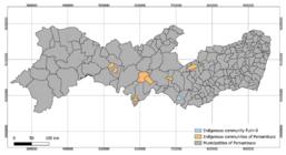
Location of the Fulni-ô indigenous community and other indigenous communities in Pernambuco state, Brazil.
The geographic coordinates of the Fulni‑ô reservation are approximately 9° 06′ 39″ S latitude and 37° 07′ 22″ W longitude, situated at the foothills of the Serra do Cumanati range in the semi-arid agreste region of the São Francisco River basin, sub-basin of the Ipanema River (Silveira et al., 2012).
The local climate is characterized by low and irregular precipitation (500–700 mm/year) and average annual temperatures around 26 °C, conditions to which the community has adapted through sustainable water management practices and subsistence agriculture (Bittencourt, 2022).
Traditional Fulni-ô dwellings, originally built with straw and wood, are now complemented by modern masonry houses; the community's "ocras" (thatched structures) remain in use as sacred spaces for ancestral rituals (Bittencourt, 2022).
The residents maintain a bilingual culture (Portuguese and Yaathe) and practice a rich repertoire of ceremonies, including the “rasgadas” dances and the “ouricuri” rites, which promote social cohesion and are periodically open to visitors through the “cafurnas” cultural festival (Silveira et al., 2012).
Their livelihoods combine small-scale agriculture, artisanal extraction of medicinal plants (Albuquerque et al., 2011), and informal trade, while domestic animals roam freely or live in semi-domicile within the village limits, living in close contact with humans, other domestic animals, and local fauna (Santana; Magalhães, 2022).
Serum samples were collected from dogs of the Fulni-ô Indigenous community during a field campaign carried august 2018, as part of a One Health surveillance initiative in northeastern Brazil.
All 66 canine sera were obtained as convenience samples, corresponding to all free-ranging or semi-domiciled dogs encountered during field visits. The Inclusion criteria were age ≥ 3 months and owner consent (when domiciled). Due to logistical constraints and the absence of a formal canine census, we sampled all accessible animals that met these criteria, aiming to capture the broadest possible representation within the Fulni-ô reserve.
2.2. Ethical considerations
This study was approved by the Ethics Committee on the Use of Animals (CEUA) of the Universidade Federal Rural de Pernambuco (128/2017). This research was conducted as recommended by the Brazilian College of Animal Experimentation.
2.3. Biological samples
Blood samples were collected from free-ranging and semi-domiciled stray dogs of different breeds, sex, and ages by cephalic vein venipuncture, and stored in Vacutainer ® tubes (New Jersey, USA). The tubes were identified and centrifuged at 1,500 ×g for 5 min. to obtain blood serum. Sera were aliquoted into 1.5mL polypropylene tubes, identified, and stored at -20 °C until serological analysis.
Prior to blood collection, each dog underwent a brief physical examination, including measurement of rectal temperature, inspection of mucous membranes, palpation of peripheral lymph nodes, and assessment of skin and gait. No overt clinical signs (fever, lymphadenopathy, dermatologic or neurologic abnormalities) were detected in any of the 66 animals, which were therefore classified as clinically healthy at the time of sampling.
2.4. Serological analysis
2.4.1. Toxoplasma gondii and Neospora caninum
The detection of IgG antibodies against T. gondii and N. caninum was performed using the indirect immunofluorescence test (IFAT), with cut-off titters of 1:16 and 1:50, respectively (Dubey, 2010). Tachyzoites of the T. gondii strain ME-49 and the N. caninum strain Nc-Sp7 were used as antigens. Serum samples were diluted in phosphate-buffered saline (PBS) and applied to 12-well slides. A fluorescein-conjugated anti-canine IgG secondary antibody (Sigma Chemical®, USA) was added to each microwell containing the processed serum samples (Dubey, 2010). Positive and negative controls were included on each slide for both pathogens to ensure test validity.
2.4.2. Leishmania infantum
The presence of antibodies against Leishmania infantum was assessed using the TR DPP® 7 Canine Visceral Leishmaniasis Kit (Bio-Manguinhos, Brazil). The kit includes a DPP support, running buffer, disposable collection handle (5 μL capacity), and a disposable lancet. All procedures were performed following the manufacturer’s instructions.
2.4.3. Leptospira spp.
The detection of antibodies against Leptospira spp. was carried out using the MAT, with a positivity threshold set at titters ≥ 50 (Bernardino et al., 2021a, b). A total of 15 serovars representing various serogroups were tested, as detailed in Table 1 (OIE, 2014).
Seroprevalence of zoonotic agents detected in dogs from the Fulni-ô Indigenous community, Pernambuco, Brazil.
The reaction was considered positive when titters equal to or greater than 1:50 were observed. The tiers obtained were classified into three categories: low (≤100), medium (200 to 400), and high (≥800). In addition, the mean titers observed for each serovar, and the frequency of different titer levels (low, medium, and high) were determined. A detailed analysis of co-positivity was also performed, identifying how many animals showed positivity for one or more serovars simultaneously.
2.5. Statistical analysis
Epidemiological and clinical information was obtained through a structured questionnaire applied to the owners or caretakers of the animals.
All statistical analyses were performed using IBM SPSS Statistics, version 26.0 (IBM Corp., Armonk, NY, USA). Associations between categorical variables (e.g., domicile status, contact with other animals, feeding type, and age category) and seropositivity to each pathogen were assessed using Pearson’s chi-square test or Fisher’s exact test when expected frequencies were <5. Statistical significance was set at p < 0.05 for all comparisons. (Thrusfield, 2007).
To identify possible associations between the different etiological agents investigated, Spearman's correlation test was used. The results of the analyses were organized into tables to facilitate visualization, interpretation, and presentation of the information obtained in this study.
3. Results
In the serological analysis of canine sera from the Fulni‑ô community, the following antibody prevalences were observed: 46.9% (31/66) were reactive for Toxoplasma gondii; 9.1% (6/66) for Neospora caninum; 22.7% (15/66) for Leishmania infantum; and 18.2% (12/66) for Leptospira spp. (Table 1).
Regarding the overall serostatus, 28.8% (19/66) of dogs were seronegative for all tested agents. Most dogs, 60.9% (42/66), were reactive to only one pathogen. Co‑reactivity to more than one agent occurred in 11.6% (8/66) of animals, seven dogs (10.6%, 7/66) were positive for two pathogens and one dog (1.5%, 1/66) for three pathogens, while none reacted to all four agents. The most frequent dual reactivity was T. gondii+Leptospira spp. (10.6% 7/66), and the sole triple reactivity involved T. gondii, Leptospira spp. and L. infantum (Table 1).
In the characterization of epidemiological variables among the study population, most dogs (23/66) reported contact with all categories of animals (domestic, synanthropic and wild), while 15 dogs had no contact with other animals. Regarding domicile status, 47 dogs were domiciled, 12 semi‑domiciled and 9 non‑domiciled. In terms of feeding, 36 dogs received a mixed diet of home‑cooked food plus commercial feed, 24 were fed exclusively home‑cooked food and 7 received only commercial feed. Age distribution peaked at 3 years (15 dogs), followed by 2 years (13 dogs) and dogs older than 10 years (10 animals).
An individual risk‑factor analysis showed that “contact with other animals” was not significantly associated with any of the diseases investigated (toxoplasmosis p = 0.224; neosporosis p = 1.000; leptospirosis p = 0.965; leishmaniasis p = 0.392). Likewise, “type of feeding” showed no significant association with any of the infectious agents evaluated (T. gondii p = 0.545; N. caninum p = 1.000; L. infantum p = 0.841; Leptospira spp. p = 0.820).
Age category also had no statistically significant influence on disease occurrence, with all p‑values above 0.05 (T. gondii p = 0.511; N. caninum p = 1.000; Leptospira spp. p = 0.576; L. infantum p = 0.224). In contrast, domicile status was significantly associated with two pathogens: L. infantum (χ2= 9.22; p = 0.027) and Leptospira spp. (χ2= 8.97; p = 0.030), whereas no association was found for T. gondii (p = 0.606) or N. caninum (p = 1.000).
Of the 66 dogs evaluated, 12 (18.2%) were reactive by MAT for Leptospira spp. Among these, the serovar Icterohaemorrhagiae (ICT) was most frequent (83.3%, 10/12), followed by Pomona (POM; 50.0%, 6/12) and Canicola (CAN; 33.3%, 4/12). Each of the other serovars, Hardjo‑prajitno (HP), Djasiman (DJA), Australis (AUS), Tarassovi (TAR), Grippotyphosa (GRI) and Bataviae (BAT), was detected in one animal (8.3%, 1/12 each) (Table 2).
The highest titer observed among reactive sera was 1:1 600 for ICT, followed by 1:800 for POM, 1:400 for TAR, 1:200 for CAN, DJA and AUS, 1:100 for HP, and 1:50 for GRI and BAT. Overall, low titers (≤ 1:100) predominated. Only ICT exhibited reactivity across all titer categories (low, medium and high), whereas DJA, AUS and TAR were exclusively medium‑titer (1:200–1:400) and elevated titers (≥ 1:800) were observed only for ICT and POM.
With respect to co‑reactivity, seven dogs (10.6%) were positive for two serovars, two dogs (3.0%) for three serovars, and one dog (1.5%) for four distinct serovars. Only two dogs showed reactivity to a single serovar. Dog no. 3 was the sole animal reactive to four serovars (HP, CAN, GRI and BAT). Dogs no. 7 and no. 19 were reactive to three serovars each: dog 7 to DJA, AUS and TAR, and dog 19 to ICT, CAN and POM. Of the seven dogs with dual reactivity, nos. 5, 44, 54, 58 and 63 were positive for ICT + POM, while nos. 47 and 48 were positive for ICT + CAN. Dogs no. 28 and no. 62 were the only animals reactive to a single serovar both to ICT, which was the most frequently detected serovar in this sample.
4. Discussion
The inclusion of domestic and production animals in serological investigations within indigenous communities is crucial, as these populations often rely on subsistence agriculture as their primary source of income and food. In this context, dogs and cats play vital roles, including pest management, property guarding, and companionship (Oliveira et al., 2011; Pulina et al., 2018).
Among the Kamentsá (Putumayo, Colombia), the canine seroprevalence of leptospirosis (80% by MAT) suggests intense environmental transmission and sanitation failures (H. Romero et al., 2018). In Manaus (urban), dogs have lower seroprevalence for Toxoplasma gondii (12.3%) and Neospora caninum (1.9%), indicating mitigation through infrastructure and management (Campos et al., 2022). In the rural Northeast (Paraíba), intermediate patterns emerge with co-exposure to Leptospira, T. gondii, and N. caninum, driven by household and wandering factors (Fernandes et al., 2018a). This contextualizes the Fulni-ô ecology, sanitation, and management determine risk.
However, these animals can also serve as reservoirs for zoonotic pathogens, contributing to the transmission of infectious diseases. The vulnerability of indigenous populations is exacerbated by poor sanitary conditions and close contact between humans, domestic animals, and wildlife, which facilitates the spread of infectious agents (Pulina et al., 2018; Romero-Sandoval et al., 2019).
The findings in dogs from the Fulni-ô community suggest that Toxoplasma gondii is circulating in the local environment. Infection with T. gondii occurs through the ingestion of infective oocysts in contaminated food or water, or through the consumption of raw meat from farm animals or wild prey (Innes et al., 2019; Awobode et al., 2020).
Indigenous contexts can also concentrate T. gondii exposure through food and landscape pathways. In Amazonian Indigenous communities, the handling and consumption of wild meat/offal have been linked to sustaining sylvatic T. gondii cycles. Proximity to forest edges and river margins has shown a spatial association with canine exposure in basin-wide studies (Fornazari et al., 2023). These determinants differ from predominantly urban exposures seen in Manaus, where contact with other dogs emerged as a key risk factor, highlighting how local subsistence practices and landscape shape infection ecology (Campos et al., 2022).
Studies conducted in indigenous communities in Brazil have shown that infection with T. gondii is associated with the consumption of water from contaminated rivers, the presence of felines, and cultural habits such as consuming raw mushrooms (Boia et al., 2008; Santos et al., 2019).
The presence of stray dogs in the village is particularly concerning, as these animals may play a role in the parasite's life cycle, potentially contaminating water and food consumed by humans and other intermediate hosts (Chalkowski et al., 2019; Arruda et al., 2021). Stray dogs act as indicators of environmental contamination (Opsteegh et al., 2019), underscoring the need for investigations in humans to estimate antibody prevalence and the health impacts of toxoplasmosis on the Fulni-ô population.
The detection of anti-Neospora caninum antibodies in dogs indicates an active transmission cycle of the protozoan. The presence of stray and semi-domiciled dogs in the community promotes the spread of N. caninum, especially given their propensity to hunt or consume raw meat—factors that have been associated with a higher risk of infection in rural areas (Sicupira et al., 2012). Previous studies have reported antibody prevalences in dogs from traditional communities, including 20.6% in semi-domiciled dogs in Australia (King et al., 2012) and 9.8% in indigenous villages in the Brazilian Amazon (Minervino et al., 2012).
In Brazil, anti-N. caninum antibodies have been detected in various domestic and wild animal species (Vianna et al., 2005; Gondim et al., 2010), reinforcing the role of dogs in maintaining the protozoan's transmission cycle. Moreover, N. caninum infection is a significant cause of abortion in cattle, particularly in regions where the disease is neglected (Abreu et al., 2014; Pinto-Ferreira et al., 2019).
Coinfections and diagnostic context warrant careful interpretation. In community surveys and necropsy-based series, co-seropositivity to protozoa is frequent, and clinical canine leishmaniasis has been associated with increased susceptibility to T. gondii in co-infected dogs, potentially modifying disease expression (Fornazari et al., 2023)
Moreover, serology panels (IFAT/ELISA) and MAT capture exposure but not necessarily active infection; cross-reactivity may occur, and confirmatory molecular testing adds clarity where feasible (Gaio et al., 2014; Fernandes et al., 2018b).
These caveats are relevant to Fulni-ô co-reactivity patterns (T. gondii and Leptospira spp.), which could reflect true co-exposure, shared environmental risks, or serologic cross-reaction.
The detection of antibodies against Leishmania infantum in dogs from the Fulni-ô community is noteworthy, especially given that the municipality of Águas Belas (PE) is classified as an endemic area for visceral leishmaniasis (VL). According to DataSUS/SINAN data, 29 cases of VL in humans were reported in the municipality between 2010 and 2024, indicating active circulation of the protozoan in the region. Additionally, one case of American tegumentary leishmaniasis (ATL) was recorded, further evidencing the disease's presence in the area (Brasil, 2025a, b).
Regionally, dog exposure profiles vary widely and frame the Fulni-ô signal. In the Xingu River Basin (Brazilian Amazon), dogs showed measurable exposure to L. infantum and T. gondii with spatial patterns linked to land cover, while a clinical cohort in Brazil’s Northeast documented high Leishmania seropositivity in symptomatic dogs, emphasizing that the sampling frame (clinical vs. community) inflates apparent prevalence (Fornazari et al., 2023). Against this backdrop, the Fulni-ô community’s 22.7% for L. infantum and 46.9% for T. gondii indicate substantial zoonotic circulation consistent with semi-arid ecology and close human–dog–wildlife interfaces.
The identification of seropositive dogs for Leishmania infantum suggests an ongoing risk of active transmission. However, the absence of confirmatory tests, such as PCR, limits the interpretation of these findings (Solano-Gallego et al., 2001). Integrated strategies, including the management of infected dogs, vector control, and health education, are essential. Prioritizing epidemiological characterization of leishmaniasis in the region is critical to protecting the Fulni-ô community and the broader population.
Leptospirosis, a widespread bacterial zoonosis, was also detected in dogs from the Fulni-ô community. The presence of various serovars reflects environmental reservoirs, such as synanthropic rodents (Cave et al., 2014; Lau et al., 2017). The natural infection of unvaccinated dogs likely occurred through exposure to contaminated water or moist soil, environments where leptospires can survive for extended periods (Thibeaux et al., 2017).
The serovar Icterohaemorrhagiae was the most prevalent (83.3%, 10/12) and is associated with high virulence, often resulting in severe cases of leptospirosis in humans and animals. This serovar is frequently linked to the presence of rats, its primary reservoirs, posing a significant risk in communities with poor sanitary conditions, such as indigenous communities (Samir et al., 2015; Riley et al., 2022).
The serovar Pomona (50%, 6/12) is highly pathogenic and frequently associated with pigs, though it is capable of infecting dogs and other species. Its detection in community dogs suggests environmental exposure or contact with sources of infection, such as contaminated water (Browne et al., 2022; Fahimipour et al., 2021).
The serovar Canicola (33.3%, 4/12), commonly found in dogs in Brazil, is linked to severe cases of renal failure. Dogs are the primary reservoirs of this serovar, and their shedding of bacteria through urine perpetuates transmission, posing a significant environmental risk (Hagiwara et al., 2015; Pellizzaro et al., 2019).
Other detected serovars, including Hardjoprajitno, Dijasiman, Australis, Tarassovi, Grippotyphosa, and Bataviae (8.3%, 1/12), highlight the diversity of Leptospira serovars circulating in the region. These are often associated with wild reservoirs and humid environments, which are common in rural areas (Thibeaux et al., 2017).
Detecting multiple serovars suggests repeated exposure or diverse sources of infection, characteristics of regions where frequent interactions occur between dogs, humans, and wild reservoirs. This underscores the complex epidemiological dynamics of leptospirosis in rural and semi-rural indigenous communities.
Actionable One Health implications follow from these patterns. In Indigenous territories with high Leptospira serogroup diversity and relevant T. gondii and L. infantum exposure, targeted steps include environmental sanitation, rodent control, safe water access, and health education on handling/cooking wild meat, alongside dog management (roaming reduction), vaccination where applicable, and periodic dog-sentinel surveillance (Campos et al., 2022; Galvão et al., 2023a, b; Khan et al., 2023).
Evidence from Indigenous and rural settings shows that sanitation and husbandry improvements correspond with lower dog seroprevalence, supporting integrated interventions with veterinary services embedded in Indigenous health systems (H. Romero et al., 2018).
We acknowledge that the limited sample size and convenience sampling approach limit the generalizability of our findings, but the detection of multiple pathogens and zoonotic agents in this population highlights the complex epidemiological dynamics at the rural-urban interface of indigenous communities, reflecting repeated exposure or multiple sources of infection caused by frequent interactions between dogs, humans, and wildlife.
5. Conclusion
Dogs in the Fulni-ô community showed extensive exposure to Toxoplasma gondii, Neospora caninum, Leishmania infantum, and Leptospira serovars, indicating an active zoonotic risk at the human-dog-environment interface. From a One Health perspective, integrated and periodic surveillance, environmental sanitation and vector control actions, and measures aimed at dogs such as reducing straying, vaccination, and health education are recommended. The proposed protocol can be replicated in other Indigenous territories, adjusted to local ecology and customs, serving as a model to strengthen zoonosis surveillance and prevention nationwide.
Acknowledgements
Thanks to the Coordination for the Improvement of Higher Education Personnel – Brazil (CAPES).
-
Data Availability Statement
The datasets generated and analyzed during the current study are not publicly available due to ethical and legal restrictions related to the conservation status and handling of wildlife species. However, anonymized data supporting the findings of this study are available from the corresponding author upon reasonable request.
References
-
ABREU, R.A., WEISS, R.R., THOMAZ-SOCCOL, V., LOCATELLI-DITTRICH, R., LASKOSKI, L.M., BERTOL, M.A., KOCH, M.O., ALBAN, S.M. and GREEN, K.T., 2014. Association of antibodies against Neospora caninum in mares with reproductive problems and presence of seropositive dogs as a risk factor. Veterinary Parasitology, vol. 202, no. 3-4, pp. 128-131. https://doi.org/10.1016/j.vetpar.2014.03.022 PMid:24731383.
» https://doi.org/10.1016/j.vetpar.2014.03.022 -
ALBUQUERQUE, U.P., SOLDATI, G.T., SIEBER, S.S., SÁ, E.M.F. and SOUZA, L.C., 2011. Use and extraction of medicinal plants by the Fulni-ô indians in northeastern Brazil: implications for local conservation. Sitientibus. Série Ciências Biológicas, vol. 11, no. 2, pp. 309-320. https://doi.org/10.13102/scb78
» https://doi.org/10.13102/scb78 -
ARRUDA, I.F., MILLAR, P.R., BARBOSA, A.D.S., ABBOUD, L.C.S., DOS REIS, I.C., MOREIRA, A.S.D.C., GUIMARÃES, M.P.P. and AMENDOEIRA, M.R.R., 2021. Toxoplasma gondii in domiciled dogs and cats in urban areas of Brazil: risk factors and spatial distribution. Parasite : Journal de la Société Française de Parasitologie, vol. 28, pp. 56. https://doi.org/10.1051/parasite/2021049 PMid:34236311.
» https://doi.org/10.1051/parasite/2021049 -
AWOBODE, H.O., OHIOLEI, J.A., ADEKEYE, T.A., ADEYI, A.O. and ANUMUDU, C.I., 2020. Shedding proportion of Toxoplasma gondii-like oocysts in feral cats and soil contamination in Oyo State, Nigeria. Parasite Epidemiology and Control, vol. 11, e00181. https://doi.org/10.1016/j.parepi.2020.e00181 PMid:32995580.
» https://doi.org/10.1016/j.parepi.2020.e00181 -
BAKER, T., ROCK, M., BROOK, R., VAN DER MEER, F. and KUTZ, S., 2020. Indigenous community perspectives on dogs in Northern Canada after 10 years of veterinary services indicates improved animal and human welfare. Preventive Veterinary Medicine, vol. 181, pp. 105061. https://doi.org/10.1016/j.prevetmed.2020.105061 PMid:32599372.
» https://doi.org/10.1016/j.prevetmed.2020.105061 -
BERNARDINO, M.G.S., COSTA, D.F., NOGUEIRA, D.B., SILVA, L.C.R. and AZEVEDO, S.S., 2021a. Cross-sectional survey for canine leptospirosis in na Atlantic Rainforest area of the semiarid of Paraíbastate, Northeastern Brazil. Pesquisa Veterinária Brasileira, vol. 41, pp. 06640. https://doi.org/10.1590/1678-5150-pvb-6640
» https://doi.org/10.1590/1678-5150-pvb-6640 -
BERNARDINO, M.G.S., COSTA, D.F., NOGUEIRA, D.B., SILVA, M.L.C.R., SILVA, E.G., CARREIRO, A.N., ALVES, C.J. and AZEVEDO, S.S., 2021b. Cross-sectional survey for canine leptospirosis in an Atlantic Rainforest area of the semiarid of Paraíba state, Northeastern Brazil. Pesquisa Veterinária Brasileira, vol. 41, e06640. https://doi.org/10.1590/1678-5150-pvb-6640
» https://doi.org/10.1590/1678-5150-pvb-6640 -
BITTENCOURT, M.C., 2022. Performance das cafurnas Fulni-ô na “Reserva Canto dos Guerreiros”. AntHropológicas Visual, vol. 8, no. 2, pp. 21. https://doi.org/10.51359/2526-3781.2022.256870
» https://doi.org/10.51359/2526-3781.2022.256870 -
BÓIA, M.N., CARVALHO-COSTA, F.A., SODRÉ, F.C., PINTO, G.M. and AMENDOEIRA, M.R., 2008. Seroprevalence of Toxoplasma gondii infection among indian people living in Iauareté, São Gabriel da Cachoeira, Amazonas, Brazil. Revista do Instituto de Medicina Tropical de São Paulo, vol. 50, no. 1, pp. 17-20. https://doi.org/10.1590/S0036-46652008000100004 PMid:18327482.
» https://doi.org/10.1590/S0036-46652008000100004 -
BRASIL, 2025a [viewed 13 January 2025]. Sistema de Informação de Agravos de Notificação – SINAN. Leishmaniose visceral - Notificações Registradas: banco de dados [online]. Available from: http://tabnet.datasus.gov.br/cgi/deftohtm.exe?sinannet/cnv/leishvpe.def
» http://tabnet.datasus.gov.br/cgi/deftohtm.exe?sinannet/cnv/leishvpe.def -
BRASIL, 2025b [viewed 13 January 2025]. Sistema de Informação de Agravos de Notificação – SINAN. Leishmaniose Tegumentar Americana - Notificações Registradas: banco de dados [online]. Available from: http://tabnet.datasus.gov.br/cgi/deftohtm.exe?sinannet/cnv/ltape.def
» http://tabnet.datasus.gov.br/cgi/deftohtm.exe?sinannet/cnv/ltape.def -
BROWNE, E.S., CALLEFE, J.L.R., JESUS, E.R.S., ZEPPELINI, C.G., CREMONESE, C. and COSTA, F., 2022. A systematic review of the geographic distribution of pathogenic Leptospira serovars in the Americas, 1930–2017. Anais da Academia Brasileira de Ciências, vol. 94, no. 3, e20201026. https://doi.org/10.1590/0001-3765202220201026 PMid:36074401.
» https://doi.org/10.1590/0001-3765202220201026 -
CAMPOS, H.G.N.D., SOARES, H.S., AZEVEDO, S.S.D. and GENNARI, S.M., 2022. Occurrence of Toxoplasma gondii and Neospora caninum antibodies and risk factors in domiciliated dogs of Manaus, Amazonas, Brazil. Revista Brasileira de Parasitologia Veterinária, vol. 31, no. 2, e020321. https://doi.org/10.1590/s1984-29612022024 PMid:35544881.
» https://doi.org/10.1590/s1984-29612022024 -
CANUTO, K., BROWN, A., WITTERT, G. and HARFIELD, S., 2018. Understanding the utilization of primary health care services by Indigenous men: a systematic review. BMC Public Health, vol. 18, no. 1, pp. 1198. https://doi.org/10.1186/s12889-018-6093-2 PMid:30352579.
» https://doi.org/10.1186/s12889-018-6093-2 -
CAVE, N.J., HARLAND, A.L. and ALLOTT, S.K., 2014. The serological response of working farm dogs to a vaccine containing Leptospira interrogans serovars Copenhageni and Pomona, and L. borgpetersenii serovar Hardjo. New Zealand Veterinary Journal, vol. 62, no. 2, pp. 87-90. https://doi.org/10.1080/00480169.2013.845072 PMid:24228620.
» https://doi.org/10.1080/00480169.2013.845072 -
CHALKOWSKI, K., WILSON, A.E., LEPCZYK, C.A. and ZOHDY, S., 2019. Who let the cats out? A global meta-analysis on risk of parasitic infection in indoor versus outdoor domestic cats (Felis catus). Biology Letters, vol. 15, no. 4, pp. 20180840. https://doi.org/10.1098/rsbl.2018.0840 PMid:30991913.
» https://doi.org/10.1098/rsbl.2018.0840 - DUBEY, J.P., 2010. Toxoplasmosis of animals and humans 2nd ed. Boca Raton: CRC Press, 313 p.
-
FAHIMIPOUR, A., KHAKI, P. and MORADI BIDHENDI, S., 2021. Seroepidemiological Analysis of Leptospiral infection using MAT in Stray Dogs in Alborz, Iran. Archives of Razi Institute, vol. 76, no. 2, pp. 391-396. https://doi.org/10.22092/ari.2020.127253.1376 PMid:34223737.
» https://doi.org/10.22092/ari.2020.127253.1376 -
FERNANDES, A.R.F., COSTA, D.F., ANDRADE, M.R., BEZERRA, C.S., MOTA, R.A., ALVES, C.J., LANGONI, H. and AZEVEDO, S.S., 2018a. Soropositividade e fatores de risco para leptospirose, toxoplasmose e neosporose na população canina do Estado da Paraíba. Pesquisa Veterinária Brasileira, vol. 38, no. 5, pp. 957-966. https://doi.org/10.1590/1678-5150-pvb-5137
» https://doi.org/10.1590/1678-5150-pvb-5137 -
FERNANDES, A.R.F., COSTA, D.F., PIMENTA, C.L.R.M., ARAÚJO, K.N., SILVA, R.B.S., MELO, M.A., LANGONI, H., MOTA, R.A. and AZEVEDO, S.S., 2018b. Occurrence and risk factors of zoonoses in dogs and owners in Sertão, Paraíba State, Northeastern Brazil. Semina: Ciências Agrárias, vol. 39, no. 3, pp. 1057. https://doi.org/10.5433/1679-0359.2018v39n3p1057
» https://doi.org/10.5433/1679-0359.2018v39n3p1057 -
FORNAZARI, F., SEVÁ, A.D.P., OLIVEIRA, K.M.M., ASSUNÇÃO, P.C.F. and GUIMARÃES, V.Y., 2023. Exposure of dogs and wild carnivores to canine distemper virus, canine parvovirus, Leishmania infantum, and Toxoplasma gondii in the Xingu River basin, Brazilian Amazon: Prevalence, spatial distribution, and association with land cover types. Acta Amazonica, vol. 53, no. 4, pp. 325-335. https://doi.org/10.1590/1809-4392202301340
» https://doi.org/10.1590/1809-4392202301340 - GAIO, F.C., SALINA, A., MENOZZI, B.D. and LANGONI, H., 2014. Anticorpos anti-Toxoplasma gondii e Neospora caninum em cães da zona rural do município de Botucatu-SP. Veterinária e Zootecnia, vol. 21, pp. 288-296.
-
GALVÃO, C.M.M.Q., OLIVEIRA, P.R.F., CAVALCANTI, A.L.A., NOGUEIRA, D.B., AZEVEDO, S.S., RAMOS, R.A.D.N. and MOTA, R.A., 2023a. Occurrence of IgG antibodies against Toxoplasma gondii, Neospora caninum, and Leptospira spp. in goats and sheep from an indigenous village in Pernambuco, Brazil. Revista Brasileira de Parasitologia Veterinária, vol. 32, no. 2, e000423. https://doi.org/10.1590/s1984-29612023022 PMid:37132735.
» https://doi.org/10.1590/s1984-29612023022 -
GALVÃO, G.R., GONÇALVES, E.C., MOURA, L.G.S., VIRGOLINO, R.R., NEVES, A.M.P. and AGUIAR, D.C.F., 2023b. First molecular description of autochthonous urban cases of canine visceral leishmaniasis in the city of Belém, Pará, Brazil. Brazilian Journal of Biology = Revista Brasileira de Biologia, vol. 83, e267617. https://doi.org/10.1590/1519-6984.267617 PMid:37311132.
» https://doi.org/10.1590/1519-6984.267617 -
GONDIM, L.S., ABE-SANDES, K., UZÊDA, R.S., SILVA, M.S., SANTOS, S.L., MOTA, R.A., VILELA, S.M. and GONDIM, L.F., 2010. Toxoplasma gondii and Neospora caninum in sparrows (Passer domesticus) in the Northeast of Brazil. Veterinary Parasitology, vol. 168, no. 1-2, pp. 121-124. https://doi.org/10.1016/j.vetpar.2009.09.055 PMid:19879051.
» https://doi.org/10.1016/j.vetpar.2009.09.055 - HAGIWARA, M.K., MIOTO, B.A. and FURLAN TOZZI, B., 2015. Canine leptospirosis in Brazil – a review. La Clinica Veterinaria, vol. 20, no. 119, pp. 86-104.
-
HARFIELD, S., DAVY, C., KITE, E., MCARTHUR, A., MUNN, Z., BROWN, N. and BROWN, A., 2015. Characteristics of Indigenous primary health care models of service delivery: a scoping review protocol. JBI Database of Systematic Reviews and Implementation Reports, vol. 13, no. 11, pp. 43-51. https://doi.org/10.11124/jbisrir-2015-2474 PMid:26657463.
» https://doi.org/10.11124/jbisrir-2015-2474 -
HENRIQUE, A.L.P., COSTA, M.R., PAIZ, L.M., YAMAKAWA, A.C., SILVA, E.C.D., TEIXEIRA, C.R., MENOZZI, B.D., SEVÁ, A.P., LANGONI, H. and FORNAZARI, F., 2025. Molecular diagnosis of Leishmania infantum in wildlife from Botucatu municipality, São Paulo state, Brazil. Brazilian Journal of Biology = Revista Brasileira de Biologia, vol. 85, e291583. https://doi.org/10.1590/1519-6984.291583 PMid:41092171.
» https://doi.org/10.1590/1519-6984.291583 -
HOHENTHAL, W.D. and MCCORKLE, T., 1955. The problem of aboriginal persistence. Southwestern Journal of Anthropology, vol. 11, no. 3, pp. 288-300. https://doi.org/10.1086/soutjanth.11.3.3629027
» https://doi.org/10.1086/soutjanth.11.3.3629027 -
INNES, E.A., HAMILTON, C., GARCIA, J.L., CHRYSSAFIDIS, A. and SMITH, D., 2019. A one health approach to vaccines against Toxoplasma gondii. Food and Waterborne Parasitology, vol. 15, e00053. https://doi.org/10.1016/j.fawpar.2019.e00053 PMid:32095623.
» https://doi.org/10.1016/j.fawpar.2019.e00053 -
KHAN, S., RAFIQ, K., KHABIR, M.N., KHAN, M.B., KHAN, S.N., KHATTAK, A. and ATTAULLAH, S., 2023. Toxoplasma gondii in lactating animals: potential risk to milk consuming population in Khyber Pakhtunkhwa. Brazilian Journal of Biology = Revista Brasileira de Biologia, vol. 83, e267369. https://doi.org/10.1590/1519-6984.267369 PMid:36790276.
» https://doi.org/10.1590/1519-6984.267369 -
KING, J.S., BROWN, G.K., JENKINS, D.J., ELLIS, J.T., FLEMING, P.J., WINDSOR, P.A. and SLAPETA, J., 2012. Oocysts and high seroprevalence of Neospora caninum in dogs living in remote Aboriginal communities and wild dogs in Australia. Veterinary Parasitology, vol. 187, no. 1-2, pp. 85-92. https://doi.org/10.1016/j.vetpar.2011.12.027 PMid:22245069.
» https://doi.org/10.1016/j.vetpar.2011.12.027 -
LAU, S.F., WONG, J.Y., KHOR, K.H., ROSLAN, M.A., ABDUL RAHMAN, M.S., BEJO, S.K., RADZI, R. and BAHAMAN, A.R., 2017. Seroprevalence of Leptospirosis in Working Dogs. Topics in Companion Animal Medicine, vol. 32, no. 4, pp. 121-125. https://doi.org/10.1053/j.tcam.2017.12.001 PMid:29525230.
» https://doi.org/10.1053/j.tcam.2017.12.001 -
MINERVINO, A.H., CASSINELLI, A.B., DE LIMA, J.T., SOARES, H.S., MALHEIROS, A.F., MARCILI, A. and GENNARI, S.M., 2012. Prevalence of anti- Neospora caninum and anti-Toxoplasma gondii antibodies in dogs from two different indigenous communities in the Brazilian Amazon Region. The Journal of Parasitology, vol. 98, no. 6, pp. 1276-1278. https://doi.org/10.1645/GE-3151.1 PMid:22551468.
» https://doi.org/10.1645/GE-3151.1 -
OLIVEIRA, C.J.B., HISRICH, E.R., MOURA, J.F.P., GIVISIEZ, P.E.N. and GEBREYES, W.A., 2011. On farm risk factors associated with goat milk quality in Northeast Brazil. Small Ruminant Research, vol. 98, no. 1-3, pp. 64-69. https://doi.org/10.1016/j.smallrumres.2011.03.020
» https://doi.org/10.1016/j.smallrumres.2011.03.020 -
OPSTEEGH, M., SPANO, F., AUBERT, D., BALEA, A., BURRELLS, A., CHERCHI, S., CORNELISSEN, J.B.W.J., DAM-DEISZ, C., GUITIAN, J., GYÖRKE, A., INNES, E.A., KATZER, F., LIMON, G., POSSENTI, A., POZIO, E., SCHARES, G., VILLENA, I., WISSELINK, H.J. and VAN DER GIESSEN, J.W.B., 2019. The relationship between the presence of antibodies and direct detection of Toxoplasma gondii in slaughtered calves and cattle in four European countries. International Journal for Parasitology, vol. 49, no. 7, pp. 515-522. https://doi.org/10.1016/j.ijpara.2019.01.005 PMid:31108097.
» https://doi.org/10.1016/j.ijpara.2019.01.005 -
ORGANIZAÇÃO MUNDIAL DE SAÚDE ANIMAL – OIE, 2014 [viewed 25 January 2022]. Manual de testes diagnósticos e vacinas para animais terrestres [online]. Available from: https://www.oie.int/fileadmin/Home/eng/Health_standards/tahm/3.01.12_LEPTO.pdf
» https://www.oie.int/fileadmin/Home/eng/Health_standards/tahm/3.01.12_LEPTO.pdf -
PELLIZZARO, M., MARTINS, C.M., YAMAKAWA, A.C., FERRAZ, D.D.C., MORIKAWA, V.M., FERREIRA, F., SANTOS, A.P.D., BIONDO, A.W. and LANGONI, H., 2019. Molecular detection of Leptospira spp. in rats as early spatial predictor for human disease in an endemic urban area. PLoS One, vol. 14, no. 5, e0216830. https://doi.org/10.1371/journal.pone.0216830 PMid:31116784.
» https://doi.org/10.1371/journal.pone.0216830 -
PINTO-FERREIRA, F., PASQUALI, A.K.S., THOMAZ-SOCCOL, V., MITSUKA-BREGANÓ, R., CALDART, E.T., LEANDRO, A.S., CHIYO, L., POZZOLO, E.M., CUBAS, P., GIORDANO, L.G.P., PETTERLE, R.R. and NAVARRO, I.T., 2019. Epidemiological relevance of dogs for the prevention of Toxoplasma gondii, Neospora caninum and Leptospira spp. Revista Brasileira de Parasitologia Veterinária, vol. 28, no. 3, pp. 383-394. https://doi.org/10.1590/s1984-29612019043 PMid:31390432.
» https://doi.org/10.1590/s1984-29612019043 -
PULINA, G., MILÁN, M.J., LAVÍN, M.P., THEODORIDIS, A., MORIN, E., CAPOTE, J., THOMAS, D.L., FRANCESCONI, A.H.D. and CAJA, G., 2018. Invited review: current production trends, farm structures, and economics of the dairy sheep and goat sectors. Journal of Dairy Science, vol. 101, no. 8, pp. 6715-6729. https://doi.org/10.3168/jds.2017-14015 PMid:29859690.
» https://doi.org/10.3168/jds.2017-14015 -
RAHMAN, M.T., SOBUR, M.A., ISLAM, M.S., IEVY, S., HOSSAIN, M.J., EL ZOWALATY, M.E., RAHMAN, A.T. and ASHOUR, H.M., 2020. Zoonotic diseases: etiology, impact, and control. Microorganisms, vol. 8, no. 9, pp. 1405. https://doi.org/10.3390/microorganisms8091405 PMid:32932606.
» https://doi.org/10.3390/microorganisms8091405 -
RILEY, T., ANDERSON, N.E., LOVETT, R., MEREDITH, A., CUMMING, B. and THANDRAYEN, J., 2021. One Health in Indigenous Communities: A Critical Review of the Evidence. International Journal of Environmental Research and Public Health, vol. 18, no. 21, pp. 11303. https://doi.org/10.3390/ijerph182111303 PMid:34769820.
» https://doi.org/10.3390/ijerph182111303 -
RILEY, T., ANDERSON, N.E., LOVETT, R., MEREDITH, A. and CUMMING, B., 2022. Zoonoses and the Aboriginal and Torres Strait Islander population: a One Health scoping review. PLOS Global Public Health, vol. 2, no. 10, e0000921. https://doi.org/10.1371/journal.pgph.0000921 PMid:36962569.
» https://doi.org/10.1371/journal.pgph.0000921 -
RILEY, T., LOVETT, R., CUMMING, B., MEREDITH, A., ANDERSON, N.E. and THANDRAYEN, J., 2023. Data analysis of zoonoses notifications in Aboriginal and Torres Strait Islander populations in Australia 1996–2021: implications for One Health. Frontiers in Public Health, vol. 11, pp. 1175835. https://doi.org/10.3389/fpubh.2023.1175835 PMid:37900024.
» https://doi.org/10.3389/fpubh.2023.1175835 -
ROMERO, H., ASTUDILLO, M., AGUILLÓN, D.M. and LUCIO, I.D., 2018. Evidencia serológica de leptospirosis canina en la comunidad indígena Kamentsá, Putumayo, Colombia. Revista de Investigaciones Veterinarias del Perú, vol. 29, no. 2, pp. 625-634. https://doi.org/10.15381/rivep.v29i2.14495
» https://doi.org/10.15381/rivep.v29i2.14495 -
ROMERO-SANDOVAL, N., CIFUENTES, L., LEÓN, G., LECARO, P., ORTIZ-RICO, C., COOPER, P. and MARTÍN, M., 2019. High Rates of Exposures to Waterborne Pathogens in Indigenous Communities in the Amazon Region of Ecuador. The American Journal of Tropical Medicine and Hygiene, vol. 101, no. 1, pp. 45-50. https://doi.org/10.4269/ajtmh.18-0970 PMid:31162016.
» https://doi.org/10.4269/ajtmh.18-0970 -
SAMIR, A., SOLIMAN, R., EL-HARIRI, M., ABDEL-MOEIN, K. and HATEM, M.E., 2015. Leptospirosis in animals and human contacts in Egypt: broad range surveillance. Revista da Sociedade Brasileira de Medicina Tropical, vol. 48, no. 3, pp. 272-277. https://doi.org/10.1590/0037-8682-0102-2015 PMid:26108004.
» https://doi.org/10.1590/0037-8682-0102-2015 -
SANTANA, P.M.S.D. and MAGALHÃES, T.Q.D., 2022. Caso Xukuru e o bem viver do povo Fulni-ô (PE). Revista Direito e Práxis, vol. 13, no. 1, pp. 607-635. https://doi.org/10.1590/2179-8966/2022/65133
» https://doi.org/10.1590/2179-8966/2022/65133 -
SANTOS, A.L.C., TERÇAS TRETTEL, A.C.P., RIBEIRO, L.J.B.B., VASCONCELLOS, M.L., ZENAZOKENAE, L.E., ATANAKA SANTOS, M., LEMOS, E.R.S. and AMENDOEIRA, M.R.R., 2019. Serological study on toxoplasmosis in the Haliti-Paresí community of the Utiariti indigenous territory, Campo Novo do Parecis, Mato Grosso, Brazil. Parasite Epidemiology and Control, vol. 5, e00097. https://doi.org/10.1016/j.parepi.2019.e00097 PMid:30886914.
» https://doi.org/10.1016/j.parepi.2019.e00097 -
SHAH, M.Z., MUSTAFA, G., IQBAL, M., QASIM, M., ABBAS, K., UMAIR, M. and BAIG, H.M.A., 2024. Prevalence of Gram positive bacteria in the affected individuals of Otitis media with effusion from the indigenous population of Southern Punjab, Pakistan: first report. Brazilian Journal of Biology = Revista Brasileira de Biologia, vol. 84, e267874. https://doi.org/10.1590/1519-6984.267874 PMid:36722679.
» https://doi.org/10.1590/1519-6984.267874 -
SHAPIRO, K., BAHIA-OLIVEIRA, L., DIXON, B., DUMÈTRE, A., WIT, L.A., VANWORMER, E. and VILLENA, I., 2019. Environmental transmission of Toxoplasma gondii: oocysts in water, soil and food. Food and Waterborne Parasitology, vol. 1, no. 15, e00049. https://doi.org/10.1016/j.fawpar.2019.e00049 PMid:32095620.
» https://doi.org/10.1016/j.fawpar.2019.e00049 -
SICUPIRA, P.M., MAGALHÃES, V.C., GALVÃO, G.S., PEREIRA, M.J., GONDIM, L.F. and MUNHOZ, A.D., 2012. Factors associated with infection by Neospora caninum in dogs in Brazil. Veterinary Parasitology, vol. 185, no. 2-4, pp. 305-308. https://doi.org/10.1016/j.vetpar.2011.09.029 PMid:22015062.
» https://doi.org/10.1016/j.vetpar.2011.09.029 - SILVEIRA, L.M.L.C.; MARQUES, L.R.; SILVA, E.H., 2012. Fulni-ô: história e educação de um povo bilingue em Pernambuco. Cadernos de Pesquisas, vol. 19, no. 1, pp. 31-41.
-
SOLANO-GALLEGO, L., MORELL, P., ARBOIX, M., ALBEROLA, J. and FERRER, L., 2001. Prevalence of Leishmania infantum infection in dogs living in an area of canine leishmaniasis endemicity using PCR on several tissues and serology. Journal of Clinical Microbiology, vol. 39, no. 2, pp. 560-563. https://doi.org/10.1128/JCM.39.2.560-563.2001 PMid:11158106.
» https://doi.org/10.1128/JCM.39.2.560-563.2001 -
SOUSA, M.C., SCATENA, J.H.G. and SANTOS, R.V., 2007. O Sistema de Informação da Atenção à Saúde Indígena (SIASI): criação, estrutura e funcionamento. Cadernos de Saúde Pública, vol. 23, no. 4, pp. 853-861. https://doi.org/10.1590/S0102-311X2007000400013 PMid:17435883.
» https://doi.org/10.1590/S0102-311X2007000400013 -
THIBEAUX, R., GEROULT, S., BENEZECH, C., CHABAUD, S., SOUPÉ-GILBERT, M.-E., GIRAULT, D., BIERQUE, E. and GOARANT, C., 2017. Seeking the environmental source of Leptospirosis reveals durable bacterial viability in river. PLoS Neglected Tropical Diseases, vol. 11, no. 2, e0005414. https://doi.org/10.1371/journal.pntd.0005414 PMid:28241042.
» https://doi.org/10.1371/journal.pntd.0005414 - THRUSFIELD, M., 2007. Veterinary epidemiology 3rd ed. Oxford: Blackwell Science Ltd.
-
VIANNA, M.C., SREEKUMAR, C., MISKA, K.B., HILL, D.E. and DUBEY, J.P., 2005. Isolation of Neospora caninum from naturally infected white-tailed deer (Odocoileus virginianus). Veterinary Parasitology, vol. 129, no. 3-4, pp. 253-257. https://doi.org/10.1016/j.vetpar.2004.02.031 PMid:15845280.
» https://doi.org/10.1016/j.vetpar.2004.02.031
Edited by
-
Editor:
Takako Matsumura Tundisi
The datasets generated and analyzed during the current study are not publicly available due to ethical and legal restrictions related to the conservation status and handling of wildlife species. However, anonymized data supporting the findings of this study are available from the corresponding author upon reasonable request.