Therapeutic potential of Cannabis sativa against tick-induced injuries (Rhipicephalus linnaei): biochemical and histopathological evaluation in rabbits
Abstract
Rhipicephalus linnaei, commonly known as the brown dog tick, are preferential hosts for dogs, to whom they also transmit various pathogens. The control of these ectoparasites is generally achieved through the use of synthetic chemical products, which can lead to the development of resistance. Bioactive compounds extracted from plants are being studied as an alternative strategy for tick control, including Cannabis sativa, which provides an extract from its flowers known for its diverse therapeutic properties, applicable to both humans and animals. Therefore, in the work that originated this project investigated the effects of C. sativa flower extract in oil dilutions of 0.2, 0.4, and 0.8 mg/mL, applied to skin lesions of rabbit hosts of R. linnaei ticks, to evaluate their healing. In addition, this procedure made it necessary to evaluate, what was done in the present work, the hepatic tissue of these animals, as the extract, while functioning as a tickicide and promoting healing of skin lesions, should also be harmless to the hosts to be considered viable for ectoparasite control. For this purpose, histological and histochemical techniques were applied to the livers of the rabbits (to identify morphological alterations), which were divided into: control group (CG), exposed to 1 mL of oil, showing no hepatic alterations; treated group 1 (TG1), exposed to 0.2 mg/mL, showing cytoplasmic vacuolization in hepatocytes; treated group 2 (TG2), exposed to 0.4 mg/mL, displaying intense morphological alterations, including disorganization of hepatocyte cords, hepatocyte hypertrophy, altered nuclei, and signs of cell death; treated group 3 (TG3), exposed to 0.8 mg/mL, also showing tissue disorganization and cellular vacuolization, but to a lesser extent than TG2. In addition to morphological evaluation, the biochemical activity of hepatic enzymes AST and ALT was assessed. An increase in ALT was observed in TG2, while no other groups showed changes in these parameters. Overall, this study concluded that although the C. sativa flower extract at various dilutions has potential as a tickicide and for healing skin lesions (personal communication), it causes hepatic tissue damage in rabbits to varying degrees, with the 0.4 mg/mL dilution showing the highest hepatotoxic potential.
Keywords:
phytoactive; tickicide; hepatic enzymes; host; brown-dog-tick; toxicity
Resumo
Rhipicephalus linnaei, conhecido como carrapato-vermelho-do-cão, têm cães como hospedeiros preferenciais, aos quais também transmitem diversos patógenos. O controle desses ectoparasitas é geralmente feito por meio do uso de produtos químicos sintéticos, que podem levar ao desenvolvimento de resistência. Compostos bioativos extraídos de plantas estão sendo estudados como uma estratégia alternativa para o controle de carrapatos, incluindo a Cannabis sativa, que fornece um extrato de suas flores conhecido por suas diversas propriedades terapêuticas, aplicáveis tanto a humanos quanto a animais. Portanto, o trabalho que originou este projeto investigou os efeitos do extrato de flores de C. sativa em diluições de óleo de 0,2, 0,4 e 0,8 mg/mL, aplicados a lesões cutâneas de coelhos hospedeiros de carrapatos R. linnaei, para avaliar sua cicatrização. Além disso, esse procedimento exigiu a avaliação, realizada no presente trabalho, do tecido hepático desses animais, já que o extrato, enquanto funciona como um carrapaticida e promove a cicatrização das lesões cutâneas, também deveria ser inofensivo para os hospedeiros, para ser considerado viável no controle de ectoparasitas. Para isso, foram aplicadas técnicas histológicas e histoquímicas nos fígados dos coelhos (para identificar alterações morfológicas), divididos em: Grupo Controle (GC), exposto a 1 mL de óleo, sem alterações hepáticas; Grupo Tratado 1 (GT1), exposto a 0,2 mg/mL, apresentando vacuolização citoplasmática nos hepatócitos; Grupo Tratado 2 (GT2), exposto a 0,4 mg/mL, mostrando intensas alterações morfológicas, incluindo desorganização dos cordões de hepatócitos, hipertrofia dos hepatócitos, núcleos alterados e sinais de morte celular; Grupo Tratado 3 (GT3), exposto a 0,8 mg/mL, também mostrando desorganização tecidual e vacuolização celular, mas em menor grau do que o GT2. Além da avaliação morfológica, foi avaliada a atividade bioquímica das enzimas hepáticas AST e ALT. Observou-se um aumento da ALT no GT2, enquanto nenhum outro grupo mostrou alterações nesses parâmetros. De maneira geral, este estudo concluiu que, embora o extrato de flores de C. sativa em diversas diluições tenha potencial como carrapaticida e para a cicatrização de lesões cutâneas (informação pessoal), ele causa danos ao tecido hepático nos coelhos em diferentes graus, sendo que a diluição de 0,4 mg/mL apresentou o maior potencial hepatotóxico.
Palavras-chave:
fitoativo; carrapaticida; enzimas hepáticas; hospedeiro; carrapato-vermelho-do-cão; toxicidade
1. Introduction
The tick species Rhipicephalus linnaei (Audouin 1826), a tropical lineage of the R. sanguineus s.l. (Šlapeta et al., 2022), known as the red dog tick, has canids as its primary host. This arthropod is widely distributed. In addition to the damage caused by blood spoliation, they are transmitting agents of pathogens such as Babesia canis (babesiosis) and Ehrlichia canis (ehrlichiosis) in dogs and humans, Rickettsia conorii and R. rickettsii, which leads to the development of spotted fever (Abreu et al., 2019; Dantas-Torres, 2008; Fonsêca et al., 2022).
These ectoparasites are currently controlled using acaricides formulated from synthetic chemical bases and with variable financial costs. Despite their efficiency, in addition to their toxicity due to continuous use, they induce the emergence of resistance in ticks, resulting in a lack of efficiency of the chemical bases after a specific time since they develop strategies to circumvent the host animal's immune system (Camargo-Mathias, 2018; Becker et al., 2019; Sunkara et al., 2022).
In the search for cleaner and more sustainable ectoparasite control strategies that do not bring so much damage to non-target organisms or the environment, plants with recognized acaricidal value have been successfully studied since they are rich sources of bioactive compounds, most of which are already known and used by local populations against a considerable range of diseases (Gonçalves et al., 2016; Mabasa et al., 2021). These bioactives have been generally studied by extracting them as crude extracts or essential oils (Mabasa et al., 2021; Camargo-Mathias, 2018).
Cannabis sativa is one of the plants considered sources of potent bioactives with diverse functions. It belongs to the Moraceae family, is an exotic plant not typical from Brazil, popularly known as “marijuana”, and has been marketed in several countries due to its numerous therapeutic properties in various sectors, including human and veterinary medicine (Gewehr, 2021; Miranda-Cortés et al., 2023). Cannabis gained its fame due to the use of hemp fibers for manufacturing nets and ropes, in addition to its seeds, from which essential oils are extracted, making it popular worldwide. In India, Cannabis has been used to reduce pain, improve anxiety, nausea, appetite, and sleep, and for presenting a relaxing and euphoric effect. The benefits of using bioactive compounds in C. sativa began to spread in Brazil in the nineteenth century after the dissemination of work developed at the Faculty of Medicine of Tours in France. However, its use in general has been controversial for ethical and legal reasons imposed in the regulations and laws of each country (Lima et al., 2021; Carlini, 2006; Miranda-Cortés et al., 2023).
The growing interest in the therapeutic use of Cannabis bioactive compounds, despite its recreational use, has attracted the attention of researchers in human and veterinary medicine due to their various beneficial properties (Pierro Neto et al., 2023; Martins et al., 2022; Chicoine et al., 2020). In the 1960s, the discovery of the endocannabinoid system revealed an intrinsic mechanism of regulation of the organism, activated by the presence of cannabinoids from Cannabis, which interact with this system. These compounds have been applied therapeutically in humans and animals due to their anti-inflammatory, analgesic, and immunomodulatory properties (Schoeman et al., 2020; Tambeli et al., 2023; Vieira et al., 2020). As a result, part of the tutors of domestic animals have sought treatments using the bioactive compounds of Cannabis, contributing to its use in veterinary medicine (Chicoine et al., 2020).
Over 100 phytocannabinoid compounds were identified in plants of the Cannabis genus. THC, terpenes, CBD, CBDA, and flavonoids are the most prominent. THC is recognized for its psychotropic and pharmacological effects (Tambeli et al., 2023; Martinez Naya et al., 2024). On the other hand, cannabidiol (CBD), another significant compound, does not produce psychotropic effects and is known for its positive pharmacological and therapeutic properties. It was recently removed from the list of prohibited substances by Anvisa (Lima et al., 2021). In addition, cannabidiolic acid (CBDA), also non-psychoactive, is found in large quantities in the flowers of various species of Cannabis, presenting antiemetic and anxiolytic properties (Carvalho et al., 2020; Elsohly et al., 2017; Miranda-Cortés et al., 2023).
In the veterinary context, cannabinoids have been widely recognized as a therapeutic tool, being prescribed for dogs and cats to treat various conditions, including contact and atopic dermatitis, epilepsy, asthma, diabetes, glaucoma, and other inflammatory diseases (Miranda-Cortés et al., 2023). These compounds have also presented appetite-stimulating properties, which may be especially beneficial to animals suffering from loss of appetite due to conditions such as tick disease and cancer, among others (Chicoine et al., 2020; Miranda-Cortés et al., 2023).
Cannabinoids are rapidly distributed throughout the body, reaching well-vascularized organs such as the lungs, heart, brain, and liver. This distribution is influenced by body size and composition and is affected by health conditions that can alter the permeability of barriers between blood and tissues. THC, one of the major cannabinoids, is metabolized in the liver, with the cytochrome P450 (CYP 450) enzymes CYP2C9, CYP2C19, and CYP3A4 playing a crucial role in this process (Martinez Naya et al., 2024; Lucas et al., 2018).
The liver is an essential organ for the human and animal bodies, composed mainly of two types of cells: hepatocytes and cholangiocytes. Hepatocytes perform various vital functions, making up most of the liver mass. Its activities include protein synthesis, body detoxification, bile production, and carbohydrate and lipid metabolism (Trefts et al., 2017; Bechmann et al., 2012). These functions are crucial for maintaining metabolic balance and overall body health. In addition, the liver plays a central role in the metabolism of exogenous substances, such as drugs and toxins, where hepatocytes process and neutralize these substances, contributing significantly to maintaining the body's homeostatic balance (Junqueira and Carneiro, 2023; Ramachandran et al., 2020).
Thus, due to the positive results described regarding the efficacy and benefits arising from the use of C. sativa, focusing on the veterinary context, this project aimed to verify if the bioactive compounds present in this plant would not act as toxic agents for the non-target organism, the host, if they were efficient in healing, through morphophysiological evaluation of the liver and clinical blood analysis (evaluation of the TGO and TGP enzyme rates).
2. Objective
This project aimed to conduct a morphohistological study of the liver and evaluate the levels of the hepatic enzymes AST (aspartate aminotransferase) and ALT (alanine aminotransferase) through biochemical blood assays to investigate the effects of topical exposure to Cannabis sativa extract on rabbits infested with the dog tick R. linnaei, simulating tick-host models. The C. sativa extract, known for its bioactive compounds with healing and anti-inflammatory properties, was diluted in extra virgin olive oil to obtain concentrations of 0.2, 0.4, 0.8 mg/mL and based on and modified from the study by Sangiovanni et al. (2019).
3. Methodology
3.1. Equipment and study locations
The study utilized equipment available at the Histology Laboratory of the Department of General and Applied Biology at UNESP in Rio Claro, SP. Within this institution, the Brazilian Central of Studies on Ticks Morphology (BCSTM) is established, where tick colonies are maintained under the coordination of Prof. Dra. Maria Izabel Souza Camargo.
3.2. Obtaining Cannabis sativa extract
The extract used for this study was provided by the Maria Flor Cannabis Association, located in Marília, São Paulo, under the technical supervision of Dr. Caroline Marroni Cremonez.
3.3. Obtaining the host rabbits
Twelve healthy female rabbits of the Botucatu Genetic Group, aged four months and weighing between 2.5 to 3.0 kg, which had no prior contact with ticks or acaricides, were acquired from the Central Animal Facility of UNESP/Botucatu, SP.
For the bioassays, the rabbits were divided into four groups (3 rabbits/group): GC (Control Group), TG1 (Treated Group 1), TG2 (Treated Group 2), and TG3 (Treated Group 3).
The procedures described here were approved by the Animal Ethics Committee (CEUA) of the Institute of Biosciences, UNESP – Rio Claro Campus, SP, decision no. 13/2023.
3.4. Ticks
Adult female Rhipicephalus linnaei ticks were used, obtained from established colonies maintained in a BOD incubator under controlled conditions (29 °C, 80% humidity, and a 12-hour light cycle) in a room at the Histology Laboratory of the Department of General and Applied Biology at UNESP in Rio Claro, SP. The ticks were kept fasting inside plastic syringes with the plunger opening sealed with lightly moistened cotton until the moment of infestation on the rabbits.
3.5. Preparation of the feeding chamber (Bechara et al., 1995)
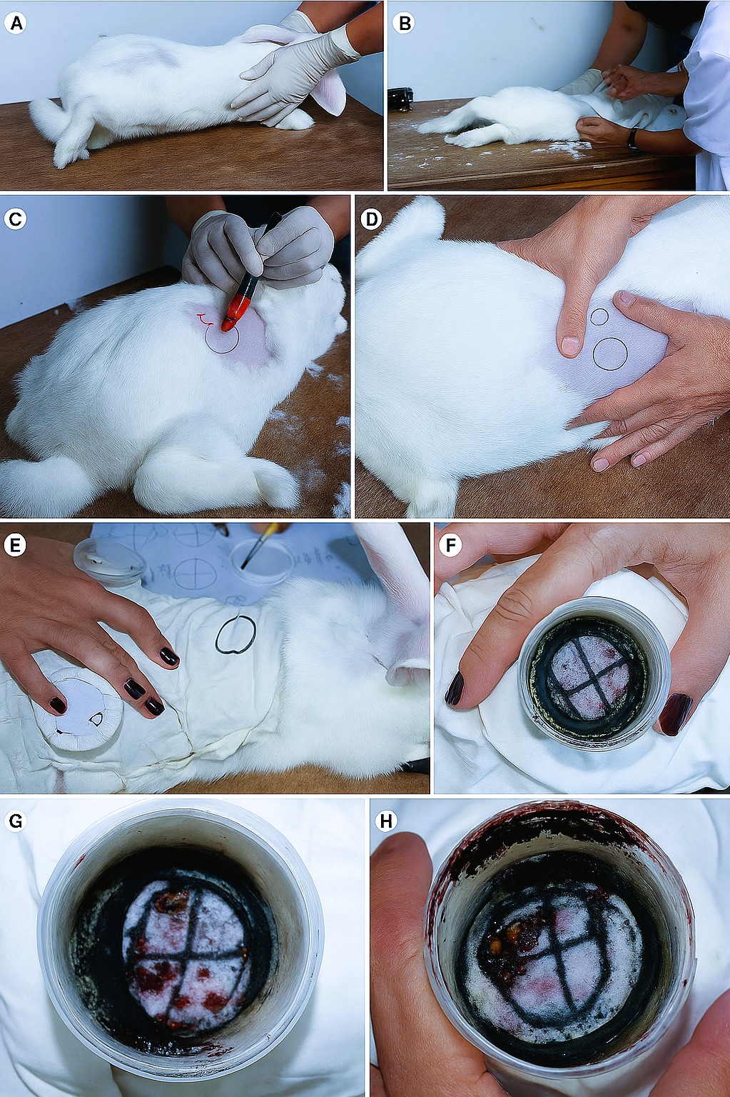
The feeding chambers were prepared following the protocol of Bechara et al. (1995). A 9 cm diameter circle of thin rubber was cut and subsequently covered with cotton fabric. Next, the fabric-covered side was adhered with non-toxic glue to the shaved dorsal skin of the host. Following this, a plastic tube (universal collector type) with a height of 2 cm and its bottom part removed, was fixed on the rabbit's back and sealed internally and externally with non-toxic glue and adhesive tape. The lid of the collector, which would be later placed on top, was perforated to allow air entry for oxygenation of the R. linnaei females (Figure 1).

(A-H): Photographs showing the various steps of the procedures conducted on the dorsal skin of the host rabbits during infestation by R. linnaei, according to Ferraz Hebling (2011).
The chambers remained uncovered for 24 hours to allow the glue to dry completely before placing the ticks inside.
3.6. Infestation of the rabbits by R. linnaei ticks
It was placed 15 pairs of R. linnaei ticks in each feeding chamber, where they remained for a period of 7 days, allowing them to attach and start the feeding process.
3.7. Preparation of Cannabis sativa extract dilutions
The crude Cannabis extract was diluted in olive oil (solvent) to achieve concentrations of THC that were less than 0.2%. The dilutions were carried out under the guidance of Prof. Dr. Sidney José Lima Ribeiro (UNESP/Araraquara) following these procedures:
-
0.2 mg/mL of concentrated Cannabis sativa extract was diluted in 100 mL of olive oil to achieve a final concentration of 0.05% THC.
-
0.4 mg/mL of concentrated Cannabis sativa extract was diluted in 100 mL of olive oil to achieve a final concentration of 0.1% THC.
-
0.8 mg/mL of concentrated Cannabis sativa extract was diluted in 100 mL of olive oil to achieve a final concentration of 0.2% THC.
After dilution, it was ensured that the final product complied with regulations set by ANVISA, which establishes a maximum permitted THC limit of 0.2%.
The dilutions of the Cannabis extract were based on those proposed by Sangiovanni et al. (2019), authors who investigated the healing and anti-inflammatory potential of C. sativa in cell culture.
It is important to emphasize that the objective of this study was not to conduct a clinical trial, as that would require approval from federal regulatory agencies. Rather, the aim was to test the proposed dilutions in a laboratory setting to confirm their effectiveness in healing skin lesions on rabbits simulating hosts of dog ticks, without causing liver damage.
3.8. Topical application of Cannabis sativa extract dilutions on the skin lesions of rabbits post-infested by R. linnaei ticks
After 7 days, when the female ticks were fully engorged and detached from the host, a lesion remained at the site of their attachment (bite). Topically, 1 mL of the prepared dilutions of Cannabis sativa extract was applied to the following groups:
-
Control (CG): Only 1 mL of olive oil (which served as the solvent) was applied.
-
Treatment 1 (TG1): 0.2 mg/mL diluted extract was applied.
-
Treatment 2 (TG2): 0.4 mg/mL diluted extract was applied.
-
Treatment 3 (TG3): 0.8 mg/mL diluted extract was applied.
The monitoring of the animals exposed in all groups was conducted daily, during which images were also recorded to track the progression of the healing process.
3.9. Clinical analysis
Clinical (biochemical) tests were conducted to evaluate the liver enzymes AST (aspartate aminotransferase) and ALT (alanine aminotransferase) after exposure to different dilutions of the extracts. Blood samples were collected on the following days:
-
D0: 1st day before the infestation of the rabbits by R. linnaei ticks;
-
D1: 7th day after the ticks' detachment and the 1st day of exposure to C. sativa dilutions;
-
D2: 14th day after the ticks' detachment and the 7th day after exposure to the C. sativa extract dilutions.
The blood collection was performed by veterinarian Brunna Fernanda Arraez Alves, CRMV nº 59838, via jugular puncture, extracting 2 mL of blood per rabbit using a 3 mL syringe and a 25x0.08mm (sterile) needle, and storing the samples in Vacutainer® tubes with a gel separator.
The laboratory analyses were conducted at CEDIVET Laboratory located in Rio Claro-SP, under the responsibility of veterinarians Tamara Maria Franzin Julio, CRMV nº 20.137, and Tassiana Ferreira de Mello, CRMV nº 31.267. They used the URIT 8031 (automatic biochemical and turbidimetric analyzer) with Bioclin® brand reagents, following the manufacturer's protocols.
3.10. Statistical analysis
The enzyme data were expressed as mean ± standard deviation and subsequently subjected to the Shapiro-Wilk test for normality verification. Then, these data were analyzed using the ANOVA/Tukey test. A p-value of <0.05 was considered statistically significant. These statistical analyses were performed using GraphPad Prism 7.0® software.
3.11. Creation of the diagrams
The diagrams were obtained from histological images and created using Graphics Suite 2023 and Canva Create 2023 software.
3.12. Morphological analyses
3.12.1. Collection of liver samples from the rabbits
After the bioassays were completed, euthanasia of the rabbits was performed by veterinarian Brunna Fernanda Arraez Alves, CRMV nº 59838, in the vivarium room of the Department of General and Applied Biology at IB/UNESP/Rio Claro, SP. Euthanasia was carried out using ketamine and xylazine, administered intraperitoneally at doses of 300 mg/kg and 30 mg/kg, respectively, to collect liver fragments.
3.12.2. Staining with Harris hematoxylin-aqueous eosin (Junqueira and Junqueira, 1983)
After collection, the tissue fragments were fixed in 4% paraformaldehyde for 72 hours at 4 °C. Subsequently, they were transferred to a phosphate buffer solution (NaCl 7.5 g/L, Na2HPO4 2.38 g/L, and KH2PO4 2.72 g/L) and kept for 24 hours. The material was then dehydrated in a graded series of ethanol (70-95%, 30 minutes each), infiltrated with Leica® Historesin for 24 hours, and embedded in plastic molds containing Historesin plus polymerizer (Leica Historesin Kit®). Following embedding, the blocks were sectioned (3 μm thickness) using a Leica RM2265 microtome (Leica®), and the sections were collected on clean glass slides. The sections were rehydrated for 1 minute in distilled water and stained with Harris hematoxylin for eight minutes. After rinsing in running water for three minutes, the sections were counterstained with aqueous eosin for five minutes and rinsed again in running water. After air-drying on wooden supports at room temperature, the sections were cleared in xylene, mounted with Entellan®, and covered with a coverslip. Permanent slides were examined and photographed using a Leica DM750 brightfield microscope (Leica®) at the Histology Laboratory of the Department of General and Applied Biology at IB/UNESP/Rio Claro - SP.
3.13. Histochemistry
3.13.1. Bromophenol Blue Technique (Pearse, 1985)
For total protein detection, liver tissue fragments from the hosts were fixed in 4% paraformaldehyde for 48 hours. They were then transferred to a phosphate buffer solution (NaCl 7.5 g/L, Na2HPO4 2.38 g/L, and KH2PO4 2.72 g/L) and incubated for 24 hours. Subsequently, the samples were dehydrated in a graded series of ethanol (70-95%, 30 minutes each), infiltrated with Leica® Historesin for 24 hours, and embedded in plastic molds containing Historesin plus polymerizer (Leica Historesin Kit®). After embedding, the blocks were sectioned (3 μm thickness) using a Leica RM2265 microtome (Leica®), and the sections were collected on clean glass slides for subsequent staining with bromophenol blue for one hour at room temperature. Following staining, they were briefly immersed in 0.5% acetic acid for 5 minutes and washed in running water for 15 minutes, then rapidly passed through tertiary butyl alcohol solution. The slides containing the sections were air-dried and cleared in xylene, mounted with Entellan®, and covered with coverslips. Permanent slides were examined and photographed using a Leica DM750 brightfield microscope (Leica®) at the Histology Laboratory of the Department of General and Applied Biology at UNESP in Rio Claro, SP.
3.13.2. Periodic Acid-Schiff (PAS) technique (Junqueira and Junqueira, 1983)
After fixation in 4% paraformaldehyde for 72 hours at 4 °C, liver tissue fragments from the hosts were transferred to a phosphate buffer solution (NaCl 7.5 g/L, Na2HPO4 2.38 g/L, and KH2PO4 2.72 g/L) and incubated for 24 hours. They were then dehydrated in increasing concentrations of alcohol (70-95%) with 30-minute baths each. Subsequently, the samples were infiltrated with Leica Historesin for 24 hours and embedded in plastic molds containing Historesin plus polymerizer (Leica Historesin Kit). After embedding, the blocks were sectioned (3 μm thickness) using a Leica RM2265 microtome (Leica®), and the sections were collected on clean glass slides. The sections were rehydrated for 1 minute in distilled water and then transferred to 4% periodic acid solution for 10 minutes. After rinsing in distilled water for 1 minute, the sections were immersed in Schiff's reagent for 1 hour. They were then rinsed in running water for 30 minutes, air-dried at room temperature, cleared in xylene, mounted with Entellan®, and covered with coverslips. Permanent slides were examined and photographed using a Leica DM750 brightfield microscope (Leica®) at the Histology Laboratory of the Department of General and Applied Biology at UNESP in Rio Claro, SP.
4. Results
4.1. Clinical analysis
The results of the activity of the enzymes AST (aspartate aminotransferase) and ALT (alanine aminotransferase) obtained from the blood analysis of rabbits exposed to Cannabis sativa flower extract diluted at concentrations of 0.2, 0.4, and 0.8 mg/mL are presented in Tables 1 and 2, respectively. The findings indicated that for the enzyme AST (TGO), there were no significant changes observed either between the groups exposed to different dilutions or between the exposure days to the extract. All values remained below the reference values established by Bioclin® reagents as recommended by the manufacturer.
Data of the enzyme aspartate aminotransferase (AST) from rabbits exposed to dilutions of Cannabis sativa flower extract.
Data of the enzyme alanine aminotransferase (ALT) from rabbits exposed to dilutions of Cannabis sativa flower extract.
However, it was observed that the enzyme ALT (TGP) showed statistically significant alterations only in the Treated Group 2 (GT2), where the results exceeded the upper limit of the pre-established reference values for this enzyme (on day D2 of exposure). Overall, the results corroborated those obtained through histological and histochemical techniques, highlighting that the 0.4 mg/mL dilution (GT2) caused the most severe damage to liver tissue. Under these conditions, there was loss of normal hepatocyte structure, characterized by vacuolization and hypertrophy, along with irregularly shaped nuclei suggesting possible cell death.
4.2. Morphological
4.2.1. Control Group (GC)
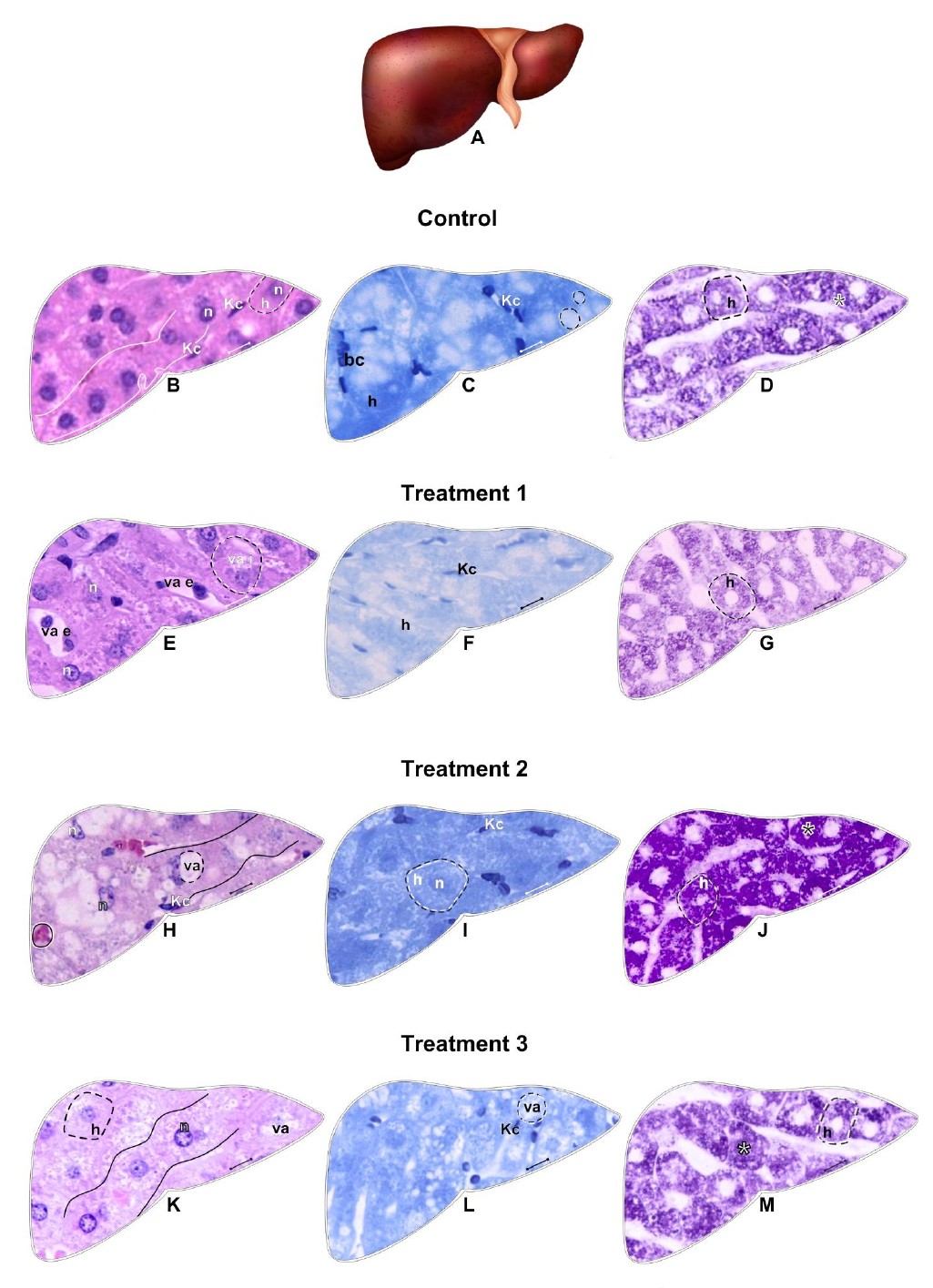
This study presents the results of hepatic tissue morpho-histology (Figure 2A-M; Table 3) of rabbit models of tick hosts allocated in different groups to develop bioassays to evaluate the effectiveness of the bioactive compounds present in the extract of C. sativa flowers in the healing process of skin lesions caused by attachment of ectoparasites.

Scheme (A) and histological sections (B-M) of rabbit liver exposed to dilutions of Cannabis sativa flower extract. Control Group-GC (B-D); Treated Group 1-GT1 (E-G); Treated Group 2-GT2 (H-J); Treated Group 3-GT3 (K-M): B, E, H, K= Hematoxylin and Eosin (HE) staining; C, F, I, L= Bromophenol Blue staining for total protein detection; D, G, J, M= Periodic Acid-Schiff (PAS) reaction for neutral polysaccharide detection. Circle = blood accumulation (congestion), n = hepatocyte nucleus, Kc = Kupffer cell, solid lines = hepatocyte cords, va e = external vacuole, va i = internal vacuole, h = hepatocyte, bc = blood cells, asterisk = polysaccharide granulation.
Summary of hepatic tissue markings in rabbit model hosts of R. linnaei ticks, exposed to Cannabis sativa flower extracts at dilutions: 0.2 mg/mL (GT1), 0.4 mg/mL (GT2), and 0.8 mg/mL (GT3).
The results obtained for the control group, that is, in the group where the rabbits were infested with R. linnaei ticks, the lesions were exposed topically to only 1 mL of olive oil, as expected, no changes were detected in the liver tissue, nor its organization or histology since the cells (hepatocytes) maintained their cubic shape, presenting clear cell boundaries and homogeneous cytoplasm and nuclei (one or two/cell) with active chromatin (Figure 2B). The organization of the cells in hepatic cords was preserved (Figure 2B).
The liver tissue also presented Kupffer cells (their nuclei and not their boundaries) between the hepatic cords, well-labeled nuclei with elongated shapes (Figure 2B).
There was a strong labeling of hepatocytes histochemically for detecting proteins by staining with bromophenol blue and polysaccharides by the SBP technique (Figures 2C and D), confirming the presence of these elements.
Figure 2C showed that the cytoplasm presented rounded structures that were suggested to be where lipid droplets would be housed, which could not be visualized/labeled due to the unspecificity of the technique. The nuclei of hepatocytes were weakly labeled, in contrast to the nuclei of Kupffer cells, which were strongly positive (Figure 2C).
The detection of polysaccharides by the PAS reaction (Figure 2D) showed that the cytoplasm of hepatocytes was strongly labeled, indicating the presence of a homogeneously distributed granulation inside the cell. The nuclei of hepatocytes and Kupffer cells could not be evidenced since this technique is specific for labeling polysaccharides.
4.2.2. Treatment Group 1 (TG1)
Disorganization in the arrangement of hepatocytes in the tissue was also observed in the liver of rabbits of treated Group 1 (GT1), which was exposed to a diluted solution of 0.2 mg/mL of C. sativa extract, which did not follow the cord structure (Figure 2E). This group also showed a tissue vacuolation between the hepatocyte cords, which prevented contact between the cells of the adjacent cords in many regions (Figure 2E).
The morphology of hepatocytes also underwent alterations since their cytoplasm indicated slight vacuolation (Figure 2E). Some hepatocyte nuclei also underwent alterations, becoming irregular and with many of them presenting marginalized chromatin, indicating a possible beginning of cell death (Figure 2E). The nuclei of Kupffer cells were preserved.
The detection of proteins by bromophenol blue confirmed the presence of vacuolization in the liver tissue. However, these vacuoles reacted to the dye applied, indicating the presence of protein elements or protein complexes (Figure 2F). The nuclei of Kupffer cells, located between the hepatocyte cords, were well labeled, thus being clearly visible.
As observed in the control group (CG), the PAS reaction for polysaccharide detection showed that the TG1 liver tissue showed a very similar result (Figures 2D and G) to that of the control group (CG).
4.2.3. Treatment Group 2 (TG2)
The liver tissue of rabbits of Treatment Group 2 (TG2) exposed to dilution of 0.4 mg/mL of the C. sativa extract suffered the most morphological changes. The arrangement of hepatocytes in the form of cords was completely lost since the cells lost their original cubic shape and underwent hypertrophy due to their extensive and intense vacuolization, which deconfigured the initial hepatic arrangement (Figure 2H).
In addition to the cell morphology, this group showed that the hepatocyte nuclei changed their shape, going from round to completely irregular (Figure 2H), also having chromatin adhered to the inner membrane of the nucleus, an evident sign that these hepatocytes would begin the cell death processes. The nuclei of Kupffer cells were apparently unchanged and strongly labeled by hematoxylin (Figure 2H). The liver tissue of this group showed congestion (accumulation of blood) in the extracellular spaces (Figure 2H).
Histochemical analysis for protein detection in the liver tissue of GT2 animals showed strong positive labelings in the cytoplasm of hepatocytes, with medially labeled nuclei, unlike those of Kupffer cells (Figure 2I).
The technique to detect polysaccharides also confirmed that the liver tissue of this group suffered the most morphological changes due to exposure to the C. sativa extract. Strong positive labeling for polysaccharides was observed in the cytoplasm of hepatocytes in more intense granulation that occupied the entire area of these cells' cytoplasm (Figure 2J). The nuclei of hepatocytes and Kupffer cells were not labeled also due to the technique's specificity (Figure 2J).
4.2.4. Treatment Group 3 (TG3)
The rabbits of Treatment Group 3 (TG3) had their lesions exposed to the C. sativa extract at a dilution of 0.8 mg/mL (Figures 2K-M). The results showed a slight alteration in the liver tissue when compared to the CG, not in its organization, but in the histology of hepatocytes, which showed cytoplasmic vacuolation and chromatin preferentially adhered to the inner membrane of the nucleus (Figure 2K).
The histochemical data for this group showed that the technique for protein detection confirmed the presence of cytoplasmic vacuolization, whose content of the vacuoles was not labeled, indicating that it was not of a protein nature (Figure 2L). The nuclei of the Kupffer cells became very evident.
The results for detecting polysaccharides were very similar to those observed in the animals of CG (Figure 2M).
5. Discussion
Controlling ticks so as not to cause damage to the environment or non-target organisms has been a persistent concern since finding efficient mechanisms that do not affect the environment, and the host has become a goal in research within this theme. Among the tick species that have been the focus of this type of control is the dog tick, Rhipicephalus linnaei, an ectoparasite widely distributed and causing damage due to blood spoliation and because it is a vector of numerous diseases (Camargo-Mathias, 2018; Silva et al., 2023). The impact of the presence of this ectoparasite currently extends to humans, with the transmission of pathogens such as Rickettsia, which is associated with the development of pathological conditions such as spotted fever (Abreu et al., 2019; Dantas-Torres, 2008; Fonsêca et al., 2022).
This urgent search for new strategies has led researchers to explore the potential of bioactive compounds produced by many plants with, among others, acaricidal properties. These bioactive compounds have been extracted from these plants and can be used as crude extracts, essential oils, or other ways (Abreu et al., 2020; Pereira et al., 2023).
Among the many studies conducted in this sense, records confirm the discovery of formulations with acaricidal properties containing bioactive compounds extracted from plants, among them neem oil (Azadirachta indica) (Remedio et al., 2016) from Euterpe oleraceae (Marques et al., 2019) and andiroba (Roma et al., 2011; Camargo-Mathias, 2018). In addition to these plants, Cannabis sativa emerges as a potential acaricide already proven in laboratory studies (personal information) since its active compounds, known as cannabinoids, in addition to the anti-tick property, have shown therapeutic efficacy in humans in the treatment of diseases such as chronic pain, epilepsy, anxiety, and even in certain types of cancer (Martins et al., 2022; Vieira et al., 2020). In veterinary medicine, it is mainly used for treating pain, contact and atopic dermatitis, epilepsy, diabetes, asthma, glaucoma, and other inflammatory diseases. It also stimulates appetite in animals affected by tick disease (Miranda-Cortés et al., 2023). Cannabinoids are lipophilic substances that, when present in the organisms, are rapidly distributed and, consequently, pass through the liver, where they are processed (Lucas et al., 2018; Martinez Naya et al., 2024).
Thus, the present study aimed to show the effects of the exposure of rabbits, simulating host models of dog ticks Rhipicephalus linnaei, at different dilutions of the extract obtained from Cannabis sativa flowers, which has been used in several areas (medical, veterinary, among others), in the treatment of diseases. Now, its potential anti-tick properties are proven.
The results of the bioassays indicated that rabbits' exposure to C. sativa did not significantly alter the values of TGO and TGP enzymes (Tables 1 and 2), except in TG2. The levels of these enzymes remained very close to those observed in the Control Group (CG). TGO and TGP are enzymes that signal the proper liver functioning since alanine aminotransferase (TGP) is found mainly in hepatocytes. In contrast, aspartate aminotransferase (TGO) is present in the myocardium, liver, kidneys, and other tissues (Luo et al., 2022). Among the several studies that analyzed the reference values of liver enzymes in organisms that were exposed to both synthetic and natural chemical agents is that of Sodelli et al. (2023), who demonstrated in a comparative clinical-morphological study that there was no change in the pattern of liver enzymes of rabbits exposed to the bioactive esters of castor oil (Ricinus communis), leaving the results within the reference values and, consequently, very close to those obtained from the control group (CG), which was corroborated by the data found in this study.
Although the results obtained in this study were very similar to those of the CG, there was only one change in the profile of liver enzymes during the exposure of rabbits to C. sativa, which was observed in the animals allocated to TG2 (0.4 mg/mL). This dose induced an increase in TGP levels, which signaled the occurrence of mild hepatic dysfunction, most likely due to the presence of the extract in the animal's system, indicating that, in a possible use of the Cannabis extracts to control R. linnaei ticks, this dosage would not be indicated although it was effective. The literature has provided information on pathological changes in various organs, including the liver, caused by exposure to bioactive agents. Studies conducted by Luo et al. (2022) and Bouassa et al. (2022) investigating the exposure of the protective effect of isoorientin (against oleic acid damage) in rats and the action of cannabinoids on chronic liver diseases in human and rat cells, respectively, also demonstrated that there was an increase in alanine aminotransferase (TGP) levels, which was interpreted as indicative of intoxication associated with liver damage, most likely due to the presence of the toxic substance, and emphasizing its origin as non-alcoholic (Nakadate et al., 2023; Marques et al., 2019).
The histopathological evaluation of the liver of rabbits exposed to different dilutions of the Cannabis extract in this study showed that the liver tissue of rabbits from the CG (exposed only to olive oil), as expected, did not undergo changes in the organization of the tissue and in cell morphology (hepatocytes and Kupffer cells), which corroborated the description of the architecture of an intact liver, found in the literature (Junqueira and Carneiro, 2023; Ramachandran et al., 2020; Marques et al., 2019).
In TG1, TG2, and TG3, histological data showed disorganization in the arrangement of hepatocytes in the tissue and cellular (and tissue) vacuolization, which were also interpreted as indicative of cell death. Cytoplasmic vacuolation is characterized by forming vacuoles or membrane-delimited compartments within the cytoplasm, usually resulting from the dilatation of the endoplasmic reticulum and mitochondria. This phenomenon can indicate cellular stress and is often associated with cell death, occurring through non-apoptotic mechanisms, such as paraptosis. Cell death is essential for maintaining tissue homeostasis and can occur by different pathways (Alberts et al., 2017; Camargo-Mathias et al., 2023). Schoeman et al. (2020) and Mokoena et al. (2024) found vacuolation in human breast cancer cells exposed to 40 µM and 5 µg/mL of CBD, respectively, in addition to cell swelling. In this study, it was inferred that this cytoplasmic vacuolation in hepatocytes could be indicating that the cells would be packaging and isolating in phagocytic or pinocytic vacuoles, cytoplasmic regions, or even organelles that were already damaged by the toxic substance in an attempt to preserve the rest of the cytoplasmic environment still intact so that the cell would not suffer more significant damage and thus could continue to perform its functions, which corroborates data from other authors (Nodari et al., 2011; Pereira et al., 2009; Erukainure et al., 2021). The presence of vacuolization areas in the hepatocyte cytoplasm was also demonstrated in other studies that exposed mice to synthetic (fipronil) and natural (thymol) chemical-based anti-tick properties (Cunha et al., 2017; Lahmi et al., 2024).
This study showed that the hepatocyte nuclei also manifested morphological changes, specifically in the TG1 and TG2 animals, characterized by the change in their shape from rounded to irregular and by the marginalization of chromatin, confirming the beginning of cell death, most often by autophagy or apoptosis. This emphasizes that autophagy is a conserved process in which the cytoplasmic content is sequestered in autophagosomes, which subsequently fuse to lysosomes so that degradation or recycling of the content occurs. Schoeman et al. (2020) showed a cell cycle arrest in the G2 phase when they exposed human cancer cell lines (breast) to the combination of C6 cannabinoids (THC, CBG, CBN, and CBD). According to several authors, the activation of autophagy induced by cannabinoids would often be mediated by the induction of endoplasmic reticulum stress (Camargo-Mathias et al., 2023; Aarestrup, 2015).
The rabbits allocated in TG2, exposed to 0.4 mg/mL of C. sativa, presented morphological changes in the liver tissue represented by the significant loss of tissue organization; that is, hepatocytes lost their arrangement in cords. The presence of extensive vacuolization in the cells and hypertrophy also occurred, which would undoubtedly contribute to modifying the hepatic circulation since this increase in cell size would alter the arrangement of hepatocytes in cords/plates, causing normal disruption of blood circulation that would cease to be efficient in removing metabolic products and performing other liver functions (Junqueira and Carneiro, 2023; Sodelli et al., 2023; Shanmuganathan et al., 2021). Thus, exposure to this dilution caused liver damage, which could be associated with a series of cellular and tissue responses, including the establishment of inflammatory processes (Devaraj et al., 2022; Ezhilarasan and Najimi, 2023; Aarestrup, 2015). The presence of hypertrophic cells and blood congestion in this group could also support the hypothesis that there was an increase in hepatic metabolism due to exposure to the extracts, characterized by increased synthesis of proteins and carbohydrates by hepatocytes, whose resulting products, or at least part of them, would require storage in the cytoplasm, which would considerably increase the size of the cells (Cunha et al., 2017; Hall and Hall, 2021).
Changes in liver tissue/cells were also detected in TG3 animals, such as cytoplasmic vacuolation and chromatin marginalized in the nuclei, in response to exposure. However, the inflammatory responses observed in this group were less intense when compared with those of TG2 animals, characterizing the known biphasic or hormetic effect, which occurs when exposure to lower doses of certain toxic substances results in more significant damage, as recorded by Ewing et al. (2019), who observed that rats exposed to lower doses of CBD indicated the expression of several genes related to hepatotoxicity, while those exposed to higher doses indicated inhibition of the expression of these same genes. In addition, Pandelides et al. (2020) also observed that lower doses of CBD would increase the incidence of senescence and inflammation markers in the livers of zebrafish (Danio rerio), as fish exposed to the lowest doses of CBD would have higher levels of indicators of cellular stress and inflammation, which suggested that the dose, in case of exposure, would be a critical factor in determining toxic/biological effects. Thus, according to the histological results obtained in the present study, the damage caused by exposure to C. sativa extracts was not dose-dependent since the dose that caused the most damage to the liver of the animals was 0.4 mg/L. These data were corroborated by histochemical analysis, which showed that the TG1 and TG3 animals did not suffer significant changes in the biosynthesis or degradation rate of polysaccharides since the results obtained were quite similar to those observed in the CG animals, indicating that the metabolic processes related to carbohydrates in the liver, crucial for the stability of blood glucose levels, were not drastically altered, corroborating other studies conducted (Zhang et al., 2020; Trefts et al., 2017; Yang et al., 2020).
Contrary to what was observed in TG1 and TG3, the GT2 group presented the most significant histochemical changes in the liver tissue, where the labeling for polysaccharides and proteins was strongly positive. Several authors who have exposed animals to synthetic chemicals or plant extracts to study toxic potential have shown changes mainly in hepatic protein synthesis (Roma et al., 2011; Ali et al., 2024). Mice exposed to permethrin and antiviral compositions sofosbuvir and ribavirin had an increase in protein synthesis, suggesting the intensification of cellular activity in the production for cell maintenance and for the export of plasma proteins such as albumin, prothrombin, fibrinogen, and lipoproteins. This phenomenon, also observed in this study, indicated a similar response when rabbits were exposed to C. sativa at 0.4 mg/mL since there was an increase in protein synthesis and degradation due to stress caused in the body. There was also an increase in the labeling of polysaccharides in liver tissue, especially in the cytoplasm of hepatocytes, probably in response to exposure to bioactive compounds present in Cannabis, suggesting an increase in the synthesis/storage of polysaccharides, specifically glycogen, considered the source of energy of animals and whose synthesis and degradation are regulated according to cellular need (Alberts et al., 2017). This regulation involves several hormones, such as insulin, glucagon, and adrenaline, which stimulate degradation. These authors exposed rabbits to Cannabis extract, corroborating the findings of other authors regarding the increase in the synthesis/storage of polysaccharides, probably glycogen, in the cytoplasm of hepatocytes, which occurred in response to chronic and prolonged stress to bioactive compounds.
The histochemical test for protein detection in hepatocytes showed extensive cytoplasmic vacuolation in TG3 animals. However, these vacuoles were not labeled by bromophenol blue or PAS, indicating that their content should probably have lipid origin (histochemical technique not performed here). This hypothesis also stems from the fact that the liver is the primary organ responsible for the homeostasis of fatty acids and cholesterol, playing an important role in regulating triacylglycerols from meals and non-esterified fatty acids (Fuhrman et al., 2004). In addition, cholesterol synthesized in the liver can be stored in droplets to be later packaged and secreted as low-density lipoproteins (LDL) or used to synthesize bile acids, steroid hormones, and vitamins.
6. Conclusion
This study evaluated the safety of the crude extract of C. sativa flowers diluted in several dilutions as an acaricide to control R. linnaei. The data found suggest that this extract may harm the liver tissue of rabbits used as host models. Analyzes showed that all dilutions tested (0.2, 0.4, and 0.8 mg/mL) caused histopathological damage to the liver. Notably, the 0.4 mg/mL dilution was the one that most altered the hepatic tissue structure and hepatocytes. These results indicate that the crude extract of C. sativa flowers, at the dilutions tested, is unsuitable for tick control in dogs as it caused significant liver damage in the exposed animals.
Data Availability Statement
The research data analyzed in this study are not publicly available by any means.
Acknowledgements
This research has been supported CAPES (Coordenação de Aperfeiçoamento de Pessoal de Nível Superior), FAPESP (Fundação de Amparo à Pesquisa do Estado de São Paulo), CNPq (Conselho Nacional de Desenvolvimento Científico e Tecnológico) and the São Paulo State University (Unesp). The authors thank Dra. Caroline Marroni Cremonez (Cannabis therapist), Fernanda Peixoto (president of Cannabis Association Maria Flor) and Prof. Dr. Sidney José Lima Ribeiro (UNESP/Araraquara) for the technical support. This work was supported by the CAPES (Coordenação de Aperfeiçoamento de Pessoal de Nível Superior) under Grant number 88887.721079/2022-00 and 88887.801974/2023-00; FAPESP (Fundação de Amparo à Pesquisa do Estado de São Paulo) under Grant number 2023/13159-0 and CNPq (Conselho Nacional de Desenvolvimento Científico e Tecnológico) under Grant number 121204/2023-5 and 301665/2019-2.
References
- AARESTRUP, B.J., 2015. Histologia essencial. Rio de Janeiro: Guanabara.
-
ABREU, M.R., DELALIBERA JUNIOR, I., PEREIRA, N.R.C. and CAMARGO-MATHIAS, M.I., 2020. Exposure of ticks Rhipicephalus sanguineus sensu lato (Acari: ixodidae) to ozonated water. Ozone Science and Engineering, vol. 42, no. 6, pp. 507-516. http://doi.org/10.1080/01919512.2020.1727310
» http://doi.org/10.1080/01919512.2020.1727310 -
ABREU, M.R., PEREIRA, M.C., SIMIONI, P.U., NODARI, E.F., PAIATTO, L.N. and CAMARGO-MATHIAS, M.I., 2019. Immunomodulatory and morphophysiological effects of Rhipicephalus sanguineus s. l. (Acari: Ixodidae) salivary gland extracts. Veterinary Immunology and Immunopathology, vol. 207, pp. 36-45. http://doi.org/10.1016/j.vetimm.2018.11.017 PMid:30593349.
» http://doi.org/10.1016/j.vetimm.2018.11.017 - ALBERTS, B., JOHNSON, A., LEWIS, J., MORGAN, D., RAFF, M., ROBERTS, K., WALTER, P., WILSON, J. and HUNT, T., 2017. Biologia molecular da célula 6. ed. Porto Alegre: Artmed.
-
ALI, R.A., AWADALLA, E.A., AMIN, Y.A., FOUAD, S.S., AHMED, M.A., HASSAN, M.H., ABDEL-KAHAAR, E. and ABDEL-AZIZ, R.H., 2024. The deleterious effects of sofosbuvir and ribavirin (antiviral drugs against hepatitis C virus) on different body systems in male albino rats regarding reproductive, hematological, biochemical, hepatic, and renal profiles and histopathological changes. Scientific Reports, vol. 14, no. 1, pp. 5. http://doi.org/10.1038/s41598-024-55950-5
» http://doi.org/10.1038/s41598-024-55950-5 - BECHARA, G., SZABÓ, M.P.J., FERREIRA, B.R. and GARCIA, M.V., 1995. Rhipicephalus sanguineus tick in Brazil: feeding and reproductive aspects under laboratorial conditions. Brazilian Journal of Medical and Biological Research, vol. 4, pp. 61-66.
-
BECHMANN, L.P., HANNIVOORT, R.A., GERKEN, G., HOTAMISLIGIL, G.S., TRAUNER, M. and CANBAY, A., 2012. The interaction of hepatic lipid and glucose metabolism in liver diseases. Journal of Hepatology, vol. 56, no. 4, pp. 952-964. http://doi.org/10.1016/j.jhep.2011.08.025 PMid:22173168.
» http://doi.org/10.1016/j.jhep.2011.08.025 -
BECKER, S., WEBSTER, A., DOYLE, R.L., MARTINS, J.R., RECK, J. and KLAFKE, G.M., 2019. Resistance to deltamethrin, fipronil and ivermectin in the brown dog tick, Rhipicephalus sanguineus sensu stricto, Latreille (Acari: ixodidae). Ticks and Tick-Borne Diseases, vol. 10, no. 5, pp. 1046-1050. http://doi.org/10.1016/j.ttbdis.2019.05.015 PMid:31175029.
» http://doi.org/10.1016/j.ttbdis.2019.05.015 -
BOUASSA, R.S.M., SEBASTIANI, G., DI MARZO, V., JENABIAN, M.A. and COSTINIUK, C.T., 2022. Cannabinoids and chronic liver diseases. International Journal of Molecular Sciences, vol. 23, no. 16, pp. 9423. http://doi.org/10.3390/ijms23169423 PMid:36012687.
» http://doi.org/10.3390/ijms23169423 -
CAMARGO-MATHIAS, M.I., 2018. Inside ticks: morphophysiology, toxicology and therapeutic perspectives. São Paulo: EDUNESP. http://doi.org/10.7476/9788595462861
» http://doi.org/10.7476/9788595462861 -
CAMARGO-MATHIAS, M.I., ABREU, M., SOCOLOWSKI, P.C., RODRIGUES, M.L., SILVA, O., GONÇALVES, M.F.C., GAINZA, Y.A., INOUE, L.V.B., BISPO, Y.M., ARAÚJO, B.J.N., SILVA, M.V.M.L. and VINHA, E.C., 2023. A morte celular como uma estratégia biológica de sobrevivência: benefícios e prejuízos Rio Claro: UNESP, 654 p. http://doi.org/10.13140/RG.2.2.33543.37288
» http://doi.org/10.13140/RG.2.2.33543.37288 -
CARLINI, E.A., 2006. The history of marijuana in Brazil. Jornal Brasileiro de Psiquiatria, vol. 55, no. 4, pp. 314-317. http://doi.org/10.1590/S0047-20852006000400008
» http://doi.org/10.1590/S0047-20852006000400008 -
CARVALHO, V., AGUIAR, A., BARATTO, L., SOUZA, F. and ROCHA, E., 2020. Quantificação de canabinoides em extratos medicinais de Cannabis por cromatografia líquida de alta eficiência. Quimica Nova, vol. 43, pp. 90-97. http://doi.org/10.21577/0100-4042.20170457
» http://doi.org/10.21577/0100-4042.20170457 -
CHICOINE, A., ILLING, K., VUONG, S., PINTO, K.R., ALCORN, J. and COSFORD, K., 2020. Pharmacokinetic and safety evaluation of various oral doses of a novel 1:20 THC. Frontiers in Veterinary Science, vol. 7, pp. 583404. http://doi.org/10.3389/fvets.2020.583404 PMid:33134364.
» http://doi.org/10.3389/fvets.2020.583404 -
CUNHA, E.L.R., MATOS, R.S., PEREIRA, N.R.C., OLIVEIRA, P.R., DAEMON, E. and CAMARGO-MATHIAS, M.I., 2017. Histopathological changes in the liver and thyroid of mice (Mus musculus) caused by the acaricides: fipronil and thymol. Histology and Histopathology, vol. 4, no. 1, pp. 9. http://doi.org/10.7243/2055-091X-4-9
» http://doi.org/10.7243/2055-091X-4-9 -
DANTAS-TORRES, F., 2008. The brown dog tick, Rhipicephalus sanguineus (Latreille, 1806) (Acari: Ixodidae): from taxonomy to control. Veterinary Parasitology, vol. 152, no. 3-4, pp. 173-185. http://doi.org/10.1016/j.vetpar.2007.12.030 PMid:18280045.
» http://doi.org/10.1016/j.vetpar.2007.12.030 -
DEVARAJ, E., PERUMAL, E., SUBRAMANIYAN, R. and MUSTAPHA, N., 2022. Liver fibrosis: extracellular vesicles mediated intercellular communication in perisinusoidal space. Hepatology, vol. 76, no. 1, pp. 275-285. http://doi.org/10.1002/hep.32239 PMid:34773651.
» http://doi.org/10.1002/hep.32239 -
ELSOHLY, M.A., RADWAN, M.M., GUL, W., CHANDRA, S. and GALAL, A., 2017. Phytochemistry of Cannabis sativa L. Progress in the Chemistry of Organic Natural Products, vol. 103, pp. 1-36. http://doi.org/10.1007/978-3-319-45541-9_1 PMid:28120229.
» http://doi.org/10.1007/978-3-319-45541-9_1 -
ERUKAINURE, O.L., MATSABISA, M.G., SALAU, V.F., OYEDEMI, S.O., OYENIHI, O.R., IBEJI, C.U. and ISLAM, M.S., 2021. Cannabis sativa L. (var. indica) exhibits hepatoprotective effects by modulating hepatic lipid profile and mitigating gluconeogenesis and cholinergic dysfunction in oxidative hepatic injury. Frontiers in Pharmacology, vol. 12, pp. 705402. http://doi.org/10.3389/fphar.2021.705402 PMid:34992528.
» http://doi.org/10.3389/fphar.2021.705402 -
EWING, L.E., SKINNER, C.M., QUICK, C.M., KENNON-MCGILL, S., MCGILL, M.R., WALKER, L.A., ELSOHLY, M.A., GURLEY, B.J. and KOTURBASH, I., 2019. Hepatotoxicity of a cannabidiol-rich cannabis extract in the mouse model. Molecules, vol. 24, no. 9, pp. 1694. http://doi.org/10.3390/molecules24091694 PMid:31052254.
» http://doi.org/10.3390/molecules24091694 -
EZHILARASAN, D. and NAJIMI, M., 2023. Intercellular communication among liver cells in the perisinusoidal space of the injured liver: pathophysiology and therapeutic directions. Journal of Cellular Physiology, vol. 238, no. 1, pp. 70-81. http://doi.org/10.1002/jcp.30915 PMid:36409708.
» http://doi.org/10.1002/jcp.30915 -
FONSÊCA, A.D.V., OLIVEIRA, L.M.B., JORGE, F.R., CAVALCANTE, R.O., BEVILAQUA, C.M.L., PINTO, F.J.M., SANTOS, J.M.L., TEIXEIRA, B.M., RODRIGUES, A.K.P.P., BRAZ, G.F., VIANA, G.A., COSTA, E.C., SERPA, M.C.A., WECK, B.C. and LABRUNA, M.B., 2022. Occurrence of tick-borne pathogens in dogs in a coastal region of the state of Ceará, northeastern Brazil. Revista Brasileira de Parasitologia Veterinária, vol. 31, no. 1, e021321. http://doi.org/10.1590/s1984-29612022010 PMid:35239843.
» http://doi.org/10.1590/s1984-29612022010 -
FUHRMAN, M.P., CHARNEY, P. and MUELLER, C.M., 2004. Hepatic proteins and nutrition assessment. Journal of the American Dietetic Association, vol. 104, no. 8, pp. 1258-1264. http://doi.org/10.1016/j.jada.2004.05.213 PMid:15281044.
» http://doi.org/10.1016/j.jada.2004.05.213 - GEWEHR, J.L.H., 2021. Guia inicial da cannabis na medicina veterinária. Santa Catarina: Edição do Autor.
-
GONÇALVES, V.M., HUERTA, M. and FREITAG, R.A., 2016. Potencial de plantas acaricidas no controle de carrapatos Rhipicephalus (Boophilus) microplus. Revista de Ciência Veterinária e Saúde Pública, vol. 3, no. 1, pp. 14-22. http://doi.org/10.4025/revcivet.v3i1.29096
» http://doi.org/10.4025/revcivet.v3i1.29096 - HALL, J.E. and HALL, M.E., 2021. Guyton & Hall: tratado de fisiologia médica 13. ed. Rio de Janeiro: Guanabara Koogan.
- HEBLING, L.M.G.F., 2011. Estudo dos efeitos de extratos de glândulas salivares de fêmeas de Rhipicephalus sanguineus (Latreille, 1806) (Acari:Ixodidae) na resposta imunológica de hospedeiros pós inoculados. Rio Claro: Universidade Estadual Paulista, 151 p. Dissertação de Mestrado em Ciências Biológicas.
- JUNQUEIRA, L.C. and CARNEIRO, J., 2023. Basic histology: text and atlas 14th ed. São Paulo: Guanabara Koogan.
- JUNQUEIRA, L.C. and JUNQUEIRA, L.M.M.S., 1983. Basic cytology and histology techniques. São Paulo: Santos.
-
LAHMI, A., ORYAN, S., EIDI, A. and ROHANI, A.H., 2024. Comparative effects of thymol and vitamin E on nonalcoholic fatty liver disease in male Wistar rats. Brazilian Journal of Biology = Revista Brasileira de Biologia, vol. 84, e268781. http://doi.org/10.1590/1519-6984.268781
» http://doi.org/10.1590/1519-6984.268781 -
LIMA, A.A., ALEXANDRE, U.C. and SANTOS, J.S., 2021. The use of marijuana (Cannabis sativa L.) in the pharmaceutical industry: a review. Research, Society and Development, vol. 10, no. 12, e46101219829. http://doi.org/10.33448/rsd-v10i12.19829
» http://doi.org/10.33448/rsd-v10i12.19829 -
LUCAS, C.J., GALETTIS, P. and SCHNEIDER, J., 2018. The pharmacokinetics and the pharmacodynamics of cannabinoids. British Journal of Clinical Pharmacology, vol. 84, no. 11, pp. 2477-2482. http://doi.org/10.1111/bcp.13710 PMid:30001569.
» http://doi.org/10.1111/bcp.13710 -
LUO, T., JIANG, S., ZHOU, B., SONG, Q., DU, J., LIU, P., WANG, X., SONG, H. and SHAO, C., 2022. Protective effect of isoorientin on oleic acid-induced oxidative damage and steatosis in rat liver cells. Frontiers in Pharmacology, vol. 13, pp. 818159. http://doi.org/10.3389/fphar.2022.818159 PMid:35185572.
» http://doi.org/10.3389/fphar.2022.818159 -
MABASA, R., MALEMELA, K., SERALA, K., KGAKISHE, M., MATSEBATLELA, T., MOKGOTHO, M. and MBAZIMA, V., 2021. Ricinus communis butanol fraction inhibits MCF-7 breast cancer cell migration, adhesion, and invasiveness. Integrative Cancer Therapies, vol. 20, pp. 1534735420977684. http://doi.org/10.1177/1534735420977684 PMid:33565349.
» http://doi.org/10.1177/1534735420977684 -
MARQUES, E.S., FRODER, J.G., OLIVEIRA, P.R., PERAZZO, F.F., ROSA, P.C.P., GAIVÃO, I.O.N., CAMARGO-MATHIAS, M.I. and MAISTRO, E.L., 2019. Cytotoxic effects of Euterpe oleraceae fruit oil (açaí) in rat liver and thyroid tissues. Revista Brasileira de Farmacognosia, vol. 29, no. 1, pp. 54-61. http://doi.org/10.1016/j.bjp.2018.12.001
» http://doi.org/10.1016/j.bjp.2018.12.001 -
MARTINEZ NAYA, N., KELLY, J., CORNA, G., GOLINO, M., POLIZIO, A.H., ABBATE, A., TOLDO, S. and MEZZAROMA, E., 2024. An overview of cannabidiol as a multifunctional drug: pharmacokinetics and cellular effects. Molecules, vol. 29, no. 2, pp. 473. http://doi.org/10.3390/molecules29020473 PMid:38257386.
» http://doi.org/10.3390/molecules29020473 -
MARTINS, A.M., GOMES, A.L., VILAS BOAS, I., MARTO, J. and RIBEIRO, H.M., 2022. Cannabis-based products for the treatment of skin inflammatory diseases: a timely review. Pharmaceuticals, vol. 15, no. 2, pp. 210. http://doi.org/10.3390/ph15020210 PMid:35215320.
» http://doi.org/10.3390/ph15020210 -
MIRANDA-CORTÉS, A., MOTA-ROJAS, D., CROSIGNANI-OUTEDA, N., CASAS-ALVARADO, A., MARTÍNEZ-BURNES, J., OLMOS-HERNÁNDEZ, A., MORA-MEDINA, P., VERDUZCO-MENDOZA, A. and HERNÁNDEZ-ÁVALOS, I., 2023. The role of cannabinoids in pain modulation in companion animals. Frontiers in Veterinary Science, vol. 9, pp. 1050884. http://doi.org/10.3389/fvets.2022.1050884 PMid:36686189.
» http://doi.org/10.3389/fvets.2022.1050884 -
MOKOENA, D., GEORGE, B.P. and ABRAHAMSE, H., 2024. Cannabidiol combination enhances photodynamic therapy effects on MCF-7 breast cancer cells. Cells, vol. 13, no. 2, pp. 187. http://doi.org/10.3390/cells13020187 PMid:38247877.
» http://doi.org/10.3390/cells13020187 -
NAKADATE, K., SONO, C., MITA, H., ITAKURA, Y. and KAWAKAMI, K., 2023. Severe acute liver dysfunction induces delayed hepatocyte swelling and cytoplasmic vacuolization and delayed cortical neuronal cell death. International Journal of Molecular Sciences, vol. 24, no. 8, pp. 7351. http://doi.org/10.3390/ijms24087351 PMid:37108515.
» http://doi.org/10.3390/ijms24087351 -
NODARI, E.F., ROMA, G.C., FURQUIM, K.C.S., BECHARA, G.H. and CAMARGO-MATHIAS, M.I., 2011. Cytotoxic effects of permethrin in salivary glands of Rhipicephalus sanguineus (Latreille, 1806) (Acari: Ixodidae) semi-engorged females. Experimental Parasitology, vol. 128, no. 2, pp. 151-158. http://doi.org/10.1016/j.exppara.2011.02.003 PMid:21352824.
» http://doi.org/10.1016/j.exppara.2011.02.003 -
PANDELIDES, Z., THORNTON, C., LOVITT, K.G., FARUQUE, A.S., WHITEHEAD, A.P., WILLETT, K.L. and ASHPOLE, N.M., 2020. Developmental exposure to Δ9-tetrahydrocannabinol (THC) causes biphasic effects on longevity, inflammation, and reproduction in aged zebrafish (Danio rerio). GeroScience, vol. 42, no. 3, pp. 923-936. http://doi.org/10.1007/s11357-020-00175-3 PMid:32227279.
» http://doi.org/10.1007/s11357-020-00175-3 - PEARSE, A.G.E., 1985. Histochemistry theoretical and applied. 3rd ed. London: Churchill Livingstone.
-
PEREIRA, C.P.M., OLIVEIRA, P.R., FURQUIM, K.C.S., BECHARA, G.H. and CAMARGO-MATHIAS, M.I., 2009. Effects of fipronil (active ingredient of Frontline®) on salivary gland cells of Rhipicephalus sanguineus females (Latreille, 1806) (Acari: ixodidae). Veterinary Parasitology, vol. 166, no. 1-2, pp. 124-130. http://doi.org/10.1016/j.vetpar.2009.08.015 PMid:19748741.
» http://doi.org/10.1016/j.vetpar.2009.08.015 -
PEREIRA, M.C., ANHOLETO, L.A., KASA, G.G., CASTRO, K.N.C., CANUTO, K.M., SOUZA, A.S.Q. and CAMARGO-MATHIAS, M.I., 2023. Efficacy of essential oils of Egletes viscosa and Lippia schaueriana on the reproductive biology of Rhipicephalus sanguineus sensu lato engorged females. Experimental Parasitology, vol. 244, pp. 108423. http://doi.org/10.1016/j.exppara.2022.108423 PMid:36403801.
» http://doi.org/10.1016/j.exppara.2022.108423 -
PIERRO NETO, P.A., PIERRO, L.M.C. and FERNANDES, S.T., 2023. Cannabis: 12,000 years of experiences and prejudices. Brazilian Journal of Pain, vol. 6, no. 2, suppl. 2, pp. 1-5. http://doi.org/10.5935/2595-0118.20230055-pt
» http://doi.org/10.5935/2595-0118.20230055-pt -
RAMACHANDRAN, P., MATCHETT, K.P., DOBIE, R., WILSON-KANAMORI, J.R. and HENDERSON, N.C., 2020. Single-cell technologies in hepatology: new insights into liver biology and disease pathogenesis. Nature Reviews. Gastroenterology & Hepatology, vol. 17, no. 8, pp. 457-472. http://doi.org/10.1038/s41575-020-0304-x PMid:32483353.
» http://doi.org/10.1038/s41575-020-0304-x -
REMEDIO, R.N., NUNES, P.H., ANHOLETO, L.A., OLIVEIRA, P.R., SÁ, I.C.G. and CAMARGO-MATHIAS, M.I., 2016. Morphological alterations in salivary glands of Rhipicephalus sanguineus ticks (Acari: Ixodidae) exposed to neem seed oil with known azadirachtin concentration. Micron, vol. 83, pp. 19-31. http://doi.org/10.1016/j.micron.2016.01.004 PMid:26852009.
» http://doi.org/10.1016/j.micron.2016.01.004 -
ROMA, G.C., FURQUIM, K.C.S., BECHARA, G.H. and CAMARGO-MATHIAS, M.I., 2011. Cytotoxic effects of permethrin in oocytes of Rhipicephalus sanguineus (Acari: Ixodidae) fully engorged females. Experimental & Applied Acarology, vol. 53, no. 3, pp. 287-299. http://doi.org/10.1007/s10493-010-9401-9 PMid:20960224.
» http://doi.org/10.1007/s10493-010-9401-9 -
SANGIOVANNI, E., FUMAGALLI, M., PACCHETTI, B., PIAZZA, S., MAGNAVACCA, A., KHALILPOUR, S., MELZI, G., MARTINELLI, G. and DELL’AGLI, M., 2019. Cannabis sativa L. extract and cannabidiol inhibit in vitro mediators of skin inflammation and wound injury. Phytotherapy Research, vol. 33, no. 8, pp. 2083-2093. http://doi.org/10.1002/ptr.6400 PMid:31250491.
» http://doi.org/10.1002/ptr.6400 -
SCHOEMAN, R., BEUKES, N. and FROST, C., 2020. Cannabinoid combination induces cytoplasmic vacuolation in MCF-7 breast cancer cells. Molecules, vol. 25, no. 20, pp. 4682. http://doi.org/10.3390/molecules25204682 PMid:33066359.
» http://doi.org/10.3390/molecules25204682 -
SHANMUGANATHAN, M., SARFARAZ, M.O., KROEZEN, Z., PHILBRICK, H., POON, R., DON-WAUCHOPE, A., PUGLIA, M., WISHART, D. and BRITZ-MCKIBBIN, P., 2021. A cross-platform metabolomics comparison identifies serum metabolite signatures of liver fibrosis progression in chronic hepatitis C patients. Frontiers in Molecular Biosciences, vol. 8, pp. 676349. http://doi.org/10.3389/fmolb.2021.676349 PMid:34414211.
» http://doi.org/10.3389/fmolb.2021.676349 -
SILVA, I.S., VALE, L., COUTINHO, A.L., SOUSA, L.J.M.P., MARCHESINI, P., MATOS, R.S., ANHOLETO, L.A., CAMARGO-MATHIAS, M.I., FERREIRA, L.L. and ARRUDA, W., 2023. Thymol and eugenol against Rhipicephalus sanguineus sensu lato engorged females: biological, histopathological and bioinformatic analysis. Veterinary Parasitology, vol. 319, pp. 109938. http://doi.org/10.1016/j.vetpar.2023.109938 PMid:37167691.
» http://doi.org/10.1016/j.vetpar.2023.109938 -
ŠLAPETA, J., HALLIDAY, B., CHANDRA, S., ALANAZI, A. and ABDEL-SHAFY, S., 2022. Rhipicephalus linnaei (Audouin, 1826) recognised as the “tropical lineage” of the brown dog tick Rhipicephalus sanguineus sensu lato: neotype designation, redescription, and establishment of morphological and molecular reference. Ticks and Tick-Borne Diseases, vol. 13, no. 6, pp. 102024. http://doi.org/10.1016/j.ttbdis.2022.102024 PMid:36063755.
» http://doi.org/10.1016/j.ttbdis.2022.102024 -
SODELLI, L.F., SILVA, O., ARAÚJO, B.J.N. and CAMARGO-MATHIAS, M.I., 2023. Liver and spleen of hosts of Rhipicephalus linnaei exposed to synthetic (afoxolaner) and natural acaricides (esters from castor oil): a comparative clinical-morphological study. Revista Brasileira de Parasitologia Veterinária, vol. 32, no. 3, e004023. http://doi.org/10.1590/s1984-29612023041 PMid:37466625.
» http://doi.org/10.1590/s1984-29612023041 -
SUNKARA, P., CHENNURU, S., KROVVIDI, S. and CHITICHOTI, J., 2022. Cypermethrin and ivermectin resistance in field populations of Rhipicephalus sanguineus sensu lato (Latrielle, 1806) collected from dogs in south India. Veterinary Research Communications, vol. 46, no. 1, pp. 67-78. http://doi.org/10.1007/s11259-021-09830-2 PMid:34535851.
» http://doi.org/10.1007/s11259-021-09830-2 -
TAMBELI, C.H., MARTINS, G.A., BARBOSA, S.L. and MACHADO, T.T., 2023. Integrative approach to the therapeutic use of cannabis for orofacial pain. Brazilian Journal of Pain, vol. 6, no. 1, pp. S49-S53. http://doi.org/10.5935/2595-0118.20230013-en
» http://doi.org/10.5935/2595-0118.20230013-en -
TREFTS, E., GANNON, M. and WASSERMAN, D.H., 2017. The liver. Current Biology, vol. 27, no. 21, pp. R1147-R1151. http://doi.org/10.1016/j.cub.2017.09.019 PMid:29112863.
» http://doi.org/10.1016/j.cub.2017.09.019 - VIEIRA, L.S., MARQUES, A.E.F. and SOUSA, V.A., 2020. The use of Cannabis sativa for therapeutic purposes in Brazil: a literature review. Science and Nature, vol. 2, no. 2, pp. 901-919.
-
YANG, Y., JI, J., DI, L., LI, J., HU, L., QIAO, H., WANG, L. and FENG, Y., 2020. Resource, chemical structure and activity of natural polysaccharides against alcoholic liver damages. Carbohydrate Polymers, vol. 241, pp. 116355. http://doi.org/10.1016/j.carbpol.2020.116355 PMid:32507196.
» http://doi.org/10.1016/j.carbpol.2020.116355 -
ZHANG, C., BJORNSON, E., ARIF, M., TEBANI, A., LOVRIC, A., BENFEITAS, R., OZCAN, M., JUSZCZAK, K., KIM, W., KIM, J.T., BIDKHORI, G., STÅHLMAN, M., BERGH, P.O., ADIELS, M., TURKEZ, H., TASKINEN, M.R., BOSLEY, J., MARSCHALL, H.U., NIELSEN, J., UHLÉN, M., BORÉN, J. and MARDINOGLU, A., 2020. The acute effect of metabolic cofactor supplementation: a potential therapeutic strategy against non-alcoholic fatty liver disease. Molecular Systems Biology, vol. 16, no. 4, e9495. http://doi.org/10.15252/msb.209495 PMid:32337855.
» http://doi.org/10.15252/msb.209495
Edited by
-
Editor:
Marcelo A. M. Esquisatto