Leachable residual monomers from conventional and 3D printable poly(meth)acrylates
ABSTRACT
Poly(meth)acrylates have been widely used in healthcare for dental prostheses, fixation of hip implants and skull reconstructions, and in cosmetics as manicure products. Unreacted monomers remain in the final product and could cause adverse reactions. The release pattern of these residual monomers are not known for all different compositions and processing methods of poly(meth)acrylates.
Objective This study aims to investigate the release patterns of residual monomers from different compositions and processing methods of poly(meth)acrylates.
Material and Methods Eluates from four representative poly(meth)acrylates (40 x 28 x 6.0 mm, height x outer radius x thickness; Vertex Self-Curing, Palacos R+G, NextDent C&B MFH, and DePuy CMW-3; n = 3) were examined by reversed-phase high-performance liquid chromatography (HPLC). The residual monomer leached into water at 37.0 ± 1.0°C and the concentration was measured at time points between one hour and 14 days.
Results The materials released concentrations of residual monomers between 22,75 ± 4,05 µg/g (NextDent C&B MFH) and 78,83 ± 7,67 µg/g (DePuy CMW-3) over 14 days. Palacos R+G released the lowest monomer concentration in the first hour (p<0.01) and potentially did not reach a plateau within the observed timeframe, but was comparable to Vertex Self-Curing after 14 days (42,54 ± 3,49 vs 46,26 ± 2,23 µg/g). NextDent C&B MFH, formed with methacrylate oligomers, released a significantly lower residual monomer concentration (p < 0.005) at 14 days compared to the other materials and did not follow pseudo-first order release kinetics in the observed period.
Conclusion Different compositions and processing methods for poly(meth)acrylates show different release patterns and quantities of residual monomers. It is therefore advisable to develop specific compositions of and processing methods for poly(meth)acrylates in the medical field that are optimized for their specific application.
KEYWORDS:
Leaching; Medical device; Poly(methyl methacrylate); Release; Residual monomer
RESUMO
Os polimetacrilatos têm sido amplamente utilizados na área da saúde para próteses dentárias, fixação de implantes de quadril e reconstruções cranianas, além de aplicações cosméticas, como produtos para manicure. Monômeros não convertidos permanecem no produto final e podem causar reações adversas.
Objetivo Este estudo teve como objetivo investigar os padrões de liberação de monômeros residuais provenientes de diferentes composições e métodos de processamento de polimetacrilatos.
Material e Métodos Os eluatos de quatro meios-cilindros representativos de polimetacrilato (40×28×6,0 mm; altura × raio externo × espessura; Vertex Self-Curing, Palacos R+G, NextDent C&B MFH e DePuy CMW-3; n = 3) foram analisados por cromatografia líquida de alta eficiência em fase reversa (HPLC). O monômero residual liberado em água a 37,0 ± 1,0 °C foi quantificado em intervalos de tempo entre 1 hora e 14 dias.
Resultados Os materiais liberaram concentrações de monômeros residuais variando entre 22,75 ± 4,05 µg/g (NextDent C&B MFH) e 78,83 ± 7,67 µg/g (DePuy CMW-3) ao longo de 14 dias. O Palacos R+G apresentou a menor concentração de monômero liberada na primeira hora (p < 0,01) e, possivelmente, não atingiu um platô dentro do período observado, mas apresentou valores comparáveis ao Vertex Self-Curing após 14 dias (42,54 ± 3,49 versus 46,26 ± 2,23 µg/g). O NextDent C&B MFH, composto por oligômeros metacrílicos, liberou uma concentração significativamente menor de monômeros residuais (p < 0,005) após 14 dias em comparação aos demais materiais e não seguiu uma cinética de liberação de pseudo-primeira ordem durante o período avaliado.
Conclusão Diferentes composições e métodos de processamento dos polimetacrilatos apresentam distintos padrões e quantidades de liberação de monômeros residuais. Portanto, é recomendável desenvolver composições e métodos de processamento específicos para os polimetacrilatos utilizados na área médica, otimizados para suas respectivas aplicações.
PALAVRAS-CHAVE:
Lixiviação; Dispositivo médico; Polimetilmetacrilato; Liberação; Monômeros residuais
INTRODUCTION
Poly(meth)acrylates are frequently used in healthcare for the creation of personalized medical devices such as dentures and craniofacial reconstructions, as bone cement in orthopedics, and in cosmetics as manicure products [1-4]. Recently, a global increase in allergic contact dermatitis (ACD), a type IV allergic reaction, to the hydrophilic hydroxyethyl methacrylate (HEMA) applied as a bonding agent in manicure products and dentistry has been reported [5]. ACD is caused by this resin being applied on nails or teeth in an unpolymerized stage, after polymerization, so-called 'residual monomers' remain in the material and could result in further sensitization [6]. The quantity of residual monomers in poly(meth)acrylates following polymerization typically varies between 2% and 6% [7] and depends on the initial polymer-to-monomer ratio [8], the method of polymerization [9-11], and the (post)processing method [12,13]. The accumulated release of monomers from eight different poly(meth)acrylates ranged approximately between 185 and 470 µg/g [7]. Although the specimen thickness has been shown to significantly influence the content of residual monomers [12], it is unknown whether this affects the leaching behavior of residual monomers. Auto-, heat-, and photopolymerization are well-known techniques to manufacture poly(meth)acrylates, each with advantages and disadvantages. Curing under increased pressure decreases porosity while heat increases the degree of conversion, reducing the amount of residual monomers [14]. There are two common processing methods: direct and indirect application. Bone cement, craniofacial reconstructions, and repairs of dentures are mainly produced by the direct method, while dentures and some craniofacial reconstructions are created indirectly using molds and post-curing. The amount of residual monomers is likely generally higher in the direct method due to its heat dissipating in situ environment. MMA concentrations above 1 µg/mL have been detected in plasma during total hip arthroplasty [1]. As there are many cross-allergies between different poly(meth)acrylates, patients who have developed an allergy to HEMA also react frequently to many other poly(meth)acrylates, such as PMMA [15,16].
ACD caused by occupational as well as non-occupational exposure to (meth)acrylates has been reported [17-19]. Canizares already described this for manicure products in 1956 [20]. Beside ACD other systemic adverse reactions such as cardiovascular dysfunction, fat-embolic events, and hypotension are described in patients who underwent a poly(meth)acrylate cemented knee or hip implant in orthopedics [1,21]. Patients with poly(meth)acrylate dentures can experience a burning sensation, redness, swelling, and pain in the palate, tongue, and oral mucosa [2]. Symptoms of neurological dysfunction are described after poly(meth)acrylate craniofacial reconstruction [22,23]. The most common (meth)acrylates, which are known to be strong sensitizers, are methyl methacrylate (MMA), 2-hydroxy-ethyl methacrylate (2-HEMA), and ethylene glycol dimethacrylate (EGDMA). MMA and 2-HEMA are both commonly used in manicure and healthcare products [15,16]. An example of sensitization by manicure products and a strong adverse reaction toward dental treatment has been reported recently [24]. The use of manicure products containing MMA and 2-HEMA could thus lead to an increased associated risk of adverse reactions towards these materials, potentially resulting in limitations in medical treatment.
The use of 3D printed medical devices is on the rise due to improved accessibility, cost-effectiveness, and the ability to create unique personalized geometries that optimize fit [25]. The materials that can be used in such applications have significantly different processing methods than conventional materials which likely also results in different material compositions and characteristics. The efficacy of the addition of antibiotics to conventional bone cements is still debated [26]. The research community started to investigate alternative antibiotic-free bone cements, containing Cu- Zn-, Mg-, and Ag-nanoparticles [27]. However, little attention is paid to the poly(meth)acrylate matrix. The amount of residual monomers in the final product depends strongly on the matrix material and processing method. Therefore, we investigated the release patterns and quantities of residual monomers during the first two weeks from cured poly(meth)acrylates with different compositions and processing methods: two conventional MMA-based bone cements, one MMA-based denture material, and a new methacrylate oligomer-based material that is suitable for 3D-printing dental structures and personalized medical devices.
MATERIALS AND METHODS
Specimen preparation
Four representative materials Palacos R+G (Heraeus, Germany; bone cement), DePuy CMW-3 (DePuy International Ltd., United Kingdom; bone cement), Vertex Self-Curing (Vertex-Dental, The Netherlands; denture-base material), and NextDent C&B MFH (NextDent, The Netherlands; dental crowns and bridges / personalized medical device material) were used (Table 1).
A half cylinder, 40 mm (height) by 28 mm (outer radius) with 6.0 mm thickness (volume: 18.9 cm3, surface area: 72.5 cm2), was designed using Netfabb software (Autodesk Inc., CA, USA), inverted to create a mold, and 3D printed out of a poly-(dimethacrylate) on a Rapidshape D30 printer (Rapidshape, Heimsheim, Germany) with curvature confined to the horizontal plane and a layer thickness of 50 µm. Vertex Self-Curing, Palacos R+G, and DePuy CMW-3 were hand-mixed following the manufacturer's instructions and were used to fill the 3D-printed mold. The design was used to 3D-print half cylinders of NextDent C&B MFH. After printing, the cylinders were immersed in ethanol twice (respectively three and two min.) under ultrasonic vibrations. The cylinders were dried for 10 minutes before a post-curing period of 30 minutes in an ultraviolet lightbox (NextDent LC3D-PrintBox, Soesterberg, The Netherlands; 5mW/cm2 (315-400nm & 400-550nm), 60-80 °C). Sharp edges were wet ground with standard metallographic grinding paper (P500, P1000, and P1200). Three cylinders of each material were prepared. All specimens were stored under standard laboratory climate conditions (22.0 ± 1.0 °C and 50 ± 10% humidity) in a dark environment for 20 ± 4 hours.
The half cylinders were each inserted into a separate pre-heated bottle containing 200 mL of distilled water and were stored in a stove at 37.0 ± 1.0°C. A 0.5 ml sample was taken from the distilled water from each bottle after 1, 2, 4, 8, 12, 24, 48, and 72 hours, 7 and 14 days. During this time, no water was added to the bottles. All the water samples were stored at 7.0 ± 1.0 °C in a dark environment until further analysis.
High-performance liquid chromatography (HPLC) and liquid chromatography/mass spectrometry (LCMS)
All samples were analyzed using a reversed-phase high-performance liquid chromatography (HPLC) system (Shimadzu LC-20AT) with Diode Array (DA) detectors and a ‘Zorbax Eclipse Plus C18 Analytical Column’. MMA stock solutions (0.41, 1.03, 2.06, 4.13, and 8.25 µg/mL) in acetonitrile were measured on the HPLC to create a calibration curve relating the area under the curve [mV*s], at the peak locations, and the concentration [µg/mL]. LC/MS analyses of the different peaks in the HPLC samples were performed using a ‘Waters XSelect HSS’ HPLC with a ‘C18 2.1x50 mm, 3.5 µm’ column (Waters Chromatography B.V., The Netherlands) and a formic acid/acetonitrile/Milli-Q running solution interfaced to a ‘Bruker Amazon 230 SL’ MS (Iontrap and Dionex Ultimate 3000 (HPLC)) using positive electrospray ionization. Spectra were scanned over a mass range of m/z 70-550, taking an average of 10 spectra and using an ion spray voltage of 4.5 kV, a source temperature of 325 °C, and a nebulizer gas flow rate of 50 L/min.
Statistical analysis
Data were statistically analyzed using one-way analysis of variance (ANOVA) followed by Tukey’s post hoc test (α = 0.05) in JASP version 0.19.3 (JASP Team).
Pseudo first-order kinetics curves were fitted to the data in GraphPad Prism 5 (GraphPad Software, Boston, MA, USA) to investigate the influence of different compositions and processing methods on the amount of released residual monomers using:
where M is the quantity of released monomer [µg/g] at time t [h], M0 is the total quantity of released monomer [µg/g], and k is the rate constant.
RESULTS
High-performance liquid chromatography (HPLC)
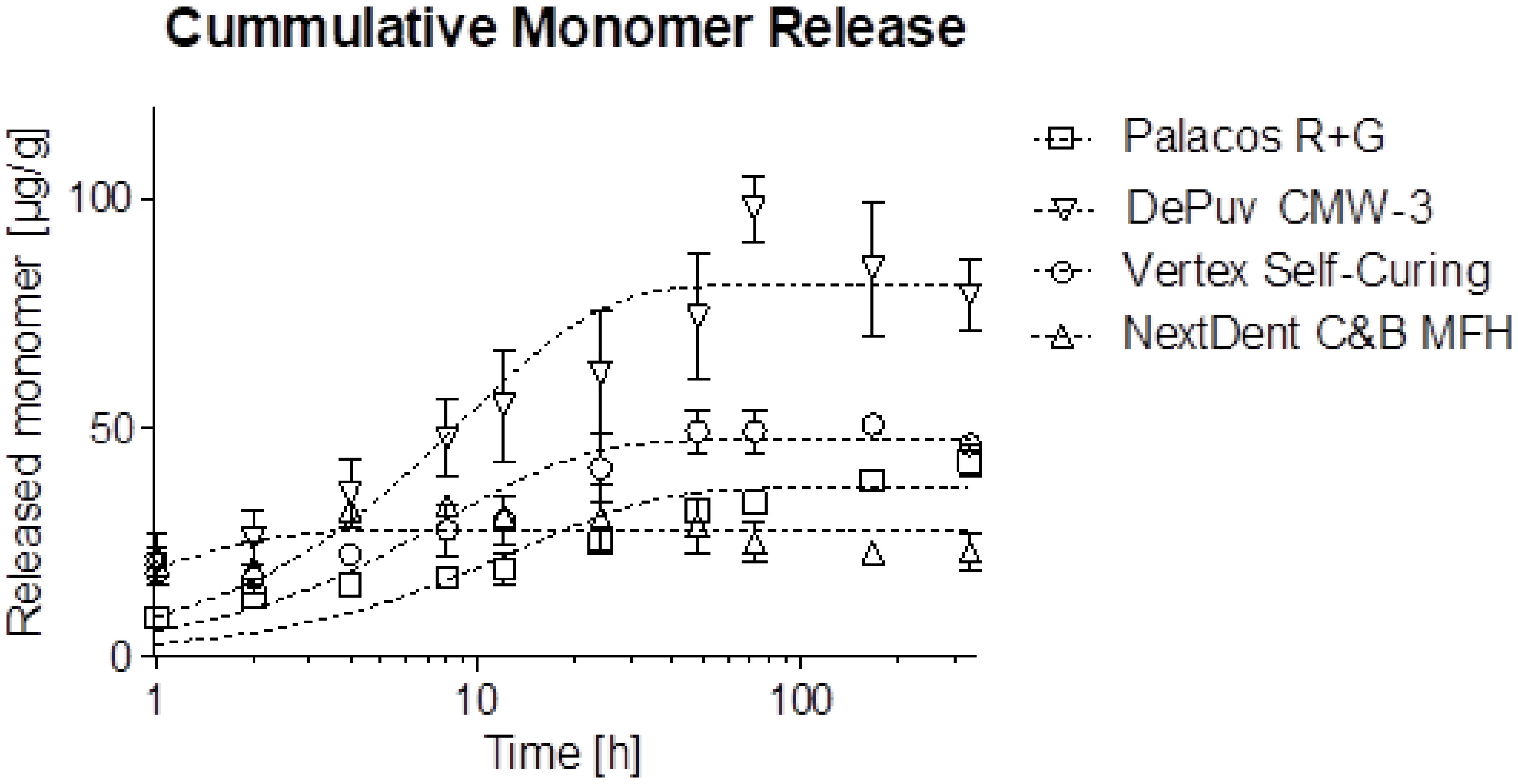
The results of the release of residual monomer from different poly(meth)acrylates over time are presented in Table 2. The results and fits of pseudo-first-order kinetics curves for the leaching process in time are graphically depicted in Figure 1. Corresponding non-linear regression variables are presented in Table 3.
- Cumulative residual monomer release in µg monomer per g product (µg/g) during 2 weeks incubation in water at 37 °C

- Cumulative residual monomer release during 2 weeks incubation in water at 37 °C with fitted pseudo first-order kinetics curves.
- Pseudo first order (M = M0 * (1 – exp(-k*t))) release profiles of Vertex Self-Curing, Palacos R+G, and NextDent C&B MFH
A majority of the released monomers from NextDent C&B MFH appear before the first time point (1 hr), whereas Vertex Self-Curing, Palacos R+G, and DePuy CMW-3 release more monomers over a longer period. NextDent C&B MFH released a similar amount of residual monomers in the first hour compared to Vertex Self-Curing and DePuy CMW-3, although all significantly more than Palacos R+G (p<0.005). This is confirmed in the pseudo first-order kinetics model where NextDent C&B MFH has a doubling time of 0.59 hrs, however, the release profile does not follow a pseudo first-order kinetics profile (R2 = 0.13). After 48 hrs all other materials surpassed the cumulative release of NextDent C&B MFH. The doubling time of Vertex Self-Curing and DePuy CMW-3 are similar at around 8 hrs, Palacos R+G is higher at approximately 13 hrs, however, this is not a significant difference. DePuy CMW-3, Vertex Self-Curing, and NextDent C&B MFH appear to reach an equilibrium within the observed time period whereas Palacos R+G may not have reached a plateau yet after 2 weeks.
Liquid chromatography/mass spectrometry (LC/MS)
Multiple peaks were visible in the HPLC spectrum of NextDent C&B MFH. LC/MS was performed to determine whether these substances are related molecules with a common precursor, which would lead to an increased concentration of released monomers (this was assumed in Tables 2, 3 and Figure 1). If this is not the case, the concentration of the released monomers is lower, but multiple molecules are being released. The MS spectra showed that these peaks contain multiple related derivatives of the monomers in NextDent C&B MFH. It is important to note that no traces of other chemicals such as the initiator system, catalyst, or colorants were detected.
DISCUSSION
This study reports on the leaching behavior of four representative poly(meth)acrylates that are used for medical devices. The following trends were observed: (i) the amount of leached monomers differed significantly for different poly(meth)acrylates, and (ii) release patterns of residual monomers of the two bone cements and the denture poly(meth)acrylates were similar but completely different from the 3D-printable material.
The different compositions of investigated poly(meth)acrylates resulted in different release patterns with different quantities of residual monomers present within the polymerized material. For the two bone cements, the amount of activator could possibly explain the different amount of residual monomers as Palacos R+G likely contains less activator than DePuy CMW-3 [7]. This typically results in a final product with longer polymer chains and thus a higher molecular weight. Due to this structure, it should be more difficult for residual monomers in Palacos R+G to release into the water, resulting in a slower release pattern and a lower cumulative concentration of leached monomers. This corresponds to our findings where Palacos R+G showed both a significantly lower initial release as well as a significantly lower overall release over two weeks compared to CMW-3. The release pattern of denture-base material Vertex Self-Curing was initially significantly higher than Palacos R+G but at two weeks there were no significant differences to Palacos R+G. NextDent C&B MFH releases fewer residual monomers, this is most probably due to the use of pre-cured oligomers and highly crosslinked materials after post-curing. NextDent C&B MFH is also the only material that is light-cured. It was shown for Bis-GMA, UDMA, and TEGDM-based materials that light-curing had a reduced amount of monomer leached out of the material compared to the chemical-cured specimens [28]. Poly(meth)acrylates which can be dual-cured (chemical- and light-curing), could therefore potentially be used to reduce the amount of free monomers in the material.
The release of some poly(meth)acrylates was reported previously. Kühn reports that Palacos R+G released approximately 185 µg MMA per g PMMA over two weeks into 5 mL of water using specimens with a volume of 0.45 cm3 and surface area of 4.5 cm2. Immediately following curing, Palacos R contained approximately 4% residual monomers; after 1 day this dropped to roughly 1.5% [7]. In our study, the specimens (volume: 18.9 cm3, surface area: 72.5 cm2) were placed in 200 mL of water after 20 ± 4 hours. The thickness of the specimens was 6 mm, comparable to the human skull [29,30]. van Landuyt et al. [31] reported a significant increase in leaching when a sample was put in a larger volume of extraction solution. Together with the difference in volume-to-surface ratio, such behavior may explain our finding of lower values than those reported in the literature [7]. The actual monomer exposure for the patient in a clinical setting, where the medical device is placed into the patient immediately upon cooling down using a directly manufactured material, is thus likely to be higher than the values reported in this study. This is especially the case when the resin is in direct contact with metal or bone while polymerizing, thereby dissipating the heat which is produced by the exothermic reaction of the polymerization reaction, and leading to a reduction of the degree of conversion. Indirectly manufactured materials should therefore in general be more likely to contain fewer residual monomers.
Biodegradable or bioinert cements based on glasses or ceramics still have to find its way from the lab to the clinic. Nevertheless, the increasing incidence of adverse reactions towards MMA stresses the need for alternative materials. For (temporary) crowns and bridges, dentures, craniofacial reconstructions, and other personalized medical devices, the amount of leached monomers can be reduced easily and effectively. Our results show that storing the material in water for 2 weeks, before placing a device in the body can reduce the amount of leached monomers significantly. For each specific combination of material composition and processing method a minimum required duration could be determined experimentally.
Medical professionals and manufacturers should strive for optimal compositions and handling for specific applications. Standards applied to assess conformity with regulations, like e.g. ISO 2017/745, should focus on describing comparable evaluation methods to enable comparison of data from multiple applications, which is now hardly possible, amongst others due to different volume-to-surface ratios.
In medicine and dentistry, an optimal implantation material is mandatory for safety, ease of use, and functional and aesthetic outcomes. As reported in this study, these materials showed significantly different quantities and release patterns of leached residual monomers in water over two weeks. The in vitro results from this study may not directly relate to the in vivo situation. As such, future research should focus on the short and long-term release into relevant physiological solutions and the resulting biological responses to elucidate the behavior of these materials at the location of their specific application within the human body. Ideally measurements at one minute, 15 minutes, one month, and two months are included to further characterize the release patterns.
The relatively small number of materials and sample size limit the generalizability of the findings. Materials were processed according to manufacturer’s instructions which gives better insight into the expected release of residual monomers in practice, however, do not relate to potential optimizations to reduce residual monomers.
CONCLUSION
Different compositions of poly(meth)acrylates showed different release patterns and quantities of residual monomers in water. The bone cement Palacos R+G showed the lowest initial monomer release after 1 hour whereas the 3D printable light curing material NextDent C&B MFH released the lowest overall quantity of monomers over two weeks. Nearly all materials have reached a release plateau within the measured time period. It is therefore advisable to develop specific compositions of and processing methods for poly(meth)acrylates in the medical field that are optimized for their specific application. Especially for applications near sensitive tissues an optimized material may prove to be advantageous. Additional post-processing could improve material characteristics, for example incubation in water as demonstrated in this study reduced the amount of leached monomers from the material.
Acknowledgements
We acknowledge the help of Brian Jacobs and his expertise in this field.
CranioSafe Group: Collaborators include: A.G. Becking, L. Dubois, L.H.E. Karssemakers, D.M.J. Milstein and S.E.C.M van de Vijfeijken, Department of Oral and Maxillofacial Surgery, Amsterdam UMC, the Netherlands; P.R.A.M. Depauw, Department of Neurosurgery, Elisabeth-Tweesteden Hospital, Tilburg, the Netherlands; F.W.A. Hoefnagels and W.P. Vandertop, Neurosurgical Center Amsterdam, Amsterdam UMC, the Netherlands; C.J. Kleverlaan and T.J.A.G. Münker, Department of Dental Material Sciences, Academic Centre for Dentistry Amsterdam, the Netherlands; T.J.J. Maal, 3D Laboratory of Oral and Maxillofacial Surgery, Amsterdam UMC, the Netherlands; E. Nout, Department of Oral and Maxillofacial Surgery, Elisabeth-Tweesteden Hospital, Tilburg, the Netherlands; M. Riool and S.A.J. Zaat, Department of Medical Microbiology, Amsterdam UMC, Amsterdam Infection and Immunity Institute, University of Amsterdam, the Netherlands.
How to cite:
Regulatory Statement
Funding
List of abbreviations
REFERENCES
-
1 Leggat PA, Smith DR, Kedjarune U. Surgical applications of methyl methacrylate: a review of toxicity. Arch Environ Occup Health. 2009;64(3):207-12. https://doi.org/10.1080/19338240903241291 PMid:19864224.
» https://doi.org/10.1080/19338240903241291 -
2 Leggat PA, Kedjarune U. Toxicity of methyl methacrylate in dentistry. Int Dent J. 2003;53(3):126-31. https://doi.org/10.1111/j.1875-595X.2003.tb00736.x PMid:12873108.
» https://doi.org/10.1111/j.1875-595X.2003.tb00736.x -
3 Ivković N, Božović D, Ristić S. The residual monomer in dental acrylic resin and its adverse effects. Contemp Mater. 2013;4(1):84-91. https://doi.org/10.7251/COMEN1301084I
» https://doi.org/10.7251/COMEN1301084I -
4 Lee SY, Lai YL, Hsu TS. Influence of polymerization conditions on monomer elution and microhardness of autopolymerized polymethyl methacrylate resin. Eur J Oral Sci. 2002;110(2):179-83. https://doi.org/10.1034/j.1600-0722.2002.11232.x PMid:12013564.
» https://doi.org/10.1034/j.1600-0722.2002.11232.x -
5 Gatica-Ortega ME, Pastor-Nieto MA, Giménez-Arnau AM, Mercader-García P, Sanz-Sánchez T, Carrascosa-Carrillo JM, et al. 2-Hydroxyethyl methacrylate (2-HEMA) sensitization, a global epidemic at its peak in Spain? Contact Dermatitis. 2024;90(5):507-13. https://doi.org/10.1111/cod.14520 PMid:38351475.
» https://doi.org/10.1111/cod.14520 -
6 Bayraktar G, Guvener B, Bural C, Uresin Y. Influence of polymerization method, curing process, and length of time of storage in water on the residual methyl methacrylate content in dental acrylic resins. J Biomed Mater Res B Appl Biomater. 2006;76(2):340-5. https://doi.org/10.1002/jbm.b.30377 PMid:16161124.
» https://doi.org/10.1002/jbm.b.30377 - 7 Kühn K-D. PMMA cements. 1st ed. Berlin: Springer; 2014.
-
8 Kedjarune U, Charoenworaluk N, Koontongkaew S. Release of methyl methacrylate from heat-cured and autopolymerized resins: cytotoxicity testing related to residual monomer. Aust Dent J. 1999;44(1):25-30. https://doi.org/10.1111/j.1834-7819.1999.tb00532.x PMid:10217017.
» https://doi.org/10.1111/j.1834-7819.1999.tb00532.x -
9 Çelebi N, Yüzügüllü B, Canay Ş, Yücel Ü. Effect of polymerization methods on the residual monomer level of acrylic resin denture base polymers. Polym Adv Technol. 2008;19(3):201-6. https://doi.org/10.1002/pat.996
» https://doi.org/10.1002/pat.996 -
10 Doğan A, Bek B, Çevik NN, Usanmaz A. The effect of preparation conditions of acrylic denture base materials on the level of residual monomer, mechanical properties and water absorption. J Dent. 1995;23(5):313-8. https://doi.org/10.1016/0300-5712(94)00002-W PMid:7560378.
» https://doi.org/10.1016/0300-5712(94)00002-W -
11 Silva TM, Petrucelli NF, Mendonça RP, Silva JP Jr, Campos TMB, Gonçalves SEP. Impact of photoinitiator quality on chemical-mechanical properties of dental adhesives under different light intensities. Braz Dent Sci. 2023;26(1):e3704. https://doi.org/10.4322/bds.2022.e3704
» https://doi.org/10.4322/bds.2022.e3704 -
12 Sadamori S, Ganefiyanti T, Hamada T, Arima T. Influence of thickness and location on the residual monomer content of denture base cured by three processing methods. J Prosthet Dent. 1994;72(1):19-22. https://doi.org/10.1016/0022-3913(94)90210-0 PMid:8083837.
» https://doi.org/10.1016/0022-3913(94)90210-0 -
13 Tsuchiya H, Hoshino Y, Tajima K, Takagi N. Leaching and cytotoxicity methacrylate from acrylic. J Prosthet Dent. 1994;71(6):618-24. https://doi.org/10.1016/0022-3913(94)90448-0 PMid:8040827.
» https://doi.org/10.1016/0022-3913(94)90448-0 -
14 Ayman A-D. The residual monomer content and mechanical properties of CAD\CAM resins used in the fabrication of complete dentures as compared to heat cured resins. Electron Physician. 2017;9(7):4766-72. https://doi.org/10.19082/4766 PMid:28894533.
» https://doi.org/10.19082/4766 -
15 Aalto-Korte K, Henriks-Eckerman ML, Kuuliala O, Jolanki R. Occupational methacrylate and acrylate allergy – cross-reactions and possible screening allergens. Contact Dermatitis. 2010;63(6):301-12. https://doi.org/10.1111/j.1600-0536.2010.01760.x PMid:20698861.
» https://doi.org/10.1111/j.1600-0536.2010.01760.x -
16 Kucharczyk M, Słowik-Rylska M, Cyran-Stemplewska S, Gieroń M, Nowak-Starz G, Krȩcisz B. Acrylates as a significant causes of allergic contact dermatitis: new sources of exposure. Postepy Dermatol Alergol. 2021;38(4):555-60. https://doi.org/10.5114/ada.2020.95848 PMid:34658693.
» https://doi.org/10.5114/ada.2020.95848 -
17 Gatica-Ortega ME, Pastor-Nieto MA, Beneyto P, Borrego L. Contact sensitization to (meth)acrylates in three construction workers. Contact Dermatitis. 2023;88(3):242-4. https://doi.org/10.1111/cod.14263 PMid:36484153.
» https://doi.org/10.1111/cod.14263 -
18 Gatica-Ortega ME, Pastor-Nieto MA. The present and future burden of contact dermatitis from acrylates in manicure. Curr Treat Options Allergy. 2020;7(3):291-311. https://doi.org/10.1007/s40521-020-00272-w
» https://doi.org/10.1007/s40521-020-00272-w -
19 Sasseville D. Acrylates in contact dermatitis. Dermatitis. 2012;23(1):6-16. https://doi.org/10.1097/DER.0b013e31823d1b81 PMid:22653063.
» https://doi.org/10.1097/DER.0b013e31823d1b81 -
20 Canizares O. Contact dermatitis due to the acrylic materials used in artificial nails. AMA Arch Derm. 1956;74(2):141-3. https://doi.org/10.1001/archderm.1956.01550080027004 PMid:13353985.
» https://doi.org/10.1001/archderm.1956.01550080027004 -
21 Yli-Kyyny T, Ojanperä J, Venesmaa P, Kettunen J, Miettinen H, Salo J, et al. Perioperative complications after cemented or uncemented hemiarthroplasty in hip fracture patients. Scand J Surg. 2013;102(2):124-8. https://doi.org/10.1177/1457496913482249 PMid:23820689.
» https://doi.org/10.1177/1457496913482249 -
22 Pikis S, Goldstein J, Spektor S. Potential neurotoxic effects of polymethylmethacrylate during cranioplasty. J Clin Neurosci. 2015;22(1):139-43. https://doi.org/10.1016/j.jocn.2014.06.006 PMid:25085727.
» https://doi.org/10.1016/j.jocn.2014.06.006 -
23 Gautschi OP, Schlett CL, Fournier JY, Cadosch D. Laboratory confirmed polymethyl-methacrylate (Palacos®)- hypersensitivity after cranioplasty. Clin Neurol Neurosurg. 2010;112(10):915-6. https://doi.org/10.1016/j.clineuro.2010.07.013 PMid:20719430.
» https://doi.org/10.1016/j.clineuro.2010.07.013 -
24 Obayashi N, Shintani T, Kamegashira A, Oka H, Ando T, Miyata R, et al. A case report of allergic reaction with acute facial swelling: a rare complication of dental acrylic resin. J Int Med Res. 2023;51(7):3000605231187819. https://doi.org/10.1177/03000605231187819 PMid:37480276.
» https://doi.org/10.1177/03000605231187819 -
25 Culmone C, Smit G, Breedveld P. Additive manufacturing of medical instruments: a state-of-the-art review. Addit Manuf. 2019;27:461-73. https://doi.org/10.1016/j.addma.2019.03.015
» https://doi.org/10.1016/j.addma.2019.03.015 -
26 Schwarz EM, McLaren AC, Sculco TP, Brause B, Bostrom M, Kates SL, et al. Adjuvant antibiotic-loaded bone cement: concerns with current use and research to make it work. J Orthop Res. 2021;39(2):227-39. https://doi.org/10.1002/jor.24616 PMid:31997412.
» https://doi.org/10.1002/jor.24616 -
27 Świeczko-Żurek B, Zieliński A, Bociąga D, Rosińska K, Gajowiec G. Influence of different nanometals implemented in pmma bone cement on biological and mechanical properties. Nanomaterials. 2022;12(5):732. https://doi.org/10.3390/nano12050732 PMid:35269220.
» https://doi.org/10.3390/nano12050732 -
28 Eliades T, Eliades G, Brantley WA, Johnston WM. Residual monomer leaching from chemically cured and visible light-cured orthodontic adhesives. Am J Orthod Dentofacial Orthop. 1995;108(3):316-21. https://doi.org/10.1016/S0889-5406(95)70027-7 PMid:7661150.
» https://doi.org/10.1016/S0889-5406(95)70027-7 - 29 Mahinda HAM, Murty OP. Variability in thickness of human skull bones and sternum: an autopsy experience. J. Forensic Med Toxicol. 2009;26:26-31.
-
30 Law SK. Thickness and resistivity variations over the upper surface of the human skull. Brain Topogr. 1993;6(2):99-109. https://doi.org/10.1007/BF01191074 PMid:8123431.
» https://doi.org/10.1007/BF01191074 -
31 van Landuyt KL, Nawrot T, Geebelen B, De Munck J, Snauwaert J, Yoshihara K, et al. How much do resin-based dental materials release? A meta-analytical approach. Dent Mater. 2011;27(8):723-47. https://doi.org/10.1016/j.dental.2011.05.001 PMid:21664675.
» https://doi.org/10.1016/j.dental.2011.05.001