Condylar fracture due to sports trauma associated with a solitary bone cyst: a case report
ABSTRACT
Background This paper presents a rare case of a 16-year-old male patient, referring pain and restricted mouth-opening following facial trauma while playing soccer. The computed tomography scan revealed hypodense lesions involving the mandibular condyle and the subcondylar region along the fracture line.
Objective The primary objective was to treat the condylar fracture, associating it to the surgical removal of the lesion and primary reconstruction with autogenous bone graft harvested from the anterior iliac crest. This study aims to determine how predictable this treatment is and while also evaluating bone remodeling.
Case report The lesion was surgically removed and part of the ascending ramus of the mandible, which includes the condyle, was replaced with a free bone graft harvested from the anterior iliac crest.
Results Analysis under the microscope confirmed the clinical hypothesis of a solitary bone cyst, as the lesion consisted of an empty cavity. A 2-year follow-up showed a satisfactory outcome, with no signs of lesion recurrence or other complaints.
Conclusion Although rarely found in the condyle, when present solitary bone cyst render patients susceptible to pathologic fractures. Requiring careful treatment planning to establish ideal rehabilitation for patients.
KEYWORDS:
Bone grafting; Iliac crest; Mandibular condyle; Mandibular fracture; Solitary bone cyst
RESUMO
Contexto Este artigo apresenta um caso raro de paciente do sexo masculino, de 16 anos de idade, referindo dor e restrição de abertura bucal após trauma na face durante o jogo de futebol. A tomografia computadorizada revelou lesões hipodensas envolvendo o côndilo mandibular e a região subcondilar ao longo de uma linha de fratura.
Objetivo O objetivo primário foi tratar a fratura condilar, associando-a à remoção cirúrgica da lesão e reconstrução primária com enxerto ósseo autógeno colhido da crista ilíaca anterior. O objetivo do estudo é determinar quão previsível esse tipo de tratamento é e também avaliar o remodelamento ósseo.
Relato do caso A lesão foi retirada cirurgicamente conjuntamente com parte do ramo ascendente da mandíbula, que incluiu o côndilo, sendo realizada a reconstrução óssea imediata utilizando-se enxerto ósseo livre colhido da crista ilíaca anterior.
Resultados A análise ao microscópio confirmou a hipótese clínica de um cisto ósseo solitário, pois a lesão consistia de uma cavidade vazia. O seguimento de 2 anos mostrou resultado satisfatório, sem sinais de recidiva da lesão ou outras queixas.
Conclusão Embora raramente encontrado no côndilo, quando presente cisto ósseo solitário torna os pacientes suscetíveis a fraturas patológicas. Requerendo um cuidadoso plano de tratamento para a reabilitação ideal dos pacientes.
PALAVRAS-CHAVE:
Enxerto ósseo; Crista ilíaca; Côndilo mandibular; Fratura mandibular; Cisto ósseo solitário
INTRODUCTION
Pathologic fractures (PF) of the mandible can be defined as fractures occurring in regions of bone weakened by an underlying pathological process and account for fewer than 2% of mandibular fractures [1-3]. PF are normally related to medication- or radiation-induced osteonecrosis, osteomyelitis, metastatic disease, and both malignant and benign lesions, including bone cysts [2,3].
The reported case has two very interesting and uncommon findings: firstly, the presence of a solitary bone cyst (SBC) in the mandibular condyle and secondly, a fracture associated with SBC. Additionally, to the best of our knowledge, this is only the second case to describe a PF associated with an SBC in the mandibular condyle and the first to be treated with a condylectomy followed by a free bone graft reconstruction (Table I).
CASE REPORT
A 16-year-old male was referred to our Oral and Maxillofacial Surgery Clinic, complaining of pain following a facial trauma sustained during a game of soccer on the same day. Clinical examination revealed restricted mouth-opening, measuring 28 mm (Figure 1A) with left-side deviation in mandibular movements, impaired occlusion, swelling in the left preauricular area, and popping of the temporomandibular joint (TMJ). The patient had not complained of any pain in the TMJ before the trauma and did not report any previous facial trauma. The patient’s medical history did not reveal any systemic, endocrine or metabolic disorders. A computed tomography (CT) scan was requested, revealing an incidental finding, namely a hypodense area involving the condyle and subcondylar area, measuring 4.22 cm in diameter in the most coronal portion and surrounded by less than 1 mm of bone. Additionally, it was possible to detect a fracture line associated with the lesion on the left mandibular condyle (Figure 1C-1D).

A, Preoperative restricted mouth opening (28 mm); B, Restoration of maximum mouth-opening amplitude on the 2-year follow-up (44 mm); C-D, Preoperative CT image showing the hypodense lesion in left mandibular condyle, circumscribed by a paper-like cortical bone and discrete facial asymmetry, coronal and sagittal sections, respectively; E-F, Immediate postoperative CT image showing the position of the bone graft, coronal and sagittal sections, respectively; G-H, 1-year postoperative CT image, showing the remarkable bone graft remodeling and integration to the native bone, coronal and sagittal sections, respectively; I-J, 2-year postoperative CT image, showing additional improvement in the bone graft remodeling resulting in a condyle-like shape, coronal and sagittal sections, respectively.
Having analyzed the signs and symptoms presented by the patient and the findings of the CT images, as well as the location of the lesion and related fracture, the recommended treatment was low condylectomy with immediate reconstruction, applying a free bone graft harvested from the anterior iliac crest. After a detailed explanation and discussion intended to clarify any doubts expressed by the patient’s parents, the treatment plan was approved by them. It is important to emphasize that they were informed that needle puncture and aspiration would be performed immediately after achieving general anesthesia and, and that depending on the outcome of this puncture, the surgical procedure could be altered or even aborted.
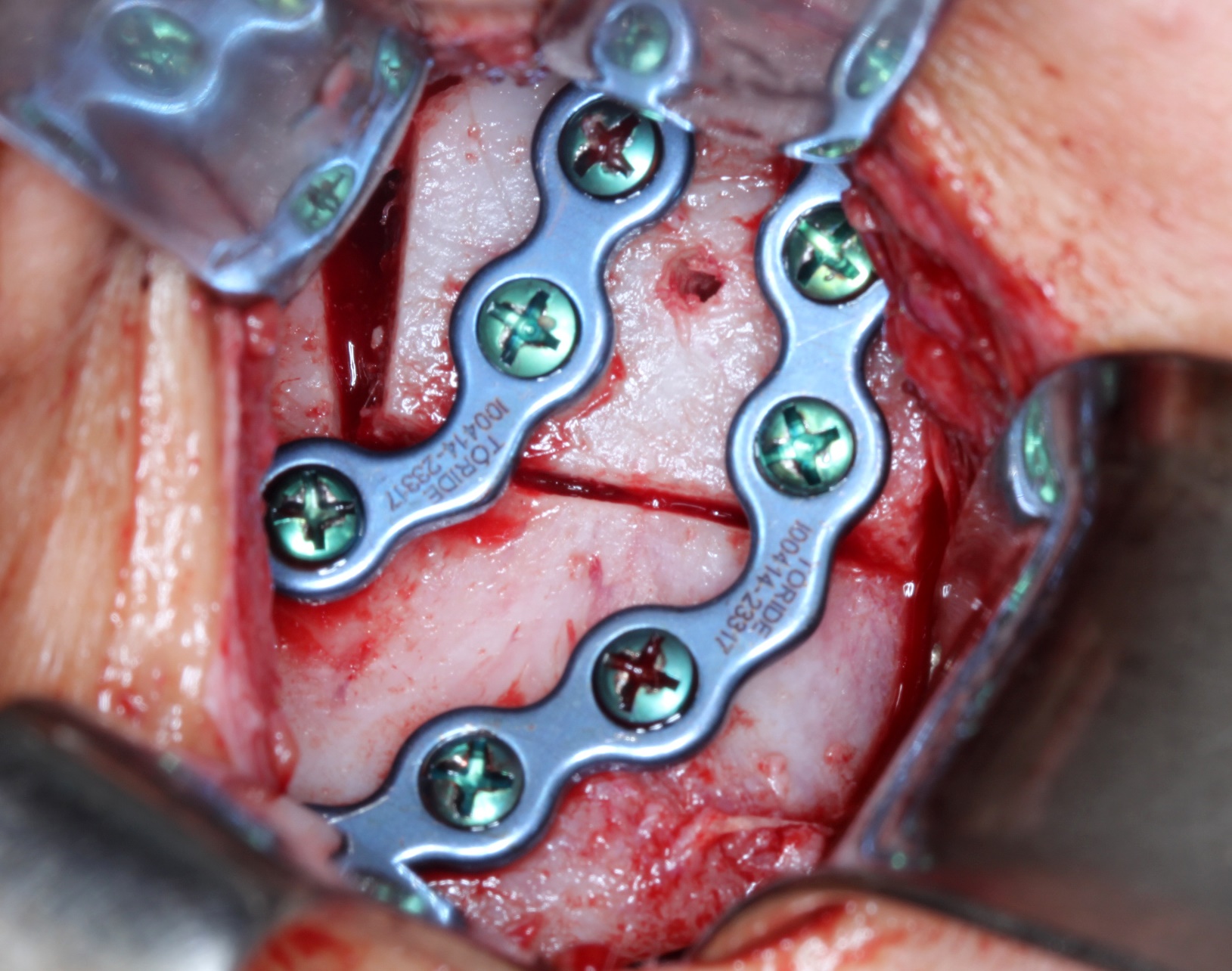
The needle puncture suction revealed an amber-colored liquid characteristic of cystic fluid, thus the initially proposed surgical plan was maintained. The lesion was properly exposed through preauricular and submandibular approaches and was removed by performing a low condylectomy which consisted of an “L” shape osteotomy, utilizing a piezoelectric tip. The biopsy specimen revealed translucid, paper-like, cortical bone that demarcated a cavity which contained amber-colored liquid and a blood clot, no soft tissue was found inside (Figure 2). The surgical dissection at the TMJ was done preserving the disc and capsule. With the intention of restoring function and facial contours, an autogenous free bone graft was harvested from the anterior iliac crest containing both cortices, measuring 45 mm in length (Figure 3). After intermaxillary fixation, the bone graft was sculpted by applying a piezoelectric tip with the aim of passively adapting it to the articular fossa of the TMJ (Figure 4) and to the bone defect created by the “L” shaped osteotomy (Figure 5). The bone graft was fixed by the 2.0 mm reconstruction locking system consisting of 2 bicortically fixed plates (Figure 5). A suction drain was inserted to reduce blood and fluid accumulation, thereby preventing subcutaneous hematoma and infection (Figure 6A). The immediate postsurgical CT confirmed the complete removal of the lesion and affected bone, as well as adequate adaptation of the graft to the native bone (Figure 1E-1F and 7A-7B). The postoperative medical prescription consisted of antibiotic agents (cephalothin – 1 g every 6 hours for 7 days), anti-inflammatory (ketoprofen 100 mg every 8 hours for 3 days), and analgesics (dipyrone – 500 mg every 6 hours, for pain relief). The suction drain was removed on postoperative day 2 and the patient was discharged the same day. The patient’s post-operative diet was exclusively liquid for 4 weeks. After this period, a pureed diet was instructed for 4 more weeks, followed by a gradual increase in consistency, avoiding tougher foods (i.e. nuts and chocolate bars) for 8 additional weeks. Physiotherapy sessions were suggested, aiming not only to optimize and restore the jaw’s full range of motion, but also facial motricity. This therapy consisted of spontaneous mandibular opening and lateral movement exercises, without resistance, and facial muscle contraction exercises. These sessions started 2 weeks after the surgical procedure, and took place twice a week, under the supervision of a physical therapist, concluding at the fourth post-operative month.

Inferior view of the removed condyle in which it is possible to observe the expansion of the paper-like, cortical bone demarcating a cavity devoid of internal lining and containing a blood clot.

The fragment of free bone graft harvested from the anterior iliac crest involving both cortical bones and measuring 45 mm in length.

The cranial portion of the condyle-shaped bone graft adapted to the TMJ, in contact with the articular disc and mandibular fossa.
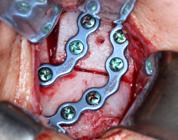
Attachment of the free bone graft to the defect created by the “L” shape osteotomy applying 2 curved reconstruction locking plate screws from the 2.0-mm system.

Closure of the surgical approaches. (A) preauricular, in which it is possible to note the flexing tube of the suction drain behind the Helix, and (B) submandibular.

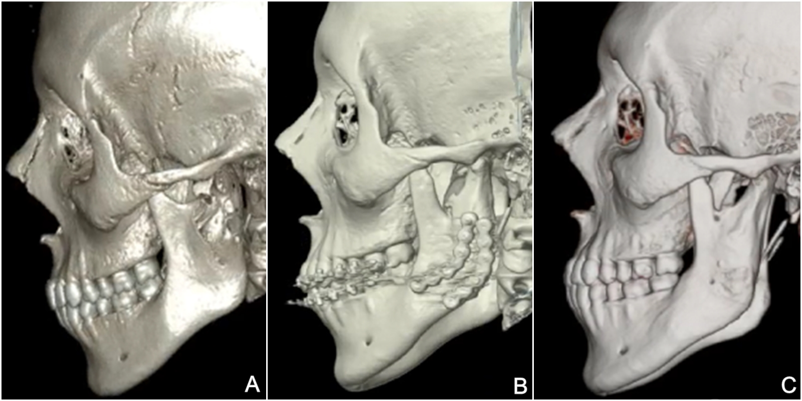
A, Three-dimensional reconstructions of the CT scans. Preoperative CT image, showing significant change in condylar anatomy and also discontinuity of the mandibular notch; B, 1-year postoperative, obtained before the removal of plates and screws; C, 2-year follow-up, showing remarkable bone remodeling, resulting in a shape similar to normal condylar anatomy.
Microscopic analysis of the biopsy specimen revealed an empty bone cavity (Figure 2 and Figure 8A) lined with an osteogenic mesenchymal proliferative process (Figure 8B) with cartilaginous clusters (Figure 8C) secondary to fracture displacement and no signs of internal epithelial or neoplastic proliferation, confirming the clinical hypothesis of a solitary bone cyst.
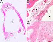
A, Low-power magnification of surgical specimen. Black star pointing to the empty cavity circumscribed by thin bone layer; B, 200x magnification of empty cavity (black star), multilayer immature bone deposition (black arrow), vascularized connective tissue (black arrowhead) and mature cortical bone (blue arrowhead); C, 400x magnification trained on the irregular cartilaginous proliferation on the condyle wall.
At the first postoperative follow-up, the preoperative dental intercuspation was maintained and mandibular movements were present, albeit with limited range. Unfortunately, a moderate impairment in facial motor function related to the Facial Nerve was noticed, specifically the Marginal Mandibular, Buccal and Temporal branches. No further complications were noted.
After 1 year of follow-up, occlusion and mandibular movements had improved significantly, as well as facial motor function related to the Facial Nerve. At this time, the patient asked for the plates and screws to be removed. The removal of plates and screws was performed under general anesthesia through submandibular and intraoral approaches, employing a trocar for the more cranially located screws. During the procedure, it was not possible to differentiate between the bone graft and the native bone, making the remarkable integration of the bone graft on to the native bone evident.
At the 2 year follow-up, the patient did not present any symptoms or complaints, nor any recurrence of the pathology. The range of mandibular movement returned to normal, including the maximum mouth-opening amplitude, which was of 44 mm (Figure 1B). The CT scan requested at this time showed remarkable integration of the graft on the native bone and bone remodeling resulting in a shape very similar to the contralateral condyle (Figure 1I-1J and Figure 7C). Nonetheless, a discrete motor deficit related to the temporal branch of the facial nerve was still evident.
DISCUSSION
Regarding cysts of the jaw, few case reports published in English are present on PubMed which cite PF facilitated by bone cysts, namely 2 odontogenic keratocysts [7,14], 2 dentigerous cysts [15,16], 1 unspecified, odontogenic cyst [3], 3 residual cysts [14,17], 3 aneurysmal bone cysts [1,18,19], 6 radicular cysts [15,20-22] and 10 instances of SBC [4-13]. Table I describes important information about those cases of SBC, such as the location, the patient’s age and gender and also the treatment applied.
SBC, also known as traumatic bone cyst, hemorrhagic bone cyst, simple bone cyst, unicameral cyst or idiopathic bone cavity, is by definition a pseudocyst, which means a pathological cavity, either empty or filled with serosanguinous fluid, without epithelial lining, and with clinical and radiographic, but no histopathological similarities to true cysts [23-25]. SBC mainly affects young adults with a mean age of 19 years, although the range is wide, from 2 to 79 years, with no gender predisposition. The mandible is affected in more than 90% of cases and few cases involving the mandibular condyle have been published [8,26,27]. The origin of SBC remains unclear, however, some authors endorse the possibility of trauma leading to SBC development [2,8,23,25]. According to these authors, trauma, including that resulting from tooth extraction, can progress to intramedullary hemorrhage. Afterwards, the blood clot liquefies and, eventually, through enzymatic activity, results in bone resorption. Other possibilities include bone growth disorders, locally abnormal calcium metabolism, ischemic medullary necrosis, aberrant synovial development and preexisting lesions [23-25].
Due to its asymptomatic profile, SBC is generally diagnosed during the planning of orthodontic treatment or from routine radiographs for other dental problems or purposes when presented with circumscribed, well-defined, radiolucent lesions, ranging from 1 to 10 cm in typical presentations [28]. The mandibular body is the most commonly affected region of the jaws, however, damage to vitality or root resorption of the involved teeth is infrequent. Most cases of SBC present as intraosseous cavities with no cortical expansion or fenestration [1,17,21]. Despite this, the volume measurement in the CT revealed a cavity of 5.9 cm3 and the analysis of the biopsy specimen showed bone expansion and some fenestration of the cortical bone.
Radiolucent images in jawbones are present in many pathologies, such as odontogenic keratocysts, ameloblastomas, Stafne bone defects, among many others [25], making the diagnosis challenging without surgical and microscopic analysis. In general, empty or fluid-filled cavities support the diagnosis of solitary bone cyst with no need for surgical resection [23,29]. For this reason, most anatomopathological reports related to SBC in which the specimens were collected from cavity curettage describe the presence of bone fragments, sometimes exhibiting myxomatous changes and often immature, lace-like osteoid or spiky collagen deposits [25]. Besides the location of the lesion and the association with bone fracture, the result of the lesion microscopy is also an important finding to be considered. The cavity was lined with mesenchymal, osteogenic and cartilaginous proliferative areas, that may result in an incorrect diagnosis of malignant osteosarcoma, since telangiectatic osteosarcomas present cavities and osteogenic/cartilaginous proliferative tissue [25]. Although this diagnostic pitfall is uncommon, close communication between the surgical team, radiologists and pathology services is crucial for the integration of clinical aspects and the imaging and microscopy findings.
Since SBC is a very benign lesion, most cases are treated with conservative techniques such as surgical access or curettage [23]. Occasionally, the SBC requires a more invasive procedure such as fenestration, cryogenic therapy, corticoids, autologous bone marrow graft, resection, with or without internal/external fixation, or a bone graft [30]. In the case described here, the quantity and quality of the remaining bone was not sufficient to preserve the condyle, contraindicating a less invasive procedure. Thus, resection and reconstruction using an anterior iliac crest free bone graft was opted for. The graft’s lack of growth potential was desired in this case, prioritizing predictability. Donor site morbidity and increased surgical time were also considered. Costochondral grafts are also commonly used for condylar reconstructions with great success [31-34], specially in younger patients due to the graft’s growth potential [35]. This characteristic, however, becomes a detriment when overgrowth occurs [35-37]. Regardless of the origin of the free bone graft, factors such as fracture, infection, ankylosis, donor site morbidity and deformity [32,34,36-40], must be considered. Sliding ramus osteotomy is another option for condylar reconstruction in smaller defects, establishing satisfactory function. Since there is no second surgical site, morbidity can also be reduced, costs are lower compared to a TMJ prosthesis, and the risk of bone necrosis and resorption is lower than for free bone grafts [38]. While vertical ramus osteotomies provide bone for the new condyle of adequate size and shape, with similar histological characteristics to the original [37], it has no growth potential, possibly a disadvantage for younger patients [32]. This treatment option alters mandibular angle contour, resulting in facial asymmetry. Although not severe enough to be of concern in most cases [37,38], the patient must be informed and secondary surgery might be required to correct the asymmetry.
Lastly, stock and custom temporomandibular joint (TMJ) prosthesis are effective alternatives, with predictable outcomes, both surgery time and morbidity are potentially reduced, since no donor site is required [41-43]. However, material cost, failure due to wear, long-term stability, manufacturing time (for custom prosthesis), and the potential growth of young patients should be considered [41-44]. Thus, revision surgery or even prosthesis replacement may be required due to the patient’s growth [45].
Regardless of the surgical treatment plan adhered to, the patient must undergo frequent examination, both clinically and by means of tomography, to ensure function is restored. Physiotherapy is highly recommended, not only to restore mandibular movement and function as quickly as possible, but also facial motor function if any branch of the Facial Nerve has been damaged, as observed in this case, probably occurring during the preauricular approach.
In summary, SBC is a benign lesion that weakens bone structure and is usually discovered incidentally during routine examination or examination for other reasons as was the case of the facial trauma presented here. Careful planning is required, and should be done according to the case’s demands, aiming for better clinical progression, and taking into consideration lesion biology, remaining bone and functional/aesthetic parameters. Finally, although very uncommon, an iliac bone graft can be used as a viable option for condyle replacement in SBC-related PF.
Acknowledgements
The authors would like to thank Dr. Manoel Rubens Porto Filho and Raimundo Nonato da Silva Pessoa, physician and nurse, respectively, for the surgical procedure and postoperative assistance related to the harvesting of the bone graft, as well all the staff at the “Hospital Irmandade da Santa Casa de Sertãozinho” involved in this case.
Data availability
The data that support the findings of this study are available from the corresponding author upon reasonable request.
-
How to cite:
Sousa LM, Sverzut TFV, Bahia MS, Santos JL, León JE, Trivellato AE et al. Condylar fracture due to sports trauma associated with a solitary bone cyst: a case report. Braz. Dent. Sci. 2026;29:e4990. https://doi.org/10.4322/bds.2026.e4990
-
Funding
This research study did not receive any specific grants from funding agencies in the public, commercial or not-for-profit domains.
-
Regulatory Statement
The present study was approved by the Research Ethics Committee of the School of Dentistry of Ribeirão Preto, University of São Paulo, Brazil, under protocol number 5.352.319.5.
REFERENCES
-
1 Choi BJ, Choi SC, Kwon YD, Drew SJ. Aneurysmal bone cyst causing a pathologic fracture of the mandibular condyle. J Oral Maxillofac Surg. 2011;69(12):2995-3000. https://doi.org/10.1016/j.joms.2010.10.058 PMid:21444139.
» https://doi.org/10.1016/j.joms.2010.10.058 -
2 Boffano P, Roccia F, Gallesio C, Berrone S. Pathological mandibular fractures: a review of the literature of the last two decades. Dent Traumatol. 2013;29(3):185-96. https://doi.org/10.1111/edt.12028 PMid:23294978.
» https://doi.org/10.1111/edt.12028 -
3 Coletti D, Ord RA. Treatment rationale for pathological fractures of the mandible: a series of 44 fractures. Int J Oral Maxillofac Surg. 2008;37(3):215-22. https://doi.org/10.1016/j.ijom.2007.09.176 PMid:18023145.
» https://doi.org/10.1016/j.ijom.2007.09.176 -
4 Baird WO, Askew PA. Traumatic mandibular bone cyst involved in line of fracture. Oral Surg Oral Med Oral Pathol. 1958;11(12):1351-6. https://doi.org/10.1016/0030-4220(58)90332-3 PMid:13613715.
» https://doi.org/10.1016/0030-4220(58)90332-3 - 5 Hughes CL. Hemorrhagic bone cyst and pathologic fracture of mandible: report of case. J Oral Surg. 1969;27(5):345-6. PMid:5253515.
-
6 Cowan CG. Traumatic bone cysts of the jaws and their presentation. Int J Oral Surg. 1980;9(4):287-91. https://doi.org/10.1016/S0300-9785(80)80036-6 PMid:6780478.
» https://doi.org/10.1016/S0300-9785(80)80036-6 -
7 Matise JL, Beto LM, Fantasia JE, Fielding AF. Pathologic fracture of the mandible associated with simultaneous occurrence of an odontogenic keratocyst and traumatic bone cyst. J Oral Maxillofac Surg. 1987;45(1):69-71. https://doi.org/10.1016/0278-2391(87)90090-5 PMid:3467041.
» https://doi.org/10.1016/0278-2391(87)90090-5 -
8 Magliocca KR, Edwards SP, Helman JI. Traumatic bone cyst of the condylar region: report of 2 cases. J Oral Maxillofac Surg. 2007;65(6):1247-50. https://doi.org/10.1016/j.joms.2005.08.026 PMid:17517316.
» https://doi.org/10.1016/j.joms.2005.08.026 -
9 Ahlers E, Setabutr D, Garritano F, Adil E, McGinn J. Pathologic fracture of the mandible secondary to traumatic bone cyst. Craniomaxillofac Trauma Reconstr. 2013;6(3):201-4. https://doi.org/10.1055/s-0033-1343782 PMid:24436760.
» https://doi.org/10.1055/s-0033-1343782 -
10 Chell M, Idle M, Green J. Case report: an unusual finding of a solitary bone cyst in a patient with a fractured mandible. Dent Update. 2015;42(10):977-8. https://doi.org/10.12968/denu.2015.42.10.977 PMid:26856006.
» https://doi.org/10.12968/denu.2015.42.10.977 -
11 Brunet-Llobet L, Lahor-Soler E, Mashala EI, Miranda-Rius J. Continuous surgical decompression for solitary bone cyst of the jaw in a teenage patient. Case Rep Dent. 2019;2019(1):9137507. https://doi.org/10.1155/2019/9137507 PMid:31110825.
» https://doi.org/10.1155/2019/9137507 -
12 Kojima I, Nishioka T, Sakamoto M, Sai Y, Ezoe Y, Iikubo M, et al. Florid cemento-osseous dysplasia-associated simple bone cyst showing marked irregular border and high apparent diffusion coefficient value. Case Rep Dent. 2020;2020(1):8854428. https://doi.org/10.1155/2020/8854428 PMid:33029434.
» https://doi.org/10.1155/2020/8854428 -
13 Pérez-Iglesias B, Sandoval-Gutiérrez J, García-Freire C, Sánchez-Torres A, Gay-Escoda C. Simple bone cyst: a case report. J Clin Exp Dent. 2021;13(2):e207-10. https://doi.org/10.4317/jced.57769 PMid:33575006.
» https://doi.org/10.4317/jced.57769 -
14 Ezsias A, Sugar AW. Pathological fractures of the mandible: a diagnostic and treatment dilemma. Br J Oral Maxillofac Surg. 1994;32(5):303-6. https://doi.org/10.1016/0266-4356(94)90051-5 PMid:7999738.
» https://doi.org/10.1016/0266-4356(94)90051-5 -
15 Gerhards F, Kuffner HD, Wagner W. Pathological fractures of the mandible. A review of the etiology and treatment. Int J Oral Maxillofac Surg. 1998;27(3):186-90. https://doi.org/10.1016/S0901-5027(98)80007-6 PMid:9662010.
» https://doi.org/10.1016/S0901-5027(98)80007-6 -
16 Kouhsoltani M, Mesgarzadeh AH, Moradzadeh Khiavi M. Mandibular fracture associated with a dentigerous cyst: report of a case and literature review. J Dent Res Dent Clin Dent Prospects. 2015;9(3):193-8. https://doi.org/10.15171/joddd.2015.035 PMid:26697153.
» https://doi.org/10.15171/joddd.2015.035 -
17 Dimitroulis G, Curtin J. Massive residual dental cyst: case report. Aust Dent J. 1998;43(4):234-7. https://doi.org/10.1111/j.1834-7819.1998.tb00165.x PMid:9775468.
» https://doi.org/10.1111/j.1834-7819.1998.tb00165.x -
18 Kalantar Motamedi MH. Aneurysmal bone cysts of the jaws: clinicopathological features, radiographic evaluation and treatment analysis of 17 cases. J Craniomaxillofac Surg. 1998;26(1):56-62. https://doi.org/10.1016/S1010-5182(98)80036-X PMid:9563596.
» https://doi.org/10.1016/S1010-5182(98)80036-X -
19 Goddard R, Patel N. Aneurysmal bone cyst masquerading as unknown mandibular metastatic deposit causing pathological fracture. Dent Update. 2007;34(4):230-2. https://doi.org/10.12968/denu.2007.34.4.230
» https://doi.org/10.12968/denu.2007.34.4.230 -
20 Marsden JL. Fracture of the mandible due to radicular and residual odontogenic cysts. Br J Oral Surg. 1964;2(1):71-5. https://doi.org/10.1016/S0007-117X(64)80013-5 PMid:5212883.
» https://doi.org/10.1016/S0007-117X(64)80013-5 - 21 Amos M, Dalghous A, Alkhabuli J, Mizen K. Massive maxillary radicular cyst presenting as facial fracture and abscess, a case report. Libyan J Med. 2007;2(4):211-3. PMid:21503246.
-
22 Xiao X, Dai JW, Li Z, Zhang W. Pathological fracture of the mandible caused by radicular cyst: a case report and literature review. Medicine (Baltimore). 2018;97(50):e13529. https://doi.org/10.1097/MD.0000000000013529 PMid:30558010.
» https://doi.org/10.1097/MD.0000000000013529 -
23 Chrcanovic BR, Gomez RS. Idiopathic bone cavity of the jaws: an updated analysis of the cases reported in the literature. Int J Oral Maxillofac Implants. 2019;48(7):886-94. https://doi.org/10.1016/j.ijom.2019.02.001 PMid:30792085.
» https://doi.org/10.1016/j.ijom.2019.02.001 - 24 Flores IL, Hamilton ME, Zanchin-Baldissera E, Uchoa-Vasconcelos AC, Chaves-Tarquinio SB, Neutzling-Gomes AP. Simple and aneurysmal bone cyst: aspects of jaw pseudocysts based on an experience of Brazilian pathology service during 53 years. Med Oral Patol Oral Cir Bucal. 2017;22(1):e64-9. PMid:27918745.
- 25 El-Naggar AK, Chan JKC, Grandis JR, Takata T, Slootweg PJ. WHO classification of head and neck tumours. 4th ed. France: International Agency for Research on Cancer (IARC); 2017. 348 p.
-
26 Kim HK, Lim JH, Jeon KJ, Huh JK. Bony window approach for a traumatic bone cyst on the mandibular condyle: a case report with long-term follow-up. J Korean Assoc Oral Maxillofac Surg. 2016;42(4):209-14. https://doi.org/10.5125/jkaoms.2016.42.4.209 PMid:27595088.
» https://doi.org/10.5125/jkaoms.2016.42.4.209 -
27 Saia G, Fusetti S, Emanuelli E, Ferronato G, Procopio O. Intraoral endoscopic enucleation of a solitary bone cyst of the mandibular condyle. Int J Oral Maxillofac Implants. 2012;41(3):317-20. https://doi.org/10.1016/j.ijom.2011.09.007 PMid:22024140.
» https://doi.org/10.1016/j.ijom.2011.09.007 -
28 Copete MA, Kawamata A, Langlais RP. Solitary bone cyst of the jaws: radiographic review of 44 cases. Oral Surg Oral Med Oral Pathol Oral Radiol Endod. 1998;85(2):221-5. https://doi.org/10.1016/S1079-2104(98)90430-9 PMid:9503460.
» https://doi.org/10.1016/S1079-2104(98)90430-9 -
29 Resnick CM, Dentino KM, Garza R, Padwa BL. A management strategy for idiopathic bone cavities of the jaws. J Oral Maxillofac Surg. 2016;74(6):1153-8. https://doi.org/10.1016/j.joms.2015.12.014 PMid:26850870.
» https://doi.org/10.1016/j.joms.2015.12.014 -
30 Zhang W, Chen M, Yang C, Han Z, Wei W, Chai Y. Does idiopathic bone cavity involving mandibular condyle need surgical intervention of bone cavity filling? J Craniofac Surg. 2017;28(6):e539-43. https://doi.org/10.1097/SCS.0000000000003851 PMid:28796103.
» https://doi.org/10.1097/SCS.0000000000003851 -
31 Lindqvist C, Pihakari A, Tasanen A, Hampf G. Autogenous costochondral grafts in temporomandibular joint arthroplasty. J Maxillofac Surg. 1986;14:143-9. https://doi.org/10.1016/S0301-0503(86)80280-6 PMid:3459791.
» https://doi.org/10.1016/S0301-0503(86)80280-6 -
32 Robert Markowitz N, Allan PG, Duffy MT. Reconstruction of the mandibular condyle using ramus osteotomies: a preliminary report. J Oral Maxillofac Surg. 1989;47(4):367-77. https://doi.org/10.1016/0278-2391(89)90338-8 PMid:2647938.
» https://doi.org/10.1016/0278-2391(89)90338-8 -
33 Poswillo DE. Biological reconstruction of the mandibular condyle. Br J Oral Maxillofac Surg. 1987;25(2):100-4. https://doi.org/10.1016/0266-4356(87)90003-9 PMid:3555611.
» https://doi.org/10.1016/0266-4356(87)90003-9 -
34 Obeid G, Guttenberg SA, Connole PW. Costochondral grafting in condylar replacement and mandibular reconstruction. J Oral Maxillofac Surg. 1988;46(3):177-82. https://doi.org/10.1016/0278-2391(88)90079-1 PMid:3280758.
» https://doi.org/10.1016/0278-2391(88)90079-1 -
35 Güzel MZ, Arslan H, Saraç M. Mandibular Condyle reconstruction with inlay application of autogenous costochondral graft after condylectomy: cerrahpaşa’s Technique. J Oral Maxillofac Surg. 2007;65(4):615-20. https://doi.org/10.1016/j.joms.2005.12.061 PMid:17368353.
» https://doi.org/10.1016/j.joms.2005.12.061 -
36 Saeed NR, Kent JN. A retrospective study of the costochondral graft in TMJ reconstruction. Int J Oral Maxillofac Surg. 2003;32(6):606-9. https://doi.org/10.1054/ijom.2003.0418 PMid:14636610.
» https://doi.org/10.1054/ijom.2003.0418 -
37 Martinez-Lage JL, Gonzalez J, Pineda A, Alvarez I. Condylar reconstruction by oblique sliding vertical–ramus osteotomy. J Craniomaxillofac Surg. 2004;32(3):155-60. https://doi.org/10.1016/j.jcms.2003.12.006 PMid:15113573.
» https://doi.org/10.1016/j.jcms.2003.12.006 -
38 Song D, Zhu S, Hu J, Li J, Luo E. Use of Ramus osteotomy for the treatment of osteochondroma in the mandibular condyle. J Oral Maxillofac Surg. 2009;67(3):676-80. https://doi.org/10.1016/j.joms.2008.07.018 PMid:19231801.
» https://doi.org/10.1016/j.joms.2008.07.018 -
39 Laurie SWS, Kaban LB, Mulliken JB, Murray JE. Donor-Site Morbidity after Harvesting Rib and Iliac Bone. Plast Reconstr Surg. 1984;73(6):933-8. https://doi.org/10.1097/00006534-198406000-00014 PMid:6374708.
» https://doi.org/10.1097/00006534-198406000-00014 -
40 Saeed N, Hensher R, McLeod NM, Kent J. Reconstruction of the temporomandibular joint autogenous compared with alloplastic. Br J Oral Maxillofac Surg. 2002;40(4):296-9. https://doi.org/10.1016/S0266-4356(02)00139-0 PMid:12175828.
» https://doi.org/10.1016/S0266-4356(02)00139-0 - 41 Morey-Mas MA, Caubet-Biayna J, Iriarte-Ortabe JI. Osteochondroma of the temporomandibular joint treated by means of condylectomy and immediate reconstruction with a total stock prosthesis. J Oral Maxillofac Res. 2011;1(4):e4. PMid:24421981.
-
42 Mercuri LG. The use of alloplastic prostheses for temporomandibular joint reconstruction. J Oral Maxillofac Surg. 2000;58(1):70-5. https://doi.org/10.1016/S0278-2391(00)80020-8 PMid:10632168.
» https://doi.org/10.1016/S0278-2391(00)80020-8 -
43 Mercuri LG, Edibam NR, Giobbie-Hurder A. Fourteen-year follow-up of a patient-fitted total temporomandibular joint reconstruction system. J Oral Maxillofac Surg. 2007;65(6):1140-8. https://doi.org/10.1016/j.joms.2006.10.006 PMid:17517298.
» https://doi.org/10.1016/j.joms.2006.10.006 -
44 Wolford LM, Pitta MC, Reiche-Fischel O, Franco PF. TMJ concepts/techmedica custom-made TMJ total joint prosthesis: 5-year follow-up study. Int J Oral Maxillofac Surg. 2003;32(3):268-74. https://doi.org/10.1054/ijom.2002.0350 PMid:12767873.
» https://doi.org/10.1054/ijom.2002.0350 -
45 Mercuri LG, Swift JQ. Considerations for the use of alloplastic temporomandibular joint replacement in the growing patient. J Oral Maxillofac Surg. 2009;67(9):1979-90. https://doi.org/10.1016/j.joms.2009.05.430 PMid:19686937.
» https://doi.org/10.1016/j.joms.2009.05.430
Edited by
-
Editor-in-chief:
Sergio Eduardo de Paiva Gonçalves
-
Editor:
Daniel Cohen Goldemberg