Characterization of glass fiber-reinforced epoxy resin materials as a substitute for human dentin for bond strength tests
ABSTRACT
Objective This in vitro study aimed to compare the bond strength (μSBS) of resin cement to human dentin and three glass fiber-reinforced epoxy resin (GFRER) formats (plate – PLATE; round-rod – RR; experimental – EXP) used as dentin-analogues in in vitro studies.
Material and Methods For this, human molar occlusal surfaces were ground to expose dentin (DENT group), while GFRER was cut into round-rod (Ø=10 mm, 2.7 mm thick) and rectangular plate (15 × 15 × 3 mm) shapes. The EXP was created from epoxy resin mixed with chopped glass fiber. Surface treatments included 10% hydrofluoric acid + silane for GFRER groups and 37% phosphoric acid + adhesive for DENT. Resin cement cylinders were built, light-cured, thermocycled (12,000 cycles), and tested for microshear bond strength (μSBS). Failure patterns, roughness, contact angle, topography, elastic modulus, sorption, and solubility were also analyzed.
Results No significant μSBS difference was found between DENT (8.40 MPa) and RR (10.24 MPa). The PLATE group had the highest bond strength (17.11 MPa), while EXP had the lowest (4.46 MPa). RR and PLATE showed the highest surface roughness, with DENT and EXP showing the lowest. GFRER groups had similar contact angles among themselves and higher values compared to DENT. The elastic modulus of PLATE (30.47 GPa) and RR (29.37 GPa) was higher than that of EXP and DENT (18 GPa). GFRER showed dimensional stability in water.
Conclusion The glass fiber-reinforced epoxy resin material in RR format can be used as an alternative substrate for bond strength in vitro studies.
KEYWORDS:
Dentin; Dental bonding; Epoxy resins; In vitro techniques; Resin cement
RESUMO
Objetivo Este estudo in vitro teve como objetivo comparar a resistência de união (μSBS) do cimento resinoso à dentina humana e a três formatos de resina epóxi reforçada com fibra de vidro (GFRER) (placa – PLATE; bastão cilíndrico – RR; experimental – EXP) utilizados como análogos da dentina em estudos laboratoriais.
Material e Métodos Para isso, as superfícies oclusais de molares humanos foram desgastadas para expor a dentina (grupo DENT), enquanto o GFRER foi cortado nas formas de bastão cilíndrico (Ø= 10 mm, 2,7 mm de espessura) e placa retangular (15 × 15 × 3 mm). O grupo EXP foi criado a partir de resina epóxi misturada com fibra de vidro picotada. Os tratamentos de superfície incluíram ácido fluorídrico 10% + silano para os grupos GFRER, e ácido fosfórico 37% + adesivo para o grupo DENT. Cilindros de cimento resinoso foram construídos, fotopolimerizados, submetidos a ciclagem térmica (12.000 ciclos) e testados quanto à resistência de união por microcisalhamento (μSBS). Também foram analisados os padrões de falha, rugosidade, ângulo de contato, topografia, módulo de elasticidade, sorção e solubilidade.
Resultados Não houve diferença significativa na μSBS entre DENT (8,40 MPa) e RR (10,24 MPa). O grupo PLATE apresentou a maior resistência de união (17,11 MPa), enquanto o grupo EXP apresentou a menor (4,46 MPa). Os grupos RR e PLATE apresentaram maior rugosidade superficial, enquanto DENT e EXP mostraram os menores valores. Os grupos GFRER apresentaram ângulos de contato semelhantes entre si e maiores em comparação ao DENT. O módulo de elasticidade do PLATE (30,47 GPa) e do RR (29,37 GPa) foi superior ao do EXP e DENT (18 GPa). Os materiais GFRER apresentaram estabilidade dimensional em meio aquoso.
Conclusão O material de resina epóxi reforçada com fibra de vidro no formato RR pode ser utilizado como substrato alternativo em estudos in vitro de resistência de união.
PALAVRAS-CHAVE:
Dentina; Adesão dentária; Resinas epóxi; Técnicas in vitro; Cimento resinoso
INTRODUCTION
Human teeth suitable for use as samples in in vitro tests are becoming increasingly difficult, as the indications for extractions are decreasing considerably, in addition to ethical issues, as there is the involvement of human tissue [1-3]. Furthermore, standardization when using human teeth becomes quite complex, since variables such as type of tooth, storage conditions, and chemical and physical modifications [4,5] can impair the specimens’ similarity for laboratory studies [6]. It is of fundamental importance that the in vitro tests present as much standardization as possible in order to limit the influence of intrinsic deviations presented by the dental substrate on the evaluated outcome.
In this sense, the literature has explored the use of a glass fiber-reinforced epoxy resin material as a substitute for dentin substrate for in vitro studies [7-10], since an industrially manufactured material can reduce the sample standardization bias considering the specimen variability [3]. Kelly et al. [7] validated an industrial material which incorporates glass fibers in an epoxy resin (NEMA G10, National Electric Manufactures Associations). According to this study, the material was similar to dentin in terms of elastic modulus (18.6 GPa) and adhesion (when the material surface was treated with 8% hydrofluoric acid followed by silane application), with a similar failure pattern of the restorative set (ceramic bonded in dentin) to that observed in clinical cases [7]. In addition, Abu-Izze et al. [10] observed through finite element analysis that the behavior of the glass fiber-reinforced epoxy resin (GFRER) material was similar to the dentin substrate regarding the stress distribution when bonded in different ceramic materials.
Despite these results, there is heterogeneity in the in vitro studies about the use of GFRER considering the format of its commercially available presentation as round-rod [8,10-13] or plate [14-20] as a substitute material for dentin. It is known that round-rod appears to have parallel fibers surrounded by an epoxy resin and that the plate format appears to have the glass fibers laid out in a blanket shape over an epoxy resin. Therefore, since the arrangement of dentinal tubules in the dental crown influences the bond strength of dentin with restorative material [21,22], could the arrangement of glass fibers in relation to the epoxy resin of a FRGR material influence its bond strength to a resin cement? If the glass fibers were simply mixed with the epoxy resin, would this condition change the adhesive capacity of the material?
There is no technical information or scientific studies regarding the differences and the possible influence of the glass fiber arrangement in relation to the epoxy resin on the adhesive performance of this material. Thus, its characterization, with an analysis of elastic modulus, sorption, solubility, wettability, adhesive performance and employability of GFRER materials are not clear in the current literature. Therefore, the aim of this study was to compare the different GFRER formats available on the market and an experimental one (epoxy resin mixed with fiberglass) with human dentine in relation to microshear bond strength, as well as analyzing the surface topography, contact angle, elastic modulus, water sorption, and solubility. The assumed null hypothesis was that the different formats of the GFRER material would not be significantly different from human dentine considering the studied aspects.
MATERIAL AND METHODS
This in vitro study consisted of 4 groups: human dentin (DENT); GFRER in plate shape (PLATE); GFRER in round-rod shape (RR); and experimental GFRER consisting of epoxy resin mixed with fiberglass (EXP). The present study was approved by the Research Ethics Committee of the Federal University of Santa Maria (CAAE: 44655321.1.00005346). Human molar teeth used in this study were obtained from the Human Teeth Bank of the Federal University of Santa Maria. The sample size for the bonding strength was determined from the study by Kelly et al. [7] through a sample calculation considering a statistical power of 80%, average standard deviation of 2.2 and a detectable difference of 2.6 MPa. The sample size was set at n= 13, and the experimental unit was considered the slice substrate, where 5 cylinders of resin cement were cemented per slice (n= 65) in order to evaluate the bond strength value. All materials used in this study are presented in Table 1.
Specimen preparation
Human Teeth - DENT
Non-carious human molar teeth were cleaned (periodontal curette McCall 11/12, Golgran, São Caetano do Sul, Brazil) and the occlusal surfaces were ground using a polishing machine (EcoMet/AutoMet 250, Buehler, Lake Bluff, USA) with #400-grit silicon carbide sandpaper for tooth enamel removal until dentin exposure and surface standardization. The human teeth surface was kept moist with gauze soaked in saline solution at room temperature in contact with the specimen during the entire preparation and analysis process prior to the bonding procedure.
Glass Fiber-Reinforced Epoxy Resin – GFRER
For the round-rod (RR group), glass fiber-reinforced epoxy resin material (Epóxi Pultrudado F, Protec, São Paulo, Brazil) in a round-rod format (1000 mm × 10 mm in diameter) was cut into 2.7 mm thick discs in a cutting machine (Isomet 1000, Buehler) with a diamond disc under constant water-cooling. For the PLATE group, glass fiber-reinforced epoxy resin material (Epóxi Pultrudado F, Protec, São Paulo, Brazil) in plate format (3 mm × 950 mm × 1000 mm) was cut into a rectangular shape (15 × 15 × 3 mm) using a handpiece (KaVo Dental, Joinville, Brazil) with a diamond disc (7047 - American Burrs, Palhoça, Brazil). To fabricate the experimental material (EXP group), an epoxy resin and catalyst (Technovit® EPOX, Kulzer GbmH, Hanau, Germany) were manipulated for 5 minutes in a 2:1 ratio, respectively, according to the manufacturer’s instructions. Chopped glass fiber (glass fiber yarn, Fibertex, Louveira Produtos Têxteis Ltda, Louveira, Brazil) (0.1 mm length) weighed on an analytical scale (FA2004, Coleman, Santo André, Brazil) in the proportion of 10% in relation to the weight of the epoxy resin mixture was added to the epoxy resin, then mixed for 10 minutes and poured into a custom-made silicon matrice (internal dimensions: 90 × 30 × 3 mm). After complete polymerization of the epoxy resin, the material was sectioned into cubes (15 × 15 × 3 mm) with a handpiece (KaVo Dental) and a diamond disc (7047 - American Burrs).
Embedding process
All specimens from the 4 groups were embedded in a cylindrical polyvinyl chloride (PVC) mold using a self-cured acrylic resin (VIPI Flash, Pirassununga, Brazil). To do so, the specimens (dentin and GFRER) were fixed on the glass plate, wrapped in a wax sheet number 7 (Lysanda, São Paulo, Brazil) with 2 mm height, leaving the top and bottom surfaces free of wax to preserve its bonding surface above the PVC level. Then, the PVC was placed over the specimen, the acrylic resin was poured into the mold and left resting until its final polymerization. All specimens were polished (#400 and #600 SiC paper [1]) in a polishing machine (EcoMet/AutoMet 250, Buehler) to remove any wax or acrylic resin residue from the bonding surface. Next, the specimens were cleaned in an ultrasonic bath with distilled water for 5 minutes (1440 D, 50/60 Hz, Odontobras, Ribeirão Preto, Brazil).
Surface treatment
The dentin surface was conditioned with 37% phosphoric acid (Acid Gel, Villevie, Joinville, Brazil) for 15 seconds, washed with air-water-spray for 30 seconds, and lightly dried with gentle oil-free air-spray. Afterwards, the dual-curing dental adhesive (ExciTE F DSC, Ivoclar AG, Schaan, Liechtenstein) was actively applied for 10 seconds, followed by an air jet, and subsequently light-cured with an LED device (Radii Cal, SDI; Bayswater, Australia) at an intensity of 1200 mW / cm2 for 10 seconds according to the manufacturer’s recommendations.
GFRER materials were etched with 10% hydrofluoric acid (Condac Porcelana 10%, FGM, Joinville, Brazil) [22] for 60 seconds and washed with air-water-spray for 30 seconds. Afterwards, the specimens were cleaned in an ultrasonic bath (1440D, Odontobras) with distilled water for 5 minutes to remove possible precipitate, and air dried for 15 seconds. Then, the silane coupling agent (Monobond N, Ivoclar AG) was actively applied for 15 seconds with a microbrush, kept intact for 45 seconds and after dried with an air jet for 15 seconds, according to the manufacturer’s recommendation for cementation of a glass ceramic.
Contact angle analysis
The contact angle was measured in the specimens (n= 13) after surface etching via the sessile drop technique using a goniometer (Drop Shape analysis, model DSA 30S, Kruss GmbH, Hamburg, Germany) connected to a computer program (DSA3, V1 .0.3-08, Kruss). Using a needle, one drop of distilled water (11 µl) was dropped at the center of the specimens, and the mean of 5 measurements per specimen was collected after 5 seconds.
Surface roughness analysis
The roughness of bonding surfaces (n= 13) was measured after etching. The measurement was carried out using a contact stylus profilometer (SJ-410, Mitutoyo, Japan). The Ra (the arithmetic mean of the absolute values of the peaks and valleys measured from a mean plane) and Rz (the mean distance between the five highest peaks and the five lowest surface valleys) were calculated by the mean of 3 measurements in each axis x and y of the specimen considering the ISO:21920-2:2021 [23] parameters.
Bonding procedure
Five starch matrices (Isabela, São Caetano do Sul, Brazil) with 1.0 mm of height and 1.2 mm of internal diameter were placed on each treated specimen surface (n=65) and fixed with wax number 7 (Lysanda) [24]. A single trained operator performed the bonding procedures at room temperature (25°C). The resin cement (Variolink N, Ivoclar AG) base and catalyst pastes were dispensed (1:1), manipulated according to the manufacturer’s recommendations, and inserted with a resin spatula (Titanium resin filled no. 6, Indusbello Company, Londrina, Brazil) into the starch matrices. The resin cement excess was carefully removed with a micro brush, and the resin cement of each starch tube was then light cured (1200 mW/cm2 of intensity; Radii Cal, SDI) for 40 seconds. The specimens were stored in distilled water at 37°C for 24 hours. After, the starch tubes were carefully removed with a clinical probe (#5, Golgran) and the resin cement cylinders were individually assessed to guarantee that no bubbles, defects, or pre-test failures were present at the interface. Specimens with any irregularity were excluded from tests. The specimens were aged by thermocycling (12,000 cycles) with baths of 30 seconds between 5°C and 55°C, and with a transfer time of 2 seconds (Nova Ética; Sao Paulo, Brazil).
Microshear bond strength test - µSBS
The PVC cylinders were placed in a jig attached to a universal testing machine (EMIC DL1000, São José dos Pinhais, Brazil) so that the resin cement cylinder was in alignment with the center of the load cell, parallel to the adhesive interface. A stainless-steel wire (Ø= 0.2 mm) was looped around the specimen cylinder parallel to and as close as possible to the cement-substrate interface. The shear load was applied (10 kgF load cell) at a rate of 1 mm/min until failure occurred, and the data were recorded in Newton (N). The bond strength (MPa) was calculated using the formula “S=L/A”, where “S” is the strength (MPa), “L” is the load at which the specimen failed (N) and “A” is the area of the adhesive interface (1.13 mm2).
Failure analysis
All tested specimens were inspected in a stereomicroscope (Discovery V20, Carl-Zeiss, Gottingen, Germany) with 10 – 50× magnification to verify the failure type. The failures were classified as adhesive (less than 50% of resin cement remained at the adhesive interface) or cohesive (more than 50% of resin cement remained in the adhesive interface).
Topographic analysis
Two additional specimens of each group were made, etched as mentioned above, cleaned and sputter-coated with gold, and analyzed under Scanning Electron Microscopy (SEM; Vega3, Tescan; Brun, Czech Republic) to assess the topography of materials at 100× and 1000× magnification.
Water sorption and solubility
Additional specimens (n= 6) of each material of glass fiber-reinforced epoxy resin material with equivalent volume were produced (169.52 mm3), regardless of the geometric shape [25]. The diameter and height of the specimens were measured with a digital caliper with 1-μm accuracy (Mitutoyo; Kanagawa, Japan) at four equidistant points from the center and the volume (V) was calculated in mm3. Specimens were then stored in a desiccator with silica gel in a laboratory kiln (EL – 1.1, Odontobrás) at 37°C for 24 hours and weighed on an analytical scale (FA2004 – Coleman). This cycle was repeated until a constant mass (variation less than ± 0.2 mg; M1) was obtained. After determining M1, all specimens were individually immersed in tubes with 10 ml distilled water and stored in a bacterial greenhouse (Orion 502 – FANEM, Guarulhos, Brazil) at 37°C for 25 days. The specimens were weighed until constant mass was reached (variation less than ± 0.2 mg; M2). Before the weighing procedures, each specimen was washed in distilled water and dried with absorbent paper. After M2, the same drying process described for M1 was repeated until a new constant mass was obtained (variation less than ± 0.2 mg; M3). The results of M1, M2, and M3, as well as volume were entered into the following equations [26]:
Water Sorption equation:
Water Solubility equation:
Elastic modulus
To characterize the materials, the elastic modulus of resin epoxy fiber-reinforced specimens was measured by the non-destructive impulse excitation technique in additional specimens (n= 5) using a Sonelastic® device (ATCP Physical Engineering, Ribeirão Preto, Brazil) with bar-shaped specimens (40 × 10 × 3 mm). The impulse excitation technique (ASTM E1876) consists in determining the elastic modulus of the material based on the natural frequency of a regular geometry sample (in this study bar-shaped samples). The frequencies were excited by a short mechanical impulse, followed by acquiring the acoustic response using a high-sensitive microphone. Next, the software program determines the frequency spectrum by a numerical calculation from the acoustic signal that was captured. Based on this, the dynamic elastic modulus was determined using the ASTM standard, which considers the specimen geometry, mass, dimensions, and frequencies obtained using the equipment [27,28].
Data analysis
The statistical analyses were performed using the SPSS statistical program (IBM SPSS Software, v.27; IBM, Armonk, NY, USA). Surface roughness and contact angle data were analyzed by One-way ANOVA and Tukey’s post-hoc tests were performed at a significance level of 0.05 since the data presented parametric distribution (Shapiro-Wilk test) and homogeneity of variance (Levene’s test).
Then, each slice of substrate (n= 13, with 5 starch tubes each slice, n= 65 starch tubes each group) was considered as an experimental unit and only the predominantly adhesive failures were included in the μSBS analysis. One-way ANOVA and Tukey’s post-hoc tests were performed at a significance level of 0.05 as the bond strength data presented parametric distribution (Shapiro-Wilk test) and homogeneity of variance (Levene’s test).
The values obtained for the elastic modulus by the specific program for acquiring the elastic modulus of commercial and experimental materials were analyzed by One-way ANOVA, and Tukey’s post-hoc tests were performed at a significance level of 0.05 since the data presented parametric distribution (Shapiro-Wilk test) and homogeneity of variance (Levene’s test). The DENT elastic modulus value was taken from previous literature [7,29]. The results of the water sorption and solubility analysis were descriptively analyzed.
RESULTS
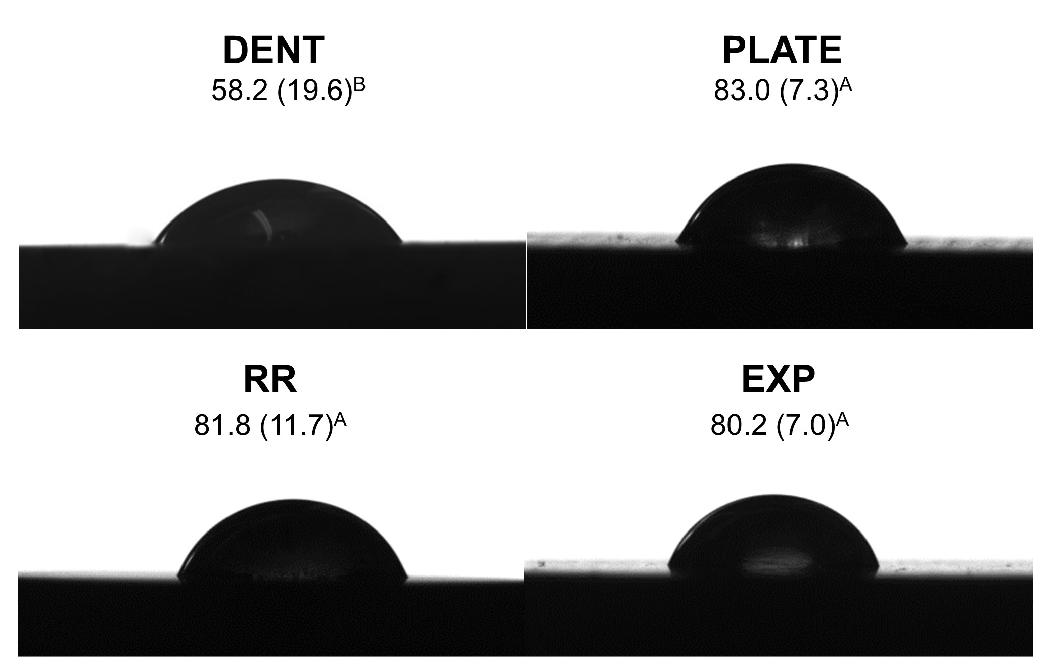
The EXP, RR and PLATE groups presented similar contact angles (p= 1.000) and higher than the DENT group (Figure 1). Considering the roughness analysis, the highest mean surface roughness was presented by the RR and PLATE groups and the lowest by the DENT and EXP groups (RR ≥ PLATE ≥ EXP ≥ DENT) (Table 2).
Representative figures, mean and standard deviation of the surfaces contact angle in each group post-etching with phosphoric acid (DENT) and 10% hydrofluoric acid (PLATE, RR and EXP).
- Mean and standard deviation of surface roughness (Ra and Rz parameters in μm), elastic modulus, sorption, and solubility (n= 6)
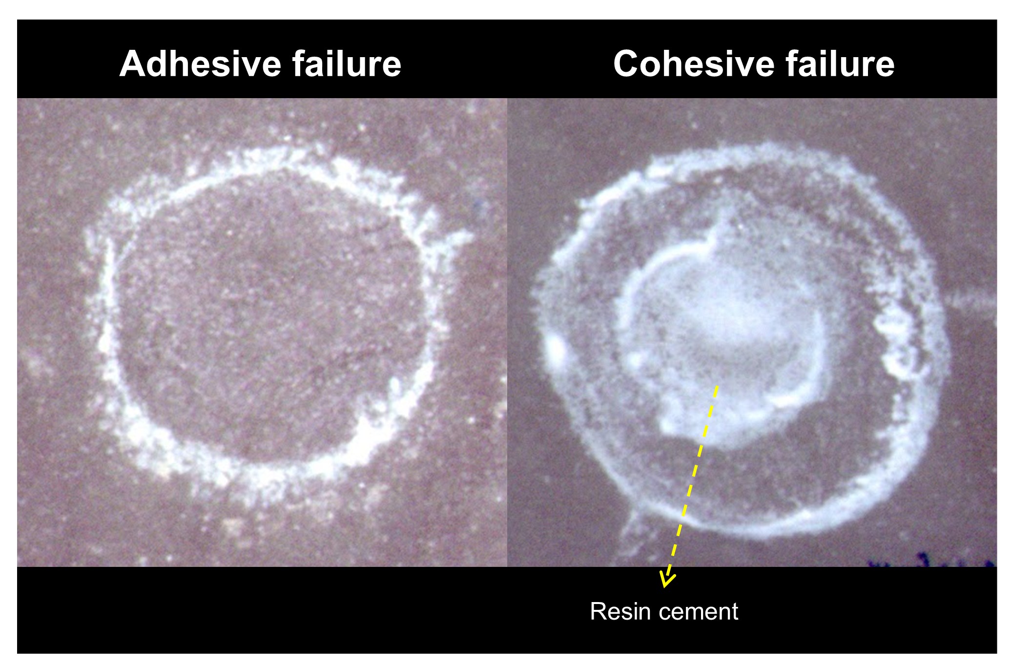
According to the one-way ANOVA and Tukey’s test, the microshear bond strength results were statistically influenced by the tested substrate (p< 0.001). There was no statistical difference of bond strength between human dentin (8.40 MPa) and GFRER material in round-rod format (10.24 MPa) (p= 0.466) (Table 3). The GFRER material in plate format promoted the highest bond strength values (17.11 MPa), while the experimental material presented the lowest values (4.46 MPa) (Table 3). The main type of failure for all the groups was adhesive (Table 3 and Figure 2), and the EXP group showed the highest number of pre-test failures (Table 3).
- Microshear bond strength (mean and standard deviation) in MPa, and types of failure evaluated after the bond strength tests – predominantly adhesive (A), predominantly cohesive (C) and the total of pre-test failures (PTF)
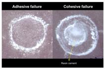
Representative imagens of failure analyses on Stereomicroscope (30× magnification) of the glass fiber-reinforced epoxy resin substrate.
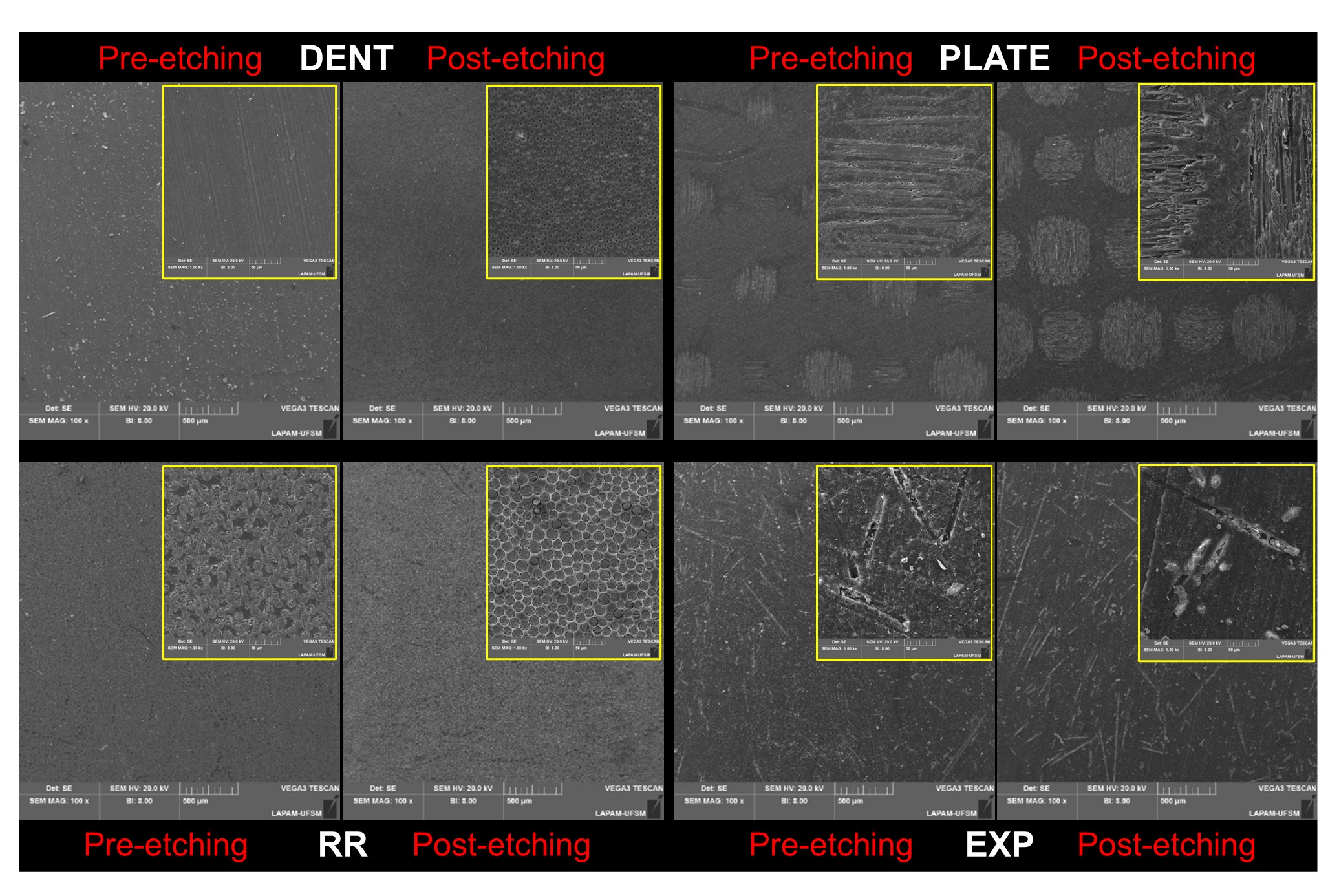
Topographic analysis showed that acid etching modified the surface of the different substrates analyzed (Figure 3). It was possible to observe removal of the surface layer of the epoxy matrix and greater exposure of the glass fibers in the RR and PLATE groups. The conditioning in the EXP group appears smoother for the surface epoxy resin matrix (Figure 3).
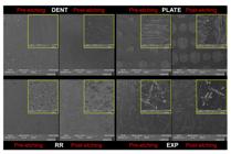
Representative topographic images on Scanning Electron Microscopy (100× and 1000× magnification) of the different materials with and without surface treatment.
The results of the sorption and solubility analyzes (Table 2) showed that the materials presented dimensional stability when immersed in water. The statistical analysis of the modulus of elasticity (Table 2) showed that the PLATE (30.47 GPa) and RR (29.37 GPa) groups were statistically similar to each other (p = 1.000) and greater than the EXP group (18.10 GPa) for the same reference value used for DENT.
DISCUSSION
This in vitro study evaluated the roughness, contact angle, microshear bond strength with resin cement, topography, water sorption and elastic modulus of GFRER with different glass fiber arrangements when compared to human dentin. The null hypothesis was rejected, since differences between dentin and the GFRER occurred in all outcomes.
In this study, both commercial GFRER materials presented similar surface roughness to each other, but higher than human dentin, which was similar to experimental epoxy resin. This may be due to the difference between the materials in terms of amount of epoxy resin and fiber. Dentin is a biological material, which is constituted by dentinal tubules that are surrounded by a highly mineralized peritubular area [30,31]. Furthermore, this area largely consists of collagen with apatite crystals and dentinal fluid [5], and the tubules are arranged in a radial arrangement [21]. The industrial material has a different composition and glass fiber arrangement. The plate material is composed of mantle layers of glass fibers intertwined throughout the material, which can be visualized through the topographic analysis (Figure 3), with the fibers in both horizontal and vertical directions incorporated into the epoxy matrix. On the other hand, the fibers in the round-rod material are presented from the outside as continuous, arranged parallel to the long axis. When the SEM image is analyzed in a cross-section along the long axis of the rod, it is observed that the glass fibers are incorporated parallel to each other in the epoxy resin matrix (Figure 3). However, the glass fibers in the experimental material were cut and incorporated into the epoxy resin matrix, presenting a more heterogeneous positioning of fibers (Figure 3). Furthermore, there is a greater amount of epoxy resin than fiber in this material compared to commercial materials, which makes this material smoother, reducing its roughness, and making it numerically closer to dentin, but not qualitatively, as shown in Figure 3.
When considering the contact angle (Figure 1), dentin presented a lower contact angle than all GFRER materials. This may have occurred due to the difference between the surface energy of the epoxy resin together with the fiber and human dentin. The wettability of human dentin is strongly dependent on roughness, chemical composition, and hydration state, and can be influenced by the density number of the tubules [32]. Acid etching is considered an important surface treatment to improve dentin adhesion, since it causes morphological, chemical, and energetic changes [33]. The etching agent can reduce or eliminate the dentin mineral content and cause changes in substrate surface and subsurface, exposing the dentinal tubules, and therefore the collagen network, which increases roughness [5,34]. Rosales-Leal [33] states that the exposure of the dentinal tubules increases the hydration and the surface roughness, and consequently the wettability, promoting a smaller contact angle in the dentin. Considering the high contact angle values presented by the GFRER materials, Hameed et al. [35] evaluated the incorporation of glass fibers in an epoxy resin matrix and observed a considerable decrease in the hydrophilic capacity of the epoxy material, decreasing its wettability due to increasing the number of non-polar groups exposed on the surface, and considerably increasing the contact angle of the material.
Topographic SEM images (Figure 3) post-etching shows a similar pattern between the DENT and RR group, and the same was observed in the study by Chen et al. [20] (in evaluating the flexural strength of the GFRER material compared to dentin). The SEM analysis (Figure 3) for the PLATE substrate shows a complex arrangement of the fibers in different directions, which seems to provide greater mechanical interlocking between its surface and the restorative material, increasing the bond strength (Table 3). The irregular disposition and smaller number of glass fibers on the surface (Figure 3), and the low interaction between the resin cement and the epoxy resin in the EXP group can explain the significant reduction in the µSBS values and the high number of pre-test failures (Table 3 and Figure 2). Thus, it can be inferred that not only the glass fiber arrangement within the epoxy resin influences the bond strength of GFRER with resin cement, but also the amount of glass fiber and its interaction with the epoxy resin, although these aspects were not measured in the present study.
The sorption and solubility analyses were conducted considering that the substrate is susceptible to volumetric expansion when exposed to water during aging or storage of the samples, which may generate damage to the bonding interface between the restorative material and the substrate, compromising the study result [36]. Furthermore, Ray and Rathore [37] state that when the GFRER material is exposed to water, moisture can be absorbed by the polymeric (epoxy resin) matrix causing it to swell, and this can induce internal stresses or relieve residual stresses caused by contraction during curing of the polymers, possibly resulting in micro voids or cracks, which would weaken the polymer or the epoxy/fiber interface bond. Berketis and Tzetzis [38] report that this material has short-term dimensional stability as a characteristic; however, exposure for long periods (3 to 12 months) can compromise its dimensional stability and its mechanical and structural properties, and this factor is highly dependent on the temperature to which the material is exposed (> 43ºC) [38-40]. The specimens in our study were exposed to moisture for a short period of time (25 days) and to low temperatures (35ºC), which may explain the dimensional stability found.
Ziskind et al. [41] pointed out that the intrinsic morphological and microstructural variations of dentin are responsible for differences in the evaluated elastic modulus, which is an important factor in dissipating the contraction stress of the material, in addition to damping and dissipating the impacts on the masticatory function [41,42]. The commercial GFRER materials in plate and round-rod formats showed statistically equal and higher elastic modulus values than the experimental material and human dentin (reference value), respectively (Table 2) [43]. Atmakuri et al. [44] states that the reinforcing material (glass fiber) works as a stiffening agent, while the matrix material (epoxy resin) not only serves as a bonding agent, but also distributes external forces (load) that act on the fiber composite. Thus, the greater number of fibers presented by commercial materials, as seen in the SEM images (Figure 3), confers greater rigidity to the material, while the experimental material has a smaller number of glass fibers and less complex structural arrangement, and so it presents a more similar elastic modulus to human dentin (Table 2).
Finding a material to be used as a substitute for human dentin substrate which has similar elastic, mechanical and adhesive behavior to that of hydrated dentin is an important step [7,8] to simplify the preparation of specimens [45], in addition to seeking a methodology with greater structural reliability [10]. Despite the differences between the roughness, contact angle and elastic modulus values between dentin and the GFRER round-rod format, the bond strength values and the topography between these substrates were statistically similar (Table 1, Figure 3). Thus, the round-rod format seems to be the most recommended material investigated in this study for human dentin substitute for microshear bond strength tests, for the adopted parameters and evaluated cementation protocol. Despite the similarity of nomenclature and generic composition, the data should be analyzed with caution due to the lack of data available from different manufacturers of glass fiber reinforced epoxy resin materials [7,8,15]. The intrinsic properties presented by each epoxy resin, classification of the reinforcement material and manufacturing methodology imply different behaviors and must be considered in any data extrapolation [37]
Future studies should seek greater knowledge and characterization of the dentin-analogue material. Different GFRER surface treatment strategies should be investigated, such as the influence of this factor on the bond and fatigue behavior. Additionally, it is important to note that, although each material was subjected to its specific recommended surface treatment prior to bonding, this may have generated different conditions among the groups, thereby potentially limiting the comparisons. Despite that, it was concluded that the glass fiber arrangement within the epoxy resin influences the bond strength results of the analog to dentin and the glass fiber reinforced epoxy resin material in the round-rod format can be used as an alternative substrate to human dentin in bond strength tests considering the cementation protocol utilized herein.
Acknowledgements
This work is part of the fulfilment for the requirements of the PhD degree (F.D.N.) in the Post Graduate Program in Oral Sciences at the Faculty of Dentistry of the Federal University of Santa Maria-RS, Brazil. We especially thank Ivoclar AG for donating the research materials, and finally we emphasize that this institution had no role in the study design, data collection or analysis, decision to publish or in preparing the manuscript.
Data availability
The data that support the findings of this study are available from the corresponding author upon reasonable request.
-
How to cite:
Dalla-Nora F, Pilecco RO, Guilardi LF, Borges ALS, Pereira GKR, Valandro LF, et al. Characterization of glass fiber-reinforced epoxy resin materials as a substitute for human dentin for bond strength tests. Braz. Dent. Sci. 2026;29:e4925. https://doi.org/10.4322/bds.2026.e4925
-
Funding
This study was partly financed by the Brazilian Federal Agency for Coordination of Improvement of Higher Education Personnel - CAPES (F.D.N. doctorate scholarship, Finance Code 001) and by the Brazilian National Council for Scientific and Technological Development – CNPq (R.O.P. doctorate scholarship, #140118/2022-5).
-
Regulatory Statement
This study was conducted in accordance with all the provisions of the local human subjects oversight committee guidelines and policies of: Federal University of Santa Maria. This study protocol was reviewed and approved by Research Ethics Committee of Federal University of Santa Maria, approval number [CAAE: 44655321.1.00005346]
REFERENCES
-
1 Lopes MB, Sinhoreti MA, Correr L So, Consani S. Comparative study of the dental substrate used in shear bond strength tests. Pesqui Odontol Bras. 2003;17(2):171-5. https://doi.org/10.1590/S1517-74912003000200014 PMid:14569362.
» https://doi.org/10.1590/S1517-74912003000200014 -
2 Rüttermann S, Braun A, Janda R. Shear bond strength and fracture analysis of human vs. bovine teeth. PLoS One. 2013;8(3):e59181. https://doi.org/10.1371/journal.pone.0059181 PMid:23527125.
» https://doi.org/10.1371/journal.pone.0059181 - 3 Facenda JC, Borba M, Benetti P, Borges ALS, Dutra MDZ, Corazza PH. Fatigue resistance of polymeric restorative materials: effect of supporting substrate. Gen Dent. 2023;71(4):24-9. PMid:37358579.
-
4 Rueggeberg FA. Substrate for adhesion testing to tooth structure - review of the literature. Dent Mater. 1991;7(1):2-10. https://doi.org/10.1016/0109-5641(91)90017-S PMid:2015994.
» https://doi.org/10.1016/0109-5641(91)90017-S - 5 Marshall GW Jr. Dentin: microstructure and characterization. Quintessence Int. 1993;24(9):606-17. PMid:8272499.
-
6 Watanabe LG, Marshall GW Jr, Marshall SJ. Dentin shear strength: effects of tubule orientation and intratooth location. Dent Mater. 1996;12(2):109-15. https://doi.org/10.1016/S0109-5641(96)80077-7 PMid:9002852.
» https://doi.org/10.1016/S0109-5641(96)80077-7 -
7 Kelly JR, Rungruanganunt P, Hunter B, Vailati F. Development of a clinically validated bulk failure test for ceramic crowns. J Prosthet Dent. 2010;104(4):228-38. https://doi.org/10.1016/S0022-3913(10)60129-1 PMid:20875527.
» https://doi.org/10.1016/S0022-3913(10)60129-1 -
8 Dibner AC, Kelly JR. Fatigue strength of bilayered ceramics under cyclic loading as a function of core veneer thickness ratios. J Prosthet Dent. 2016;115(3):335-40. https://doi.org/10.1016/j.prosdent.2015.09.017 PMid:26681598.
» https://doi.org/10.1016/j.prosdent.2015.09.017 -
9 Alessandretti R, Borba M, Benetti P, Corazza PH, Ribeiro R, Della Bona A. Reliability and mode of failure of bonded monolithic and multilayer ceramics. Dent Mater. 2017;33(2):191-7. https://doi.org/10.1016/j.dental.2016.11.014 PMid:27986280.
» https://doi.org/10.1016/j.dental.2016.11.014 -
10 Abu-Izze FO, Ramos GF, Borges ALS, Anami LC, Bottino MA. Fatigue behavior of ultrafine tabletop ceramic restorations. Dent Mater. 2018;34(9):1401-9. https://doi.org/10.1016/j.dental.2018.06.017 PMid:29934124.
» https://doi.org/10.1016/j.dental.2018.06.017 -
11 Scherer MM, Prochnow C, Venturini AB, Pereira GKR, Burgo TAL, Rippe MP, et al. Fatigue failure load of an adhesively-cemented lithium disilicate glass-ceramic: conventional ceramic etching vs etch & prime one-step primer. Dent Mater. 2018;34(8):1134-43. https://doi.org/10.1016/j.dental.2018.04.012 PMid:29789162.
» https://doi.org/10.1016/j.dental.2018.04.012 -
12 Andrade GS, Diniz V, Datte CE, Pereira GKR, Venturini AB, Campos TMB, et al. Newer vs. older CAD/CAM burs: influence of bur experience on the fatigue behavior of adhesively cemented simplified lithium-disilicate glass-ceramic restorations. J Mech Behav Biomed Mater. 2019;95:172-9. https://doi.org/10.1016/j.jmbbm.2019.04.002 PMid:31009901.
» https://doi.org/10.1016/j.jmbbm.2019.04.002 -
13 Guilardi LF, Pereira GKR, Giordani JC, Kleverlaan CJ, Valandro LF, Rippe MP. Effect of zirconia surface treatment, resin cement and aging on the load-bearing capacity under fatigue of thin simplified full-contour Y-TZP restorations. J Mech Behav Biomed Mater. 2019;97:21-9. https://doi.org/10.1016/j.jmbbm.2019.04.050 PMid:31085457.
» https://doi.org/10.1016/j.jmbbm.2019.04.050 -
14 Chen C, Trindade FZ, De Jager N, Kleverlaan CJ, Feilzer AJ. The fracture resistance of a CAD/CAM Resin Nano Ceramic (RNC) and a CAD ceramic at different thicknesses. Dent Mater. 2014;30(9):954-62. https://doi.org/10.1016/j.dental.2014.05.018 PMid:25037897.
» https://doi.org/10.1016/j.dental.2014.05.018 -
15 Kok P, Pereira GKR, Fraga S, de Jager N, Venturini AB, Kleverlaan CJ. The effect of internal roughness and bonding on the fracture resistance and structural reliability of lithium disilicate ceramic. Dent Mater. 2017;33(12):1416-25. https://doi.org/10.1016/j.dental.2017.09.018 PMid:29032826.
» https://doi.org/10.1016/j.dental.2017.09.018 -
16 Monteiro JB, Riquieri H, Prochnow C, Guilardi LF, Pereira GKR, Borges ALS, et al. Fatigue failure load of two resin-bonded zirconia-reinforced lithium silicate glass-ceramics: effect of ceramic thickness. Dent Mater. 2018;34(6):891-900. https://doi.org/10.1016/j.dental.2018.03.004 PMid:29588077.
» https://doi.org/10.1016/j.dental.2018.03.004 -
17 Rodrigues CS, Guilardi LF, Follak AC, Prochnow C, May LG, Valandro LF. Internal adjustments decrease the fatigue failure load of bonded simplified lithium disilicate restorations. Dent Mater. 2018;34(9):e225-e235. https://doi.org/10.1016/j.dental.2018.05.015 PMid:29853196.
» https://doi.org/10.1016/j.dental.2018.05.015 -
18 Machry RV, Cadore-Rodrigues AC, Borges ALS, Pereira GKR, Kleverlaan CJ, Venturini AB, et al. Fatigue resistance of simplified CAD-CAM restorations: foundation material and ceramic thickness effects on the fatigue behavior of partially- and fully-stabilized zirconia. Dent Mater. 2021;37(4):568-77. https://doi.org/10.1016/j.dental.2021.01.001 PMid:33541727.
» https://doi.org/10.1016/j.dental.2021.01.001 -
19 Dalla-Nora F, Guilardi LF, Zucuni CP, Valandro LF, Rippe MP. Fatigue behavior of monolithic zirconia-reinforced lithium silicate ceramic restorations: effects of conditionings of the intaglio surface and the resin cements. Oper Dent. 2021;46(3):316-26. https://doi.org/10.2341/20-025-L PMid:34251409.
» https://doi.org/10.2341/20-025-L -
20 Chen Y, Maghami E, Bai X, Huang C, Pow EH, Tsoi JKH. Which dentine analogue material can replace human dentine for crown fatigue test? Dent Mater. 2023;39(1):86-100. https://doi.org/10.1016/j.dental.2022.11.020 PMid:36503862.
» https://doi.org/10.1016/j.dental.2022.11.020 -
21 Carvalho RM, Tjäderhane L, Manso AP, Carrilho MR, Carvalho CAR. Dentin as a bonding substrate. Endod Topics. 2009;21(1):62-88. https://doi.org/10.1111/j.1601-1546.2012.00274.x
» https://doi.org/10.1111/j.1601-1546.2012.00274.x -
22 Marshall GW Jr, Marshall SJ, Kinney JH, Balooch M. The dentin substrate: structure and properties related to bonding. J Dent. 1997;25(6):441-58. https://doi.org/10.1016/S0300-5712(96)00065-6 PMid:9604576.
» https://doi.org/10.1016/S0300-5712(96)00065-6 - 23 ISO: International Organization for Standardization. ISO 428 (2021) Geometrical product specifications (GPS) — surface texture: profile method — terms, definitions and surface texture parameters. Geneva: ISO; 2021.
-
24 Tedesco TK, Montagner AF, Skupien JA, Soares FZ, Susin AH, Rocha RO. Starch tubing: an alternative method to build up microshear bond test specimens. J Adhes Dent. 2013;15(4):311-5. https://doi.org/10.3290/j.jad.a28602 PMid:23534009.
» https://doi.org/10.3290/j.jad.a28602 - 25 ISO: International Organization for Standardization. ISO 4049 (2000) Dentistry — Polymer-based filling, restorative and luting materials. Geneva: ISO; 2000.
-
26 Albuquerque PPAC, Duarte MFB, Moreno MBP, Schneider LFJ, Moraes RR, Cesar PF, et al. Physicochemical properties and microshear bond strength of experimental self-adhesive resin cements to dentin or yttria-stabilized tetragonal zirconia. J Adhes Dent. 2019;21(2):133-41. https://doi.org/10.3290/j.jad.a42363 PMid:30949626.
» https://doi.org/10.3290/j.jad.a42363 -
27 Borges ALS, Dal Piva AMO, Moecke SE, de Morais RC, Tribst JPM. Polymerization shrinkage, hygroscopic expansion, elastic modulus and degree of conversion of different composites for dental application. J Compos Sci. 2021;5(12):322. https://doi.org/10.3390/jcs5120322
» https://doi.org/10.3390/jcs5120322 -
28 Packaeser MG, Aragonez GC, Soares PM, Borges ALS, Pereira GKR, Valandro LF, et al. High-versus low-viscosity resin cements: its effect on the load-bearing capacity under fatigue of a translucent zirconia. J Mech Behav Biomed Mater. 2023;142:105829. https://doi.org/10.1016/j.jmbbm.2023.105829 PMid:37068429.
» https://doi.org/10.1016/j.jmbbm.2023.105829 -
29 Sabbagh J, Vreven J, Leloup G. Dynamic and static moduli of elasticity of resin-based materials. Dent Mater. 2002;18(1):64-71. https://doi.org/10.1016/S0109-5641(01)00021-5 PMid:11740966.
» https://doi.org/10.1016/S0109-5641(01)00021-5 -
30 Cevik P, Yildirim AZ, Artvin Z, Özcan M. Microtensile bond strength and failure type analysis of self‑etch adhesive systems on superficial and deep dentin after long‑term water storage. Braz Dent Sci. 2020;23(4):1-12. https://doi.org/10.14295/bds.2020.v23i4.2072
» https://doi.org/10.14295/bds.2020.v23i4.2072 -
31 Anido-Anido A, Amore R, Lewgoy HR, Anauate‑Netto C, Silva TM, Paiva Gonçalves SE. Comparative study of bond strength to human and bovine dentine at three different depths. Braz Dent Sci. 2012;15(2):56-62. https://doi.org/10.14295/bds.2012.v15i2.786
» https://doi.org/10.14295/bds.2012.v15i2.786 -
32 Rosales JI, Marshall GW, Marshall SJ, Watanabe LG, Toledano M, Cabrerizo MA, et al. Acid-etching and hydration influence on dentin roughness and wettability. J Dent Res. 1999;78(9):1554-9. https://doi.org/10.1177/00220345990780091001 PMid:10512390.
» https://doi.org/10.1177/00220345990780091001 -
33 Rosales-Leal JI, Osorio R, Holgado-Terriza JA, Cabrerizo-Vílchez MA, Toledano M. Dentin wetting by four adhesive systems. Dent Mater. 2001;17(6):526-32. https://doi.org/10.1016/S0109-5641(01)00014-8 PMid:11567691.
» https://doi.org/10.1016/S0109-5641(01)00014-8 -
34 Nakabayashi N, Nakamura M, Yasuda N. Hybrid layer as a dentin-bonding mechanism. J Esthet Dent. 1991;3(4):133-8. https://doi.org/10.1111/j.1708-8240.1991.tb00985.x PMid:1817582.
» https://doi.org/10.1111/j.1708-8240.1991.tb00985.x -
35 Hameed N, Thomas SP, Abraham R, Thomas S. Morphology and contact angle studies of poly (styrene-co-acrylonitrile) modified epoxy resin blends and their glass fiber reinforced composites. Express Polym Lett. 2007;1(6):345-55. https://doi.org/10.3144/expresspolymlett.2007.49
» https://doi.org/10.3144/expresspolymlett.2007.49 -
36 Armstrong S, Geraldeli S, Maia R, Raposo LH, Soares CJ, Yamagawa J. Adhesion to tooth structure: a critical review of “micro” bond strength test methods. Dent Mater. 2010;26(2):e50-62. https://doi.org/10.1016/j.dental.2009.11.155 PMid:20045179.
» https://doi.org/10.1016/j.dental.2009.11.155 -
37 Ray BC, Rathore D. Environmental damage and degradation of FRP composites: a review report. Polym Compos. 2015;36(3):410-23. https://doi.org/10.1002/pc.22967
» https://doi.org/10.1002/pc.22967 -
38 Berketis K, Tzetzis D. Long-term water immersion ageing characteristics of GFRP composites. J Mater Sci. 2009;44(13):3578-88. https://doi.org/10.1007/s10853-009-3485-9
» https://doi.org/10.1007/s10853-009-3485-9 -
39 Grammatikos SA, Zafari B, Evernden MC, Mottram JT, Mitchels JM. Moisture uptake characteristics of a pultruded fibre reinforced polymer flat sheet subjected to hot/wet aging. Polym Degrad Stabil. 2015;121:407-19. https://doi.org/10.1016/j.polymdegradstab.2015.10.001
» https://doi.org/10.1016/j.polymdegradstab.2015.10.001 -
40 Fan Y, Gomez A, Ferraro S, Pinto B, Muliana A, La Saponara V. The effects of temperatures and volumetric expansion on the diffusion of fluids through solid polymers. J Appl Polym Sci. 2017;134(31):45151. https://doi.org/10.1002/app.45151
» https://doi.org/10.1002/app.45151 -
41 Ziskind D, Hasday M, Cohen SR, Wagner HD. Young’s modulus of peritubular and intertubular human dentin by nano-indentation tests. J Struct Biol. 2011;174(1):23-30. https://doi.org/10.1016/j.jsb.2010.09.010 PMid:20850543.
» https://doi.org/10.1016/j.jsb.2010.09.010 -
42 Fuentes Fuentes M. Propiedades mecánicas de la dentina humana. Av Odontoestomatol. 2004;20(2):79-83. https://doi.org/10.4321/S0213-12852004000200003
» https://doi.org/10.4321/S0213-12852004000200003 -
43 Lima LC, Miranda JS, Carvalho RLA, Barcellos ASPB, Amaral M, Kimpara ET. Influence of substrate, cement and aging on the biaxial flexural strength of lithium disilicate. Braz Dent Sci. 2023;26(4):e3923. https://doi.org/10.4322/bds.2023.e3923
» https://doi.org/10.4322/bds.2023.e3923 -
44 Atmakuri A, Palevicius A, Siddabathula M, Vilkauskas A, Janusas G. Analysis of mechanical and wettability properties of natural fiber-reinforced epoxy hybrid composites. Polymers. 2020;12(12):2827. https://doi.org/10.3390/polym12122827 PMid:33261200.
» https://doi.org/10.3390/polym12122827 -
45 Dal Piva AO, Tribst JP, Borges ALS, Melo RM, Bottino MA. Influence of substrate design for in vitro mechanical testing. J Clin Exp Dent. 2019;11(2):e119-25. https://doi.org/10.4317/jced.55353 PMid:30805115.
» https://doi.org/10.4317/jced.55353
Edited by
-
Editor-in-chief:
Sergio Eduardo de Paiva Gonçalves
-
Section Editor:
Amanda Maria de Oliveira Dal Piva