An in vitro analysis of titanium aging surface optimization through variations in UV-C photofunctionalization and SLA: impact on titanium disk wettability
ABSTRACT
Objective This study aimed to evaluate the effects of UV-C duration and SLA surface treatment on the wettability of aged titanium disk surfaces.
Material and Methods Twenty-four titanium disks (10 mm diameter, 1 mm thickness) underwent a 30-day aging and were divided into eight subgroups (n = 3) based on surface treatment (non SLA or SLA) and UV-C irradiation duration (non UV, 10-minute, 30-minute, and 48-hour) at 90 W and 254 nm. Wettability was evaluated by water contact angle (WCA) measurements. Statistical analysis employed the Scheirer-Ray-Hare test with Dunn’s post hoc test Bonferroni correction.
Results UV-C significantly affected surface wettability (p < 0.001). WCA decreased progressively from 73.15 ± 5.35° (non SLA non UV) to 16.39 ± 0.49° (non SLA 48-hour UV). Surface treatment type (p = 1) and its interaction with UV duration (p = 0.89) were not significant.
Conclusion UV-C photofunctionalization effectively restored the wettability of aged titanium surfaces. Neither surface treatment type nor its interaction with UV-C irradiation duration demonstrated a statistically significant effect on wettability.
KEYWORDS:
Dental implants; Surface properties; Titanium; Ultraviolet light; Wettability
RESUMO
Objetivo O presente estudo teve como objetivo avaliar os efeitos da duração da irradiação UV-C e do tratamento de superfície do implante na molhabilidade de superfícies de discos de titânio envelhecidos.
Material e Métodos Vinte e quatro discos de titânio (10 mm de diâmetro, 1 mm de espessura) foram submetidos a envelhecimento por 30 dias e divididos em oito subgrupos (n = 3) com base no tratamento de superfície (sem e com tratamento de superfície) e na duração da irradiação UV-C (sem UV-C, 10 minutos, 30 minutos e 48 horas) a 90 W e 254 nm. A molhabilidade foi avaliada através de medições do ângulo de contato com a água. A análise estatística utilizou o teste de Scheirer-Ray-Hare, com o teste post hoc de Dunn e correção de Bonferroni.
Resultados A radiação UV-C afetou significativamente a molhabilidade da superfície (p < 0,001). O ângulo de contato diminuiu progressivamente de 73,15 ± 5,35° (sem tratamento de superfície e sem UV-C) para 16,39 ± 0,49° (sem tratamento de superfície e com 48 horas de UV-C). O tipo de tratamento de superfície (p = 1) e a sua interação com a duração da irradiação UV (p = 0,89) não foram significativos.
Conclusão A fotofuncionalização por UV-C restaurou de forma eficaz a molhabilidade de superfícies de titânio envelhecidas. O tipo de tratamento de superfície e a sua interação com a duração da irradiação UV-C não demonstraram efeito estatisticamente significativo na molhabilidade.
PALAVRAS-CHAVE:
Implantes dentais; Propriedades de superfície; Titânio; Luz ultravioleta; Molhabilidade
INTRODUCTION
The success of dental implants depends fundamentally on osseointegration, defined as a direct structural and functional connection between bone and the implant surface [1]. A key determinant is surface wettability, which governs early biological interactions at the implant-tissue interface. Surfaces with lower water contact angles exhibit greater wettability, enhancing protein adsorption and osteoblast adhesion during initial stages of healing [2,3].
Although titanium and its alloys are widely recognized for their biocompatibility due to the spontaneous formation of a stable TiO2 layer, their surface bioactivity deteriorates over time through biological aging [4]. This phenomenon arises from progressive accumulation of hydrocarbon contaminants during manufacturing, storage, and distribution, increasing surface carbon content, decreasing surface free energy, and shifting the surface from hydrophilic to hydrophobic [5-7]. The resulting 4-5 nm hydrocarbon pellicle has been reported to impair osteoblast attachment and reduce osteoconductive potential [4].
UV-C irradiation (200-280 nm, peak 254 nm) has demonstrated superior efficacy in surface decontamination compared to UV-A and UV-B through photofunctionalization, reducing surface carbon content from over 50% to below 20% atomic percentage and markedly decreasing water contact angle on titanium surfaces [5,8-10].
SLA treatment generating micro-rough topography with 10-30 μm valleys and 2-5 μm craters, also enhances early bone-implant interactions [11,12]. However, the SLA process also alters the native TiO2 layer and may modify surface chemistry in a manner that could influence the photocatalytic response to UV-C. It remains unclear whether surface topography modulates the hydrophilic response to UV-C treatment and whether a significant interaction exists between these factors. The present study aimed to evaluate the effects of UV-C irradiation duration and SLA surface treatment, as well as their interaction, on the wettability of aged titanium disk surfaces, as assessed by water contact angle measurements.
MATERIAL AND METHODS
SLA surface preparation
Twenty-four Ti6Al4V titanium disks (10 mm diameter, 1 mm thickness; Shandong Zehao Metal Materials Co. Ltd, Shandong, China) were evenly distributed into eight subgroup (n = 3) based on surface treatment and UV-C duration. SLA preparation involved polishing, ultrasonic cleaning in acetone, alcohol, and distilled water (15 minutes each), sandblasting with 50 μm alumina (Al2O3) particles, and acid etching in 8 ml of sulfuric acid (H2SO4) and 4 ml of hydrochloric acid (HCl) for 30 minutes. Pre-experimental surface roughness measurement (Starrett SR Series, USA) confirmed a mean Ra of 0.2 μm, establishing a uniform baseline prior to UV-C irradiation. All specimens were autoclaved and stored in dark conditions for 30 days.
UV irradiation of titanium disks
Following aging, the titanium disks were exposed to UV-C light (90 W, 254 nm) (Philips UVC TUV 254nm, Philips, Amsterdam, Netherlands) for non-UV (control), 10-minute UV, 30-minute UV, and 48-hour UV in a room devoid of light exposure.
Surface morphology analysis
Dry titanium disks were placed in the chamber of a Scanning Electron Microscope (SEM) (JEOL JSM-IT 200, JEOL Ltd., Tokyo, Japan). The intensity was set at an accelerating voltage of 10.0 kV, a working distance of 10.7 mm, and a magnification of 1000x. Carbon content (C 1s) was analyzed on one disk (n = 1 per group) using an X-Ray Photoelectron Spectroscope (XPS) (XPS Kratos AXIS Supra+, Kratos Analytical Ltd., Manchester, United Kingdom) employing a linear algorithm in percentage (%) and reported descriptively.
Wettability assessment
The sessile drop method was used under controlled conditions (25 °C, 62% humidity). A 2 μL distilled water droplet was deposited on each disk surface using a micropipette and imaged after 10 seconds using water contact angle measurement device (Fumalife, Indonesia) expressed in degrees (°) and analyzed with ImageJ (version 1.53, National Institutes of Health, USA).
Statistical analysis
Normality was conducted using the Shapiro-Wilk test and variance homogeneity using Brown-Forsythe's test. The Scheirer-Ray-Hare non-parametric test evaluated main effects of UV irradiation duration and surface type, and their interaction. Pairwise comparison used Dunn’s test with Bonferroni correction (GraphPad Prism version 10.0, GraphPad Software Inc., San Diego, CA, USA).
RESULTS
Surface characteristics
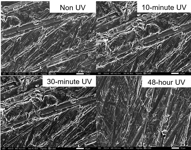
SEM images across all UV-C durations revealed no observable morphological changes or topographical damage, confirming that UV-C irradiation did not alter surface structure (Figure 1). This was consistent with the pre-experimental Ra value of 0.2 μm across all non-SLA specimens.
- SEM images of non-SLA titanium disks surface following non-UV, 10-minute UV, 30-minute UV, and 48-hour UV at x1000 magnification. SEM analysis was conducted to assess potential UV-C–induced morphological changes on the titanium surface.
Carbon composition characteristics
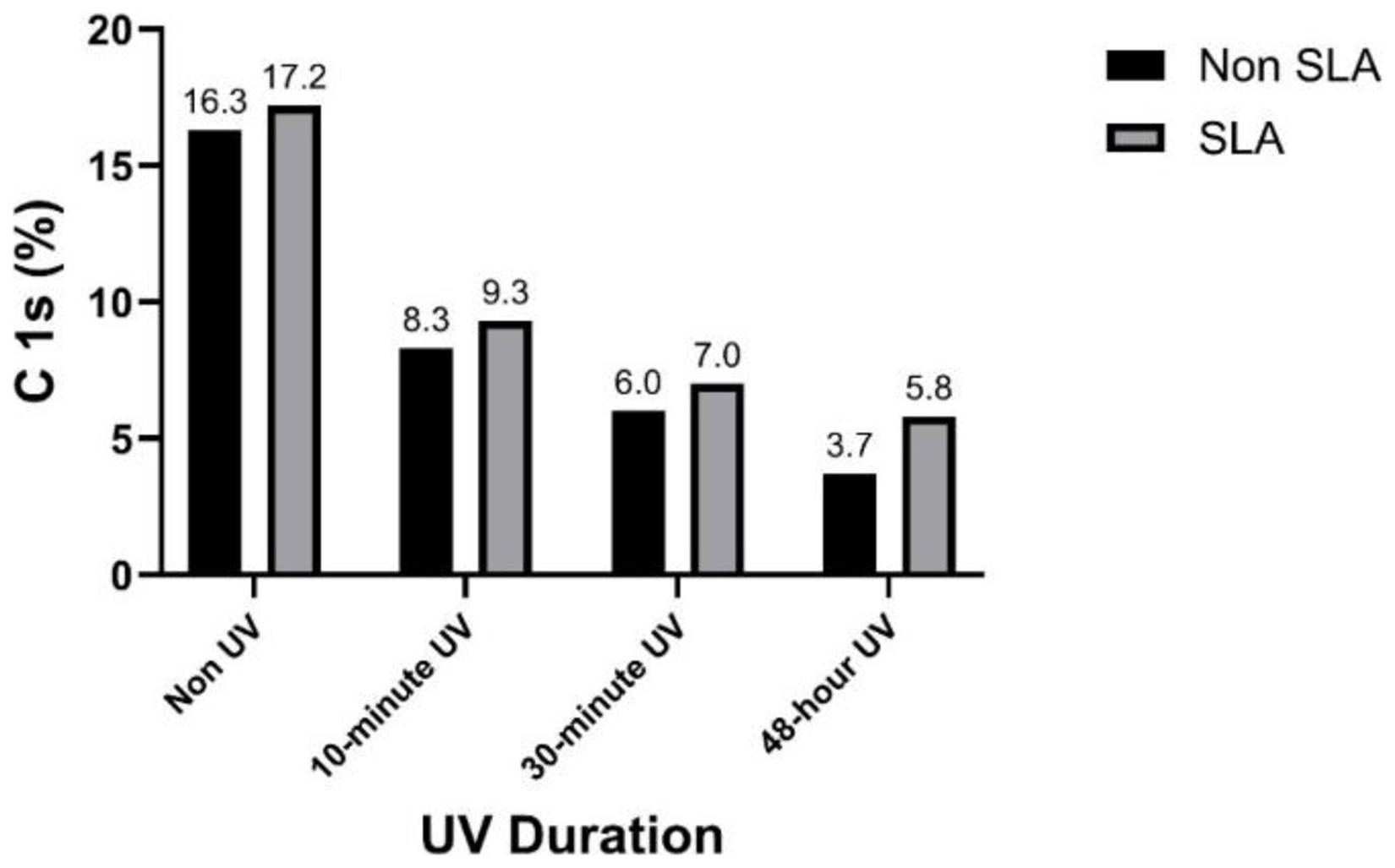
XPS analysis demonstrated a consistent reduction in C 1s content with increasing UV-C irradiation duration on both groups (Figure 2). Baseline values were 16.3% (non-SLA) and 17.2% (SLA). Following 10-minute UV, carbon decreased to 8.3% (non-SLA) and 9.3% (SLA). At 30-minute UV, values fell to 6.0% (non-SLA) and 7.0% (SLA), and 48-hour UV reached the lowest levels of 3.7% (non-SLA) and 5.8% (SLA). These data are interpreted as exploratory findings that provide mechanistic context for the observed changes in surface wettability.
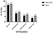
- The percentage (%) of carbon (C 1s) on non-SLA and SLA titanium disk, as determined by XPS, is presented following 30 days of aging and UV-C irradiation (non UV, 10-minute UV, 30-minute UV, and 48-hour UV). Data represent individual measurements (n = 1 per subgroup) and are reported descriptively. A progressive reduction in surface carbon content was observed with increasing UV-C irradiation duration for both surface types.
Wettability
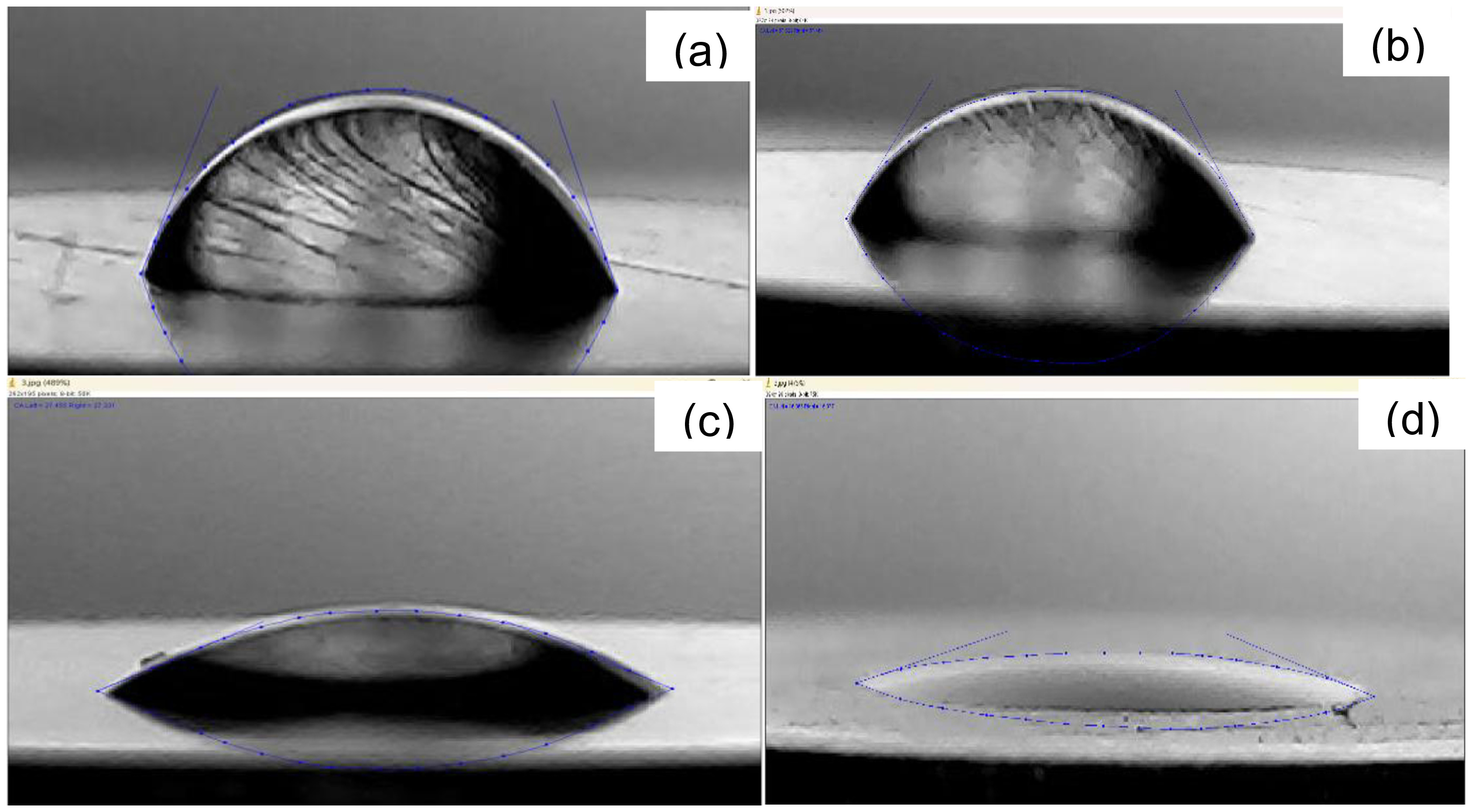
In Figure 3a, the non-UV control surface exhibits a prominently hemispherical water droplet profile, characteristic of a hydrophobic surface with a large contact angle, resulting from hydrocarbon accumulation during the 30-day aging period. Following 10-minute UV (Figure 3b), a modest reduction in droplet height is observed, indicating an early-stage improvement in surface wettability. A more pronounced flattening of the droplet profile is evident after 30-minute UV (Figure 3c). The most substantial change is observed in Figure 3d, corresponding to 48-hour UV. The water droplet exhibits a markedly flattened profile with a very low contact angle. This finding suggests enhanced wettability of the titanium surface, associated with reduced surface carbon contamination following UV-C irradiation.
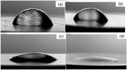
- (a) Representative water contact angle images of non-SLA titanium disk surfaces following 30 days of aging are presented, illustrating the hydrophobic condition in the non-UV; (b) A progressive increase in surface wettability is observed at 10-minute UV; (c) 30-minute UV; and (d) 48-hour UV.
A descriptive analysis of each irradiation time and type of surface modification is provided in Table I. The assumption of normality was not fully met in the Shapiro-Wilk test as the non-SLA groups of 10-minute UV (p = 0.765) and 30-minute UV (p = 0.293) irradiation did not follow a normal distribution. This may be due to the relatively large variability within the data, as indicated by the high standard deviation values, which likely contributed to deviations from a normal distribution. In contrast, all SLA groups can be considered normally distributed (p > 0.05). Brown-Forsythe test also confirmed homogeneity of variance (p = 0.579).
- Results of descriptive analysis of titanium disk wettability by UV irradiation time and surface type groups
Scheirer-Ray-Hare test revealed a significant main effect of UV duration (p < 0.001), while surface treatment (p = 1) and interaction effect (p = 0.89) were not significant (Table II).
- Results of Scheirer-Ray-Hare test of the effect and interaction between UV irradiation time and SLA surface on the wettability of titanium disk
Post hoc Dunn’s test revealed significant differences between: non-UV and 30 minutes (p < 0.01); non-UV and 48 hours (p < 0.001); and 10 minutes and 48 hours (p < 0.05). No statistically significant differences were observed between non UV and 10-minute UV; 10-minute and 30-minute UV; and 30-minute and 48-hour UV (Table III).
- Results of post hoc with the Dunn’s test of titanium disk wettability by UV irradiation time groups
DISCUSSION
As the specimen ages, the water contact angle tends to increase naturally due to the formation and accumulation of a hydrocarbon molecule pellicle on the surface [13]. In the present study, titanium disks subjected to SLA treatment initially exhibited superhydrophilicity (day 1-3) to hydrophobicity (day 7, mean 36.68°) and ultimately hydrorepellent (day 30, mean 103.99°).
UV irradiation induces wettability on titanium surfaces through two physicochemical mechanisms. The first is photocatalytic properties of TiO2. UV-C photons, interacting with the titanium oxide passive layer, provide sufficient energy to break the bonds of hydrocarbon contaminants adsorbed on the titanium surface through a process known as photocatalytic decomposition. This results in a reduction in surface carbon content, as evidenced by the decreased intensity of the C 1s peak observed in the XPS analysis [14]. Second, the formation of a hydrophilic surface is attributed to light-induced modifications in the surface structure of the TiO2 layer. Specifically, UV treatment promotes the formation of oxygen vacancies at bridge sites, leading to the reduction of Ti4+ to Ti3+ states, which are more favorable for dissociative water adsorption and leads to an increased density of surface hydroxyl (–OH) groups. The accumulation of these hydroxyl groups is directly responsible for the transition from hydrophobic to superhydrophilic surface behavior [7,15]. Consequently, this transformation is reflected by a significant reduction in the water contact angle values [16].
Neither surface treatment type (p = 1) nor its interaction with UV-C irradiation (p = 0.89) was significant, indicating wettability is primarily governed by UV-C photochemical activity rather than surface microtopography. This is consistent with pre-irradiation baseline Ra value of 0.2 μm and absence of morphological changes on SEM. SLA surfaces exhibited slightly higher baseline carbon prior to UV irradiation (17.2%) compared to non-SLA surfaces (16.3%), likely due to increased surface area providing more hydrocarbon adsorption sites.
48 hours was identified as the optimal duration, achieving an average decrease of 75% in the water contact angle in both surface groups and representing the peak effect of 90 W and 254 nm wavelength light on both SLA and non-SLA surfaces for wettability. This is in line with the previous study of Ogawa (2014) which stated that a superhydrophilic surface on titanium disks has been reported following 48-hour UV irradiation at an intensity of 2 mW/cm2 (λ = 250 ± 20 nm). Similarly, Lee et al. [17,18] demonstrated that machined titanium disk + UV achieved superhydrophilicity, exhibiting a water contact angle of approximately 0° after 48-hour UV exposure.
Although this finding about 48-hour UV-C irradiation duration is consistent with established protocols and results in the previous studies, it may have limited applicability in routine clinical settings. However, the duration is best regarded as a scientific benchmark defining the maximum photofunctionalization endpoint for the titanium disk wettability. In contrast, shorter UV durations of 10 and 30 minutes may represent more clinically feasible parameters which warrant further investigation as one of limitations of this study.
This study also possesses other limitations. Sample size of n = 3 per subgroup was limited. XPS measurements were conducted on n = 1 per subgroup, precluding statistical analysis of the chemical composition data. Contact angle was performed once per disk, limiting intra-specimen variability assessment. Future studies are recommended to incorporate repeated measurements for each specimen to enhance the data reliability. Next, SEM analysis was restricted to non-SLA surfaces. The subdivision of a single titanium disk into multiple UV measurement area may introduces a theoretical risk of cross-contamination between adjacent area. Besides, no biological assays or in vivo evaluations were conducted. Future studies incorporating a broader range of titanium materials, irradiation parameters (including light sources and intensities), and evaluations of wettability durability are warranted to further optimize UV-C photofunctionalization protocols for clinical applications.
CONCLUSION
UV-C photofunctionalization effectively restored the wettability of aged titanium disk surfaces (p < 0.001), with 48-hour UV irradiation yielding the most significant effect. Neither surface treatment type (p = 1) nor its interaction with UV-C duration (p = 0.89) demonstrated a significant effect on wettability.
Acknowledgements
The authors wished to express gratitude to the Advanced Physics and Imaging Instrumentation Laboratory, National Research and Innovation Agency (BRIN); the Integrated Research Laboratory, Faculty of Dentistry; and the Material Physics and Instrumentation Laboratory, Faculty of Mathematics and Sciences, Gadjah Mada University; and all the staff for their provision of research facilities, assistance with the sample preparation, and support in data collection.
How to cite:
Funding
Regulatory Statement
Data availability
The data supporting the findings of this study are available from the corresponding author upon reasonable request.
REFERENCES
-
1 Huang Y, Zhang H, Chen Z, Wang Y, Yang X, Yu H. Improvement in osseointegration of titanium dental implants after exposure to ultraviolet-c light for varied durations: an experimental study in beagle dogs. J Oral Maxillofac Surg. 2022;80(8):1389-97. https://doi.org/10.1016/j.joms.2022.04.013 PMid:35623451.
» https://doi.org/10.1016/j.joms.2022.04.013 -
2 Vieira GM, Almeida TCS, Oliveira FP, Azzi PC, Rodrigues CF, Souza RL, et al. Comparative study of acid etching and SLA surface modification for titanium implants. Materials. 2025;18(7):1632. https://doi.org/10.3390/ma18071632 PMid:40271856.
» https://doi.org/10.3390/ma18071632 -
3 Eun S-M, Son K, Hwang S-M, Son Y-T, Kim Y-G, Suh J-Y, et al. The impact of mechanical debridement techniques on titanium implant surfaces: a comparison of sandblasted, acid-etched, and femtosecond laser-treated surfaces. J Funct Biomater. 2023;14(10):502. https://doi.org/10.3390/jfb14100502 PMid:37888167.
» https://doi.org/10.3390/jfb14100502 -
4 Matsuura T, Komatsu K, Cheng J, Park G, Ogawa T. Beyond microroughness: novel approaches to navigate osteoblast activity on implant surfaces. Int J Implant Dent. 2024;10(1):35. https://doi.org/10.1186/s40729-024-00554-x PMid:38967690.
» https://doi.org/10.1186/s40729-024-00554-x -
5 Park G, Matsuura T, Komatsu K, Ogawa T. Optimizing implant osseointegration, soft tissue responses, and bacterial inhibition: a comprehensive narrative review on the multifaceted approach of the UV photofunctionalization of titanium. J Prosthodont Res. 2025;69(2):136. https://doi.org/10.2186/jpr.JPR_D_24_00086 PMid:38853001.
» https://doi.org/10.2186/jpr.JPR_D_24_00086 -
6 Jacobs TW, Dillon JT, Cohen DJ, Boyan BD, Schwartz Z. Different methods to modify the hydrophilicity of titanium implants with biomimetic surface topography to induce variable responses in bone marrow stromal cells. Biomimetics. 2024;9(4):227. https://doi.org/10.3390/biomimetics9040227 PMid:38667238.
» https://doi.org/10.3390/biomimetics9040227 -
7 Nakhaei K, Ishijima M, Ikeda T, Ghassemi A, Saruta J, Ogawa T. Ultraviolet light treatment of titanium enhances attachment, adhesion, and retention of human oral epithelial cells via decarbonization. Materials (Basel). 2020;14(1):151. https://doi.org/10.3390/ma14010151 PMid:33396339.
» https://doi.org/10.3390/ma14010151 -
8 Suzumura T, Matsuura T, Komatsu K, Ogawa T. A novel high-energy vacuum ultraviolet light photofunctionalization approach for decomposing organic molecules around titanium. Int J Mol Sci. 2023;24(3):1978. https://doi.org/10.3390/ijms24031978 PMid:36768297.
» https://doi.org/10.3390/ijms24031978 -
9 Arroyo-Lamas N, Arteagoitia I, Ugalde U. Surface activation of titanium dental implants by using UVC-LED irradiation. Int J Mol Sci. 2021;22(5):2597. https://doi.org/10.3390/ijms22052597 PMid:33807532.
» https://doi.org/10.3390/ijms22052597 -
10 Botta SB, Teixeira FDS, Hanashiro FS, de Araújo WWR, Cassoni A, Salvadori MCBS. Ultraviolet-C decontamination of a dental clinic setting: required amount of UV light. Braz Dent Sci. 2020;23(2). https://doi.org/10.14295/bds.2020.v23i2.2275
» https://doi.org/10.14295/bds.2020.v23i2.2275 -
11 Chauhan P, Koul V, Bhatnagar N. Critical role of etching parameters in the evolution of nano micro sla surface on the ti6al4v alloy dental implants. Materials. 2021;14(21):6344. https://doi.org/10.3390/ma14216344 PMid:34771869.
» https://doi.org/10.3390/ma14216344 - 12 Hurtado A, Portacio B, Cruz C, Rivera. Effect of ultraviolet photofunctionalization on the hydrophilic capacity of implants with different titanium grades and surface treatments. Genesis J Dent Rep. 2025;1(1):1-9.
-
13 Kido D, Komatsu K, Suzumura T, Matsuura T, Cheng J, Kim J, et al. Influence of surface contaminants and hydrocarbon pellicle on the results of wettability measurements of titanium. Int J Mol Sci. 2023;24(19):14688. https://doi.org/10.3390/ijms241914688 PMid:37834133.
» https://doi.org/10.3390/ijms241914688 -
14 Pan H, Li X, Wang Z, Guo X, Wang Y. Effect of chair-side treatment with 172 nm vacuum ultraviolet light on the surface properties of three different implant materials. Mater Res Express. 2025;12(4):045401. https://doi.org/10.1088/2053-1591/adc787
» https://doi.org/10.1088/2053-1591/adc787 -
15 Pacheco VN, Nolde J, de Quevedo AS, Visioli F, Ponzoni D. Improvement in the chemical structure and biological activity of surface titanium after exposure to UVC light. Odontology. 2021;109(1):271-8. https://doi.org/10.1007/s10266-020-00540-w PMid:32978637.
» https://doi.org/10.1007/s10266-020-00540-w -
16 Aita H, Hori N, Takeuchi M, Suzuki T, Yamada M, Anpo M, et al. The effect of ultraviolet functionalization of titanium on integration with bone. Biomaterials. 2009;30(6):1015-25. https://doi.org/10.1016/j.biomaterials.2008.11.004 PMid:19042016.
» https://doi.org/10.1016/j.biomaterials.2008.11.004 -
17 Ogawa T. Ultraviolet photofunctionalization of titanium implants. Int J Oral Maxillofac Implants. 2014;29(1):e95-102. https://doi.org/10.11607/jomi.te47 PMid:24451893.
» https://doi.org/10.11607/jomi.te47 -
18 Lee JB, Jo YH, Choi JY, Seol YJ, Lee YM, Ku Y, et al. The effect of ultraviolet photofunctionalization on a titanium dental implant with machined surface: an in vitro and in vivo study. Materials. 2019;12(13):2078. https://doi.org/10.3390/ma12132078 PMid:31261627.
» https://doi.org/10.3390/ma12132078
Conflict of Interest
Correspondence
Kwartarini Murdiastuti (Corresponding address) Universitas Gadjah Mada, Fakultas Kedokteran Gigi, Departemen Periodonsia, Yogyakarta, Indonesia. Email: kmurdiastuti@ugm.ac.idEditor-in-chief:
Editor: