The effect of additive, subtractive, and conventional manufacturing methods on the flexural strength of occlusal splints: an in vitro study
ABSTRACT
Objective This study evaluated and compared the flexural strength of occlusal splints manufactured using conventional, subtractive, and additive manufacturing methods.
Material and Methods A total of 60 specimens were prepared: 45 rectangular for uniaxial flexural strength (64 × 10 × 3.3 mm) and 15 disc-shaped for biaxial flexural strength (12.5 mm diameter, 1.5 mm thickness). The specimens were divided into 3 groups according to their manufacturing methods: conventional (heat-polymerized resin), subtractive (pre-polymerized resin block), and additive (light-polymerized liquid resin). In the additive group, both uniaxial and biaxial flexural strengths were evaluated. Rectangular specimens were tested using a three-point bending test and disc-shaped specimens were used for a piston-on-three-ball test. All measurements were performed using a universal testing machine. One-way ANOVA, Weibull analysis, and Chi-square tests were used for statistical analyses (α = 0.05).
Results The mean uniaxial flexural strength was 54.33 MPa for the additive group, 132.77 MPa for the conventional group, and 133.71 MPa for the subtractive group. The additive group also demonstrated a mean biaxial strength of 82.01 MPa. There are statistically significant differences among the groups (p < 0.001).
Conclusion Within the limitations of this study, the flexural strength of occlusal splints was influenced by the manufacturing methods. The conventional and subtractive manufacturing methods were prone to have high flexural strength values. Although additive methods showed lower flexural strength, variations in printing and post-curing parameters reported in the literature may improve performance. Further studies under simulated oral conditions are required to evaluate their long-term clinical applicability.
KEYWORDS:
Computer-aided design; Dental materials; Flexural strength; Occlusal splints; Three-dimensional printing
RESUMO
Objetivo Este estudo avaliou e comparou a resistência à flexão de placas oclusais fabricadas por métodos convencional, subtrativo e aditivo.
Material e Métodos Um total de 60 espécimes foi preparado: 45 retangulares para ensaio de resistência à flexão uniaxial (64 × 10 × 3,3 mm) e 15 em formato de disco para ensaio de resistência à flexão biaxial (12,5 mm de diâmetro e 1,5 mm de espessura). Os espécimes foram divididos em três grupos de acordo com o método de fabricação: convencional (resina termopolimerizável), subtrativo (bloco de resina pré-polimerizada) e aditivo (resina líquida fotopolimerizável). No grupo aditivo, foram avaliadas as resistências à flexão uniaxial e biaxial. Os espécimes retangulares foram submetidos ao teste de flexão em três pontos, enquanto os espécimes em disco foram avaliados pelo teste pistão sobre três esferas. Todas as medições foram realizadas em máquina universal de ensaios. Para análise estatística, foram utilizados ANOVA de uma via, análise de Weibull e teste do qui-quadrado (α = 0,05).
Resultados A resistência média à flexão uniaxial foi de 54,33 MPa para o grupo aditivo, 132,77 MPa para o grupo convencional e 133,71 MPa para o grupo subtrativo. O grupo aditivo também apresentou resistência biaxial média de 82,01 MPa. Foram observadas diferenças estatisticamente significativas entre os grupos (p < 0,001).
Conclusão Dentro das limitações deste estudo, a resistência à flexão das placas oclusais foi influenciada pelos métodos de fabricação. Os métodos convencional e subtrativo apresentaram maiores valores de resistência à flexão. Embora os métodos aditivos tenham demonstrado menor resistência, variações nos parâmetros de impressão e pós-cura descritas na literatura podem melhorar o desempenho. Estudos adicionais sob condições orais simuladas são necessários para avaliar a aplicabilidade clínica em longo prazo.
PALAVRAS-CHAVE:
Projeto assistido por computador; Materiais dentários; Resistência à flexão; Placas oclusais; Impressão tridimensional
INTRODUCTION
Temporomandibular disorders (TMD) are defined as a group of the clinical conditions affecting the masticatory muscles, temporomandibular joints (TMJ), and associated anatomical structures [1]. Their etiology is considered multifactorial: genetic, physiological, psychological, and environmental factors [2]. Among these factors, bruxism is recognized as a significant key factor for TMD. It is frequently associated with dental wear, masticatory muscle discomfort, and occlusal trauma [3]. The occlusal splints are used as a non-invasive treatment method to reduce muscle hyperactivity, protect dental structures, and relieve stress on the TMJ [4,5].
The material properties and manufacturing methods of occlusal splints may influence their effectiveness in managing bruxism-related consequences. Several conventional manufacturing methods have been applied to fabricate occlusal splints, most commonly by applying polymethyl methacrylate (PMMA) in powder-liquid form to a gypsum model derived from an intraoral impression. Although this approach is widely used, it involves multiple manual steps and chairside adjustments. Moreover, it results in porosity, polymerization shrinkage, and residual monomer content, all which compromise material quality [6]. Digital workflows have been increasingly adopted for their ability to accelerate production and minimize material-related inconsistencies [7]. These systems integrate intraoral or extraoral scanning, computer-aided design (CAD), virtual articulation, and computer-aided manufacturing (CAM). The digital fabrication is carried out using either subtractive or additive manufacturing [8,9].
In subtractive manufacturing, occlusal splints are milled from pre-polymerized blocks of PMMA, polycarbonate, or polyetheretherketone (PEEK). These materials exhibit favorable mechanical properties and low monomer release. However, the milling process inherently leads to substantial material waste and overall production inefficiency [10]. In contrast, additive manufacturing involves the layer-by-layer fabrication of occlusal splints, typically using Digital Light Processing (DLP) technology with photopolymer resins. These materials enable precise polymerization and improve internal homogeneity for minimizing structural defects. The method has some advantages such as occurring the polymerization only where needed, minimal reducing material waste and promoting resource-efficient production [11].
The acrylic and the resin-based dental materials, which have been used for a long time conventionally, require adaptation to digital manufacturing methods. Therefore, it is essential to evaluate whether such materials are compatible with the mechanical requirements for clinical situations. In bruxism-related TMD cases, occlusal splints are exposed to high occlusal forces that may exceed 770 N [12]. For this reason, these materials need adequate flexural strength to endure functional stress [13]. The in vitro studies are vital for the assessment of the dental materials because it is not possible to represent the clinical situation in a standardized way.
In this context, this study aims to evaluate and compare the flexural strength of occlusal splints fabricated using conventional, subtractive, and additive manufacturing methods. The first null hypothesis states that there are no statistically significant differences among the manufacturing methods in terms of flexural strength. The second null hypothesis states that there are no statistically significant differences between the uniaxial and biaxial testing methods for the additive manufacturing group.
MATERIAL AND METHODS
A total of 45 rectangular specimens were prepared for uniaxial flexural testing (conventional: 15, subtractive: 15, additive: 15), and 15 disc-shaped specimens were fabricated by additive manufacturing for biaxial flexural testing. The 3 groups were defined as conventional (CON), subtractive (SUB), and additive (ADD), with the additive group further subdivided into uniaxial (ADD-U) and biaxial (ADD-B) subgroups. The materials used for each manufacturing method are listed in Table I. Rectangular specimens were used for uniaxial three-point bending tests for all manufacturing groups in accordance with ISO 20795-1 to enable standardized comparison. In addition, disc-shaped specimens were prepared for the additively manufactured materials and evaluated using biaxial flexural testing (ISO 6872) to assess its behavior under multidirectional loading conditions, considering its layered and potentially anisotropic structure [14].
The material names, processing methods, manufacturers, and material specifications used in this study
Specimen preparation
The conventional specimens were manufactured using a compression molding technique. The wax patterns were shaped into rectangular forms (64 × 10 × 3.3 mm) in accordance with ISO 20795-1:2013 [15], and embedded in the condensation silicone (Zetalabor, Zhermack, Italy) inside dental flasks. The flasks were isolated with separating solution (Isolant, Dentsply, Germany), and Type IV dental stone (Fujirock EP, GC Europe, Belgium) was poured. After the plaster hardened, the plaster surfaces of the flask facing each other were smoothed with 600-grit abrasive paper. The flasks were completed with Type II dental plaster (Moldano, Heraeus Kulzer, Germany). And then these flasks were compressed and polymerized in a water bath at 70°C for 20 minutes. After wax elimination, acrylic dough (Promolux HC, Merz Dental, Germany) at a 3:1 polymer-to-monomer ratio was packed into a mold and polymerized at 100°C for 30 minutes in a curing unit (C-11, Ermetal Dental, Turkey). The specimens were finished with silicon carbide papers (400–1500 grit) and polished using pumice, plaster powder, and polishing paste (Ivoclar Vivadent AG, Schaan, Liechtenstein) with a cotton polishing wheel.
The subtractive specimens were digitally designed in a digital software (Exocad, exocad GmbH, Germany) and milled from pre-polymerized PMMA blocks (M-PM Disc, Merz Dental, Germany) using a five-axis milling machine (M30, CAMCube, Canada) The milling procedure was performed under wet milling conditions using tungsten carbide flat-end CAD/CAM milling burs with diameters of 2.5 mm and 1.0 mm (CAMCube milling tools, CAMCube, Canada). The 2.5 mm bur was used for roughing, whereas the 1.0 mm bur was used for finishing procedures. These specimens were polished using the same procedures as in the conventional group.
The additive specimens, including the disc-shaped forms (12.5 × 1.5 mm, in accordance with ISO 6872:2015 [16] and the rectangular ones, were digitally designed and fabricated layer by layer using DLP technology with a 3D printer (Dental Wings Inc., Montreal, Canada), at a 0° build orientation, using a light-cured resin (Freeprint Splint 2.0, DETAX GmbH & Co. KG, Germany) with a layer thickness of 61 µm. Each manufacturing procedure required approximately 7–11 minutes. After fabrication, the specimens were post-processed following the manufacturer’s recommendations and previously described protocols in the literature [17-19] for additively manufactured dental appliances. The specimens were ultrasonically cleaned in 91% isopropyl alcohol (Eurosonic Energy, Euronda, Italy) to remove uncured resin residues. Subsequently, post-curing was performed using a xenon flash polymerization unit (SHERA Flashlight Plus, SHERA GmbH, Germany) with a total of 4000 flashes (2000 flashes per side), in accordance with previously reported post-processing protocols for additively manufactured occlusal devices [20]. The finishing and polishing procedures were then performed using the same protocol applied to the other groups. The finishing and polishing procedures were performed like in the other groups.
Flexural strength testing
All specimens were stored in distilled water at 37°C for 48 hours and then they were held at room temperature for an hour before testing. The mechanical tests were performed using the universal testing machine (Instron 3344, Instron, USA). The uniaxial flexural strength was measured using a three-point bending test in accordance with ISO 20795-1:2013 [15]. Each specimen was placed on two 3.2 mm diameter supports spaced 50 mm apart, and a 3.2 mm diameter loading pin was applied centrally at a crosshead speed of 5 mm/min until it fractured. The maximum load (N) was recorded for each specimen, and axial flexural strength (MPa) was calculated using the ISO-specified equation:
The biaxial flexural strength was assessed using the piston-on-three-ball test, as defined by ISO 6872:2015 [16]. Each specimen was placed on three 3.2 mm diameter steel balls arranged at 120° intervals within a 5 mm radius support circle. A centrally directed load was applied using a 3.2 mm diameter piston at a crosshead speed of 1 mm/min until fracture occurred. The biaxial flexural strength (MPa) was calculated using the ISO-specified equation:
Statistical analysis
A sample size of 8 specimens per group was estimated with 95% confidence level (1-α), 95% test power (1-β), and an effect size of f = 0.40 using PASS 15 software (Power Analysis and Sample Size Software, 2017, NCSS LLC., Kaysville, Utah, USA). To enhance the statistical power and account for possible specimen loss or damage, the number of specimens was increased to 15 in each group. Data were analyzed using SPSS version 23 (IBM Corp., Armonk, NY, USA). The normality of the data distribution was assessed using the Shapiro–Wilk test. The uniaxial flexural strength values of the CON, SUB, and ADD-U groups were compared using one-way ANOVA followed by Tukey’s HSD post hoc test. Additionally, the flexural strength values of the ADD-U and ADD-B groups were compared using the Independent-Samples T-test to evaluate the effect of the testing configuration. The results were reported as mean ± standard deviation (SD) and median (minimum–maximum). A p-value of < 0.05 was considered statistically significant for all comparisons. Weibull analysis was conducted using Minitab version 17 (Minitab Inc., State College, PA, USA) with the maximum likelihood estimation method. Differences in Weibull modulus and scale parameters were evaluated using the Chi-square test, with significance set at p < 0.05.
RESULTS
The flexural strength was assessed using the uniaxial testing for the CON, SUB, and ADD-U groups, and the biaxial testing for the ADD-B group. The mean uniaxial flexural strength values were as follows: CON=132.77 ± 10.46 MPa (range: 118.25–148.56), SUB=133.71 ± 9.50 MPa (113.50–148.48), and ADD-U=54.33 ± 10.19 MPa (42.28–72.02). The ADD-B group exhibited the mean biaxial flexural strength of 82.01 ± 10.93 MPa (62.48–98.49). Descriptive statistics are summarized in Table II.
Although there were statistically significant differences in the uniaxial flexural strength among the groups (p < 0.001), there were no statistically significant differences between CON and SUB (p > 0.05). ADD-U had lower strength values than both CON and SUB, and there were highly statistically significant differences between these groups (p < 0.001). Additionally, within the additive group, the difference between the ADD-U and ADD-B groups also showed a highly statistically significant difference (p < 0.001). Table II summarizes the descriptive statistics, Table III presents the comparison of flexural strength for the additive groups, and Figure 1 presents the flexural strength results with error bars and significance letters.

- Comparison of flexural strength values among the groups, presented as mean ± standard deviation (SD). Error bars indicate the SD, and different lowercase letters above the bars denote statistically significant differences between groups according to Tukey’s HSD test (p < 0.05).
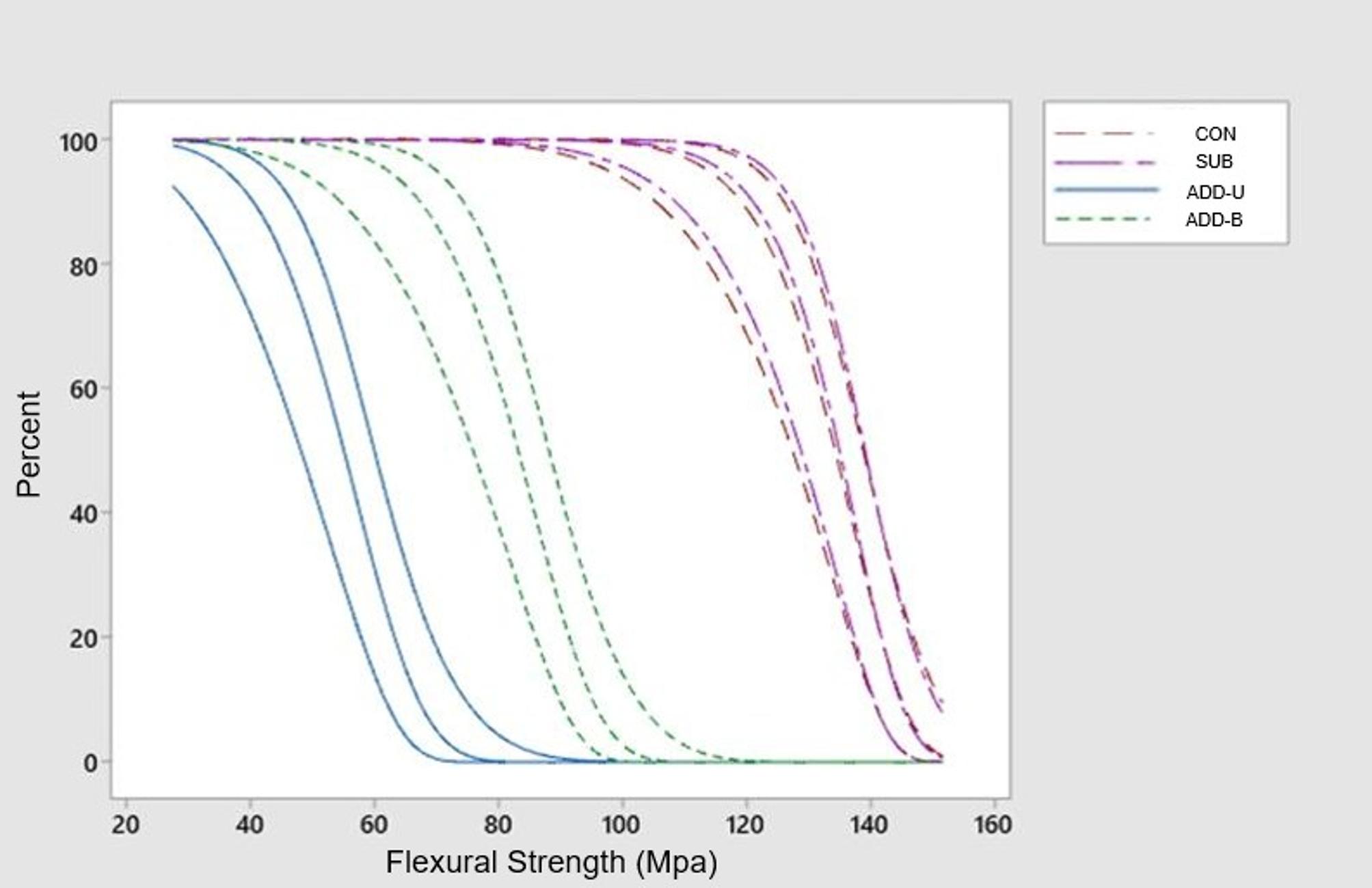
The Weibull analysis was used to assess variability and reliability through modulus (m) and scale (σ0) parameters. The modulus indicates the degree of data homogeneity, with higher values reflecting more consistent performance. The scale parameter denotes the flexural strength at which 63.2% of samples are expected to fail. The highest modulus was observed in the ADD-B group (17.31), indicating the most consistent results, while the lowest was found in the SUB group (6.10). The modulus of ADD-B differed significantly from all other groups (p = 0.001), whereas there was no significant difference between CON and SUB (p > 0.05). Scale values were CON=137.37 MPa, ADD-B=137.87 MPa, ADD-U=86.63 MPa, and SUB = 58.51 MPa. The difference among groups was statistically significant (p < 0.001). These findings suggest that CON and ADD-B exhibited not only a higher characteristic of flexural strength but also a superior reliability and performance consistency. The survival plot of Weibull analyzes are presented in Figure 2.

- Weibull survival plots of flexural strength (MPa) for all groups with 95% confidence intervals. The curves illustrate the probability of survival as a function of flexural strength, allowing comparison of the reliability and variability of the materials.
DISCUSSIONS
In this study, the flexural strengths of occlusal splints manufactured by conventional, subtractive, and additive methods were comparatively evaluated. The first null hypothesis of this study was that the manufacturing methods have no effect on the flexural strength values. The second null hypothesis was that there were no statistically significant differences between the uniaxial and biaxial testing methods for additive manufacturing. Although there were no statistically significant differences between conventional and subtractive methods, there were statistically significant differences among additive, conventional and subtractive groups for uniaxial flexural strength. There were statistically significant differences between uniaxial and biaxial testing values for additive manufacturing. Based on these findings, the first and second null hypothesis were rejected.
The presence of residual monomers in conventionally polymerized PMMA may influence the mechanical behavior of the material by acting as plasticizing agents within the polymer matrix. Residual monomers can increase polymer chain mobility and reduce intermolecular interactions, which may enhance the plastic deformation capacity of the material. This plasticizing effect may reduce stiffness while increasing the capacity for deformation before fracture, thereby potentially influencing flexural strength depending on the degree of polymerization and cross-link density. A previous study [21] reported that, despite similar chemical compositions, conventionally polymerized PMMA specimens exhibited a lower elastic modulus but higher flexural strength than milled PMMA specimens. These findings suggest that the mechanical response of PMMA-based materials is influenced not only by residual monomer content but also by polymerization conditions, cross-link density, and the internal microstructure of the material. In contrast, Arslan et al. [22] reported that conventionally polymerized PMMA exhibited a lower flexural strength than milled PMMA. These findings may be explained by the high degree of polymer conversion in pre-polymerized PMMA blocks, which are industrially processed under optimized conditions in subtractive manufacturing. It has been established by Al-Dwairi et al. [23] and Perea-Lowery et al. [24] that the use of high-temperature and high-pressure polymerization protocols in the fabrication of pre-polymerized PMMA blocks leads to improvements in flexural strength, surface hardness, and elastic modulus. The applied conditions contribute to improved polymer cross-linking, decreased monomer residue, and reduced porosity, which collectively strengthen the mechanical integrity of the material. According to Murakami et al. [25], although high-pressure polymerization contributes to improved toughness through increased polymer chain alignment, it may adversely affect the elastic modulus and flexural strength of the material. This dual effect emphasizes that optimal polymerization alone is not a definitive predictor of enhanced mechanical properties in prosthetic resin materials. In the present study, the subtractive and conventional materials exhibited similar flexural strength values. These values were also higher than those reported in previous studies, which may be attributed to differences in the polymerization pressure applied in the conventional manufacturing.
In the present study, no statistically significant difference was found between the CON and SUB groups. However, both groups had significantly higher flexural strength value compared to the ADD group [26]. In consistent with the findings of the present study, Berli et al. [27] and Wesemann et al. [28] reported that occlusal splint specimens manufactured using conventional and subtractive methods exhibited similar mean values. However, the CON group values (132 MPa) in the current study were higher than those reported for the conventional group (between 85-99 MPa) in these previous studies. The literature indicates that compression molding results in greater mechanical strength than injection molding, as evidenced by prior studies [29,30]. The higher CON group values observed in this study may be attributed to the use of compression molding, whereas previous studies [27,28] employed the injection molding technique.
In the current study, the flexural strength of specimens in the ADD group was found to be statistically significantly lower than that of the CON and SUB groups. Wesemann et al. [28] reported that one additively manufactured material demonstrated lower flexural strength than conventionally and subtractively fabricated materials, in agreement with the present findings. Nevertheless, another printed material in their study exhibited comparable values to the conventional and subtractive groups. The observed difference may be attributed to the type of printer used, as a DLP printer was employed in both the low-value group and our study, whereas the higher values were obtained using an SLA (Stereolithography) printer [31]. In the study conducted by Berli et al. [27] and Prpic et al. [32], different printing materials were compared with the materials produced by subtractive and conventional methods. In previous studies [27,32], production methods like the current study showed lower flexural strength in the additive materials compared to subtractive and conventional materials. However, in both studies, an additive material was found to have a flexural strength close to the materials produced by subtractive and conventional methods, which is incompatible with this study. This may be due to the content of the materials, wash and cure procedures, production angles and aging process.
The consistently lower flexural strength observed in the additively manufactured (ADD) specimens underscores the predominance of the material’s intrinsic chemistry and polymer network architecture over the manufacturing technique itself. Whereas the CON and SUB groups benefit from highly converted, homogeneous PMMA networks produced through heat-polymerization or industrial high-pressure polymerization, the ADD material is formulated from light-cured urethane-modified acrylates, which are inherently more susceptible to oxygen inhibition, post-curing variability, and the anisotropy arising from layer-by-layer fabrication. These factors collectively reduce cross-link density and promote localized polymerization heterogeneity. Furthermore, as demonstrated by Wulff et al. [33], DLP-printed photopolymers exhibit localized curing heterogeneity and layer interface irregularities, generating thin interstitial zones and micro-structural discontinuities between printed layers—features that persist even after standardized finishing and polishing. Such subtle interlayer discontinuities constitute potential initiation sites for surface-driven crack propagation, rendering additively manufactured specimens more vulnerable than the structurally uniform PMMA materials. Taken together, these material- and process-related characteristics provide a coherent mechanistic explanation for the reduced flexural strength recorded in the ADD group.
Beyond intrinsic material considerations, the testing configuration exerted a decisive influence on the mechanical behavior of the ADD specimens. In the present study, the ADD-B subgroup demonstrated significantly higher flexural strength than the ADD-U subgroup, a finding that is methodologically consistent with differences in stress distribution between the two test configurations. Pick et al. [34] demonstrated that uniaxial bar specimens concentrate maximum tensile stresses along their edges, making the results highly sensitive to minor surface defects, machining artifacts, or unchamfered borders—conditions particularly relevant for layered photopolymer resins. In contrast, biaxial disc specimens generate a uniform multidirectional tensile field and position the specimen periphery in a low-stress region, thereby reducing the influence of edge flaws and yielding values that more closely reflect the intrinsic strength of the material. Given that the printed rectangular bars in the present study were not beveled, stress intensification at unprocessed edges likely contributed to the lower and more variable flexural strength observed in the ADD-U subgroup. Collectively, these methodological factors indicate that three-point bending tends to underestimate the strength of printed photopolymers, whereas biaxial testing provides a more robust and reliable assessment. The observation that the ADD-B subgroup exceeded the 65-MPa minimum flexural strength requirement of ISO 20795-1 further supports the appropriateness of the biaxial test configuration for evaluating additively manufactured occlusal splint materials.
There are several limitations associated with this study. One notable limitation of this study is the absence of water sorption and aging evaluations, which are essential for assessing the long-term clinical behavior of additively manufactured materials. Since this study is limited to short-term in vitro data, additional research is necessary to confirm the materials' long-term clinical performance. Also, further studies are needed to determine the optimal universal or biaxial test method for assessing the flexural performance of additively manufactured resin-based materials. Additionally, the absence of complementary analyses such as SEM examination of fractured surfaces and quantitative surface roughness measurements constitutes a methodological limitation, as these techniques could have provided deeper insight into failure origins and surface characteristics; therefore, future studies should incorporate these evaluations to enhance the interpretation of mechanical behavior. Finally, the rectangular bar specimens used in the uniaxial test were not beveled (chamfered) at their edges as recommended by ISO 20795-1, which may have introduced localized stress concentrations and contributed to greater variability in the flexural strength results. Future studies should incorporate these considerations to enhance the accuracy and interpretability of mechanical assessments. In addition, although the sample size used in this study is consistent with similar in vitro studies, the relatively limited number of specimens may influence the precision of Weibull reliability modeling.
CONCLUSIONS
The flexural strength of occlusal splints is significantly influenced by the manufacturing technique. In the present study, conventional and subtractive methods produced comparable and consistently high flexural strength values, whereas additive manufacturing showed comparatively lower mechanical performance. Nevertheless, with continued advancements in material formulation and printing parameters, additive manufacturing demonstrates strong potential for achieving clinically acceptable and reproducible results. Considering the high functional demands associated with bruxism related TMD, conventional and subtractive fabrication methods appear to be the most reliable options at present, while additive approaches require further optimization before they can fully match the reliability of traditional techniques.
Acknowledgements
The authors declare that they have no acknowledgments to make.
Data availability
No research data was used in this study.
How to cite:
Funding
Regulatory Statement
REFERENCES
- 1 Okeson JP. Management of temporomandibular disorders and occlusion. 7th ed. St. Louis (MO): Elsevier Mosby; 2013.
- 2 Poveda Roda R, Bagán JV, Díaz Fernández JM, Hernández Bazán S, Jiménez Soriano Y. Review of temporomandibular joint pathology. Part I: Classification, epidemiology and risk factors. Med Oral Patol Oral Cir Bucal. 2007;12(4):E292-8. PMid:17664915.
-
3 Lobbezoo F, Ahlberg J, Glaros AG, Kato T, Koyano K, Lavigne GJ, et al. Bruxism defined and graded: an international consensus. J Oral Rehabil. 2013;40(1):2-4. https://doi.org/10.1111/joor.12011 PMid:23121262.
» https://doi.org/10.1111/joor.12011 -
4 Guaita M, Högl B. Current treatments of bruxism. Curr Treat Options Neurol. 2016;18(2):10. https://doi.org/10.1007/s11940-016-0396-3 PMid:26897026.
» https://doi.org/10.1007/s11940-016-0396-3 -
5 List T, Axelsson S. Management of TMD: evidence from systematic reviews and meta-analyses. J Oral Rehabil. 2010;37(6):430-51. https://doi.org/10.1111/j.1365-2842.2010.02089.x PMid:20438615.
» https://doi.org/10.1111/j.1365-2842.2010.02089.x -
6 Nekora A, Evlioglu G, Ceyhan A, Keskin H, Issever H. Patient responses to vacuum formed splints compared to heat cured acrylic splints: pilot study. J Maxillofac Oral Surg. 2009;8(1):31-3. https://doi.org/10.1007/s12663-009-0008-9 PMid:23139466.
» https://doi.org/10.1007/s12663-009-0008-9 -
7 Pereira ER, Sichi LGB, Coelho MS, Lopes GC, Araújo RM. Dimensional accuracy of provisional complete crown made by the 3D printing method. Braz Dent Sci. 2024;27(2):e3761. https://doi.org/10.4322/bds.2024.e4366
» https://doi.org/10.4322/bds.2024.e4366 -
8 Berntsen C, Kleven M, Heian M, Hjortsjö C. Clinical comparison of conventional and additive manufactured stabilization splints. Acta Biomater Odontol Scand. 2018;4(1):81-9. https://doi.org/10.1080/23337931.2018.1497491 PMid:30128331.
» https://doi.org/10.1080/23337931.2018.1497491 -
9 Pho Duc JM, Hüning SV, Grossi ML. Parallel randomized controlled clinical trial in patients with temporomandibular disorders treated with a CAD/CAM versus a conventional stabilization splint. Int J Prosthodont. 2016;29(4):340-50. https://doi.org/10.11607/ijp.4711 PMid:27479339.
» https://doi.org/10.11607/ijp.4711 - 10 Edelhoff D, Schweiger J, Prandtner O, Trimpl J, Stimmelmayr M, Güth JF. CAD/CAM splints for the functional and esthetic evaluation of newly defined occlusal dimensions. Quintessence Int. 2017;48(3):181-91. PMid:28232961.
-
11 Salmi M, Paloheimo KS, Tuomi J, Ingman T, Mäkitie A. A digital process for additive manufacturing of occlusal splints: a clinical pilot study. J R Soc Interface. 2013;10(84):20130203. https://doi.org/10.1098/rsif.2013.0203 PMid:23614943.
» https://doi.org/10.1098/rsif.2013.0203 -
12 Lutz AM, Hampe R, Roos M, Lümkemann N, Eichberger M, Stawarczyk B. Fracture resistance and 2-body wear of 3-dimensional-printed occlusal devices. J Prosthet Dent. 2019;121(1):166-72. https://doi.org/10.1016/j.prosdent.2018.04.007 PMid:30647000.
» https://doi.org/10.1016/j.prosdent.2018.04.007 -
13 Lima LC, Miranda JS, Carvalho RL, Barcellos ASP, Amaral M, Kimpara ET. Influence of substrate, cement and aging on the biaxial flexural strength of lithium disilicate. Braz Dent Sci. 2023;26(4):e3476. https://doi.org/10.4322/bds.2023.e3923
» https://doi.org/10.4322/bds.2023.e3923 -
14 Choi BJ, Yoon S, Im YW, Lee JH, Jung HJ, Lee HH. Uniaxial/biaxial flexure strengths and elastic properties of resin-composite block materials for CAD/CAM. Dent Mater. 2019;35(2):389-401. https://doi.org/10.1016/j.dental.2018.11.032 PMid:30527587.
» https://doi.org/10.1016/j.dental.2018.11.032 - 15 International Organization for Standardization. ISO 20795-1:2013: Dentistry — Base polymers — Part 1: Denture base polymers. Geneva: ISO; 2013.
- 16 International Organization for Standardization. ISO 6872:2015: Dentistry — Ceramic materials. Geneva: ISO; 2015.
-
17 Çakmak G, Wiegner S, Sabatini GP, Kahveci Ç, Fonseca M, Pieralli S, et al. Influence of cleaning solutions and hydrothermal aging on the flexural strength and microhardness of resins for additively manufactured definitive fixed restorations. J Prosthet Dent. 2026;135(1):158-64. https://doi.org/10.1016/j.prosdent.2025.03.006 PMid:40251109.
» https://doi.org/10.1016/j.prosdent.2025.03.006 -
18 Espinar C, Della Bona A, Pérez MM, Tejada-Casado M, Pulgar R. The influence of printing angle on color and translucency of 3D printed resins for dental restorations. Dent Mater. 2023;39(4):410-7. https://doi.org/10.1016/j.dental.2023.03.011 PMid:36914433.
» https://doi.org/10.1016/j.dental.2023.03.011 -
19 Messer-Hannemann P, Rabl A, Kessler A, Reymus M, Hickel R, Stawarczyk B, et al. Residual TPO content of photopolymerized additively manufactured dental resins. Biomedicines. 2024;13(1):44. https://doi.org/10.3390/biomedicines13010044 PMid:39857628.
» https://doi.org/10.3390/biomedicines13010044 -
20 Sabatini GP, Yoon HI, Orgev A, Fonseca M, Molinero-Mourelle P, Yilmaz B, et al. Complete digital workflow for occlusal device fabrication using artificial intelligence–powered design software and additive manufacturing. Int J Prosthodont. 2024;37:S275-84. https://doi.org/10.11607/ijp.8941
» https://doi.org/10.11607/ijp.8941 -
21 Ayman AD. Residual monomer content and mechanical properties of CAD/CAM vs heat cured resins. Electron Physician. 2017;9(7):4766-72. https://doi.org/10.19082/4766 PMid:28894533.
» https://doi.org/10.19082/4766 - 22 Arslan M, Murat S, Alp G, Zaimoglu A. Evaluation of flexural strength and surface properties of prepolymerized CAD/CAM PMMA-based polymers used for digital 3D complete dentures. Int J Comput Dent. 2018;21(1):31-40. PMid:29610779.
-
23 Al-Dwairi ZN, Tahboub KY, Baba NZ, Goodacre CJ. A comparison of the flexural and impact strengths and flexural modulus of CAD/CAM and conventional heat-cured polymethyl methacrylate (PMMA). J Prosthodont. 2020;29(4):341-9. https://doi.org/10.1111/jopr.12926 PMid:29896904.
» https://doi.org/10.1111/jopr.12926 -
24 Perea-Lowery L, Minja IK, Lassila L, Ramakrishnaiah R, Vallittu PK. Assessment of CAD-CAM polymers for digitally fabricated complete dentures. J Prosthet Dent. 2021;125(1):175-81. https://doi.org/10.1016/j.prosdent.2019.12.008 PMid:32063383.
» https://doi.org/10.1016/j.prosdent.2019.12.008 -
25 Murakami N, Wakabayashi N, Matsushima R, Kishida A, Igarashi Y. Effect of high-pressure polymerization on mechanical properties of PMMA. J Mech Behav Biomed Mater. 2013;20:98-104. https://doi.org/10.1016/j.jmbbm.2012.12.011 PMid:23455166.
» https://doi.org/10.1016/j.jmbbm.2012.12.011 -
26 Abdullah HA, Al-Ibraheemi ZA, Majeed M, Al-Nasrawi S. Evaluation of flexural strength and degree of conversion of temporary crown materials at different aging periods in artificial saliva. Braz Dent Sci. 2024;27(4):e3897. https://doi.org/10.4322/bds.2024.e4368
» https://doi.org/10.4322/bds.2024.e4368 -
27 Berli C, Thieringer FM, Sharma N, Müller JA, Dedem P, Fischer J, et al. Comparing mechanical properties of pressed, milled, and 3D-printed resins for occlusal devices. J Prosthet Dent. 2020;124(6):780-6. https://doi.org/10.1016/j.prosdent.2019.10.024 PMid:31955837.
» https://doi.org/10.1016/j.prosdent.2019.10.024 -
28 Wesemann C, Spies BC, Sterzenbach G, Beuer F, Kohal R, Wemken G, et al. Polymers for conventional, subtractive, and additive manufacturing of occlusal devices differ in hardness and flexural properties but not in wear resistance. Dent Mater. 2021;37(3):432-42. https://doi.org/10.1016/j.dental.2020.11.020 PMid:33288324.
» https://doi.org/10.1016/j.dental.2020.11.020 -
29 Aguirre BC, Chen JH, Kontogiorgos ED, Murchison DF, Nagy WW. Flexural strength of denture base acrylic resins: conventional vs CAD-CAM. J Prosthet Dent. 2020;123(4):641-6. https://doi.org/10.1016/j.prosdent.2019.03.010 PMid:31353106.
» https://doi.org/10.1016/j.prosdent.2019.03.010 -
30 Pacquet WW, Benoit A, Hatège-Kimana C, Wulfman C. Mechanical properties of CAD/CAM denture base resins. Int J Prosthodont. 2019;32(1):104-6. https://doi.org/10.11607/ijp.6025 PMid:30677121.
» https://doi.org/10.11607/ijp.6025 -
31 Batista LMB, Santana YVS, Borba MTLM, Silva TKA, Silva CML, Torres Neto AJ, et al. Effect of angulation of 3D printed resin provisional bridges: an in vitro study on hardness and fracture loading. Braz Dent Sci. 2025;28(1):e4023. https://doi.org/10.4322/bds.2025.e4581
» https://doi.org/10.4322/bds.2025.e4581 -
32 Prpic V, Slacanin I, Schauperl Z, Catic A, Dulcic N, Cimic S. Flexural strength and surface hardness of materials for occlusal devices. J Prosthet Dent. 2019;121(5):955-9. https://doi.org/10.1016/j.prosdent.2018.09.022 PMid:30711296.
» https://doi.org/10.1016/j.prosdent.2018.09.022 -
33 Wulff J, Rauch A, Schmidt MB, Rosentritt M. Biaxial flexural strength of printed splint materials. Materials. 2024;17(5):1112. https://doi.org/10.3390/ma17051112 PMid:38473585.
» https://doi.org/10.3390/ma17051112 -
34 Pick B, Meira JBC, Driemeier L, Braga RR. A critical view on biaxial and short-beam uniaxial flexural strength tests applied to resin composites using Weibull, fractographic and finite element analyses. Dent Mater. 2010;26(1):83-90. https://doi.org/10.1016/j.dental.2009.09.002 PMid:19819002.
» https://doi.org/10.1016/j.dental.2009.09.002
Conflict of Interest
Suzan Eda Eyyupoğlu (Corresponding address)
Istanbul Medipol University, Faculty of Dentistry, Department of Prosthodontics, Istanbul, Turkey. Email: suzanedaeyyupoglu@gmail.comEditor-in-chief:
Editor: