Addition of silver nanoparticle solutions to alginate: microbiological and mechanical analysis
Abstract
This study assesses the antimicrobial activity and physical alterations of alginate impression material in the presence of silver nanoparticle (AgNP) solutions. The samples were assigned to five groups according to the type of alginate and solution: (JW) Jeltrate + distilled water; (JCHX) Jeltrate + 0.2% chlorhexidine digluconate; (JAgNP_0.2%) Jeltrate + 0.2% AgNP; (JAgNP_1%) Jeltrate + 1% AgNP; and (AW) Avagel + distilled water. AgNPs were synthesized from the fungus Trichoderma reesei. Fifty impressions were made (n=10) from a matrix, from which plaster models were fabricated. Surface detail reproduction and dimensional stability were analyzed under a light microscope. Surface detail reproduction was achieved by replicating a 50µm line on the plaster model. Dimensional stability was calculated from measurements of X’ and X’’ in the matrix and plaster models. The antimicrobial effect was assessed independently by measuring the formation of zones of inhibition around the alginate molds (n=3) in agar medium containing Streptococcus mutans and Candida albicans. Dimensional stability values were subjected to the ANOVA and Tukey’s post-hoc test (α=0.05). JCHX showed the highest dimensional stability and differed significantly from the other groups (p<0.05). A 50µm line was reproduced in the plaster for all tested groups, regardless of the impression material and antimicrobial solution. AgNPs incorporated into the alginate mixture did not produce dimensional changes in the alginate mold. AW and JCHX exhibited antimicrobial potential, as evidenced by bacterial inhibition. Jeltrate or Avagel, regardless of AgNP concentration, did not demonstrate antimicrobial activity.
Key Words:
Alginate; Chlorhexidine; Antibacterial Agents; Streptococcus mutans; Candida albicans
Resumo
Este estudo avaliou a atividade antimicrobiana e as alterações físicas do material de moldagem alginato com soluções de nanopartículas de prata (NPAg). As amostras foram distribuídas em cinco grupos de acordo com o tipo de alginato e solução: (JA) Jeltrate + água destilada; (JCHX) Jeltrate + digluconato de clorexidina a 0,2%; (J NPAg _0,2%) Jeltrate + NPAg a 0,2%; (J NPAg _1%) Jeltrate + 1% NPAg e (AA) Avagel + água destilada. NPAg foram sintetizadas a partir do fungo Trichoderma reesei. Cinquenta impressões foram realizadas (n=10) de uma matriz metálica, a partir da qual foram confeccionados modelos de gesso. A reprodução dos detalhes da superfície e a estabilidade dimensional foram analisadas através da microscópia óptica. A reprodução de detalhes da superfície foi obtida pela replicação da linha de 50 μm no modelo de gesso. A estabilidade dimensional foi calculada pelas medidas entre X' e X'' da matriz e do modelo de gesso. O efeito antimicrobiano foi avaliado de forma independente com base na formação de zonas de inibição entre os moldes de alginato (n=3) e o meio de ágar contendo Streptococcus mutans e Candida albicans. Os valores de estabilidade dimensional foram submetidos à ANOVA e ao teste post-hoc de Tukey (α= 0,05). JCHX apresentou maior estabilidade dimensional e apresentou diferenças significativas em relação aos demais grupos (p<0,05). A linha de 50 μm foi reproduzida no gesso para todos os grupos testados, independentemente do material de moldagem e da solução antimicrobiana. NPAg incorporadas na mistura de alginato não produziram alterações dimensionais no molde de alginato. AA e JCHX exibiram potencial antimicrobiano, evidenciado pela inibição bacteriana. Jeltrate ou Avagel, independentemente da concentração da solução de nanopartículas de prata, não demonstraram atividade antimicrobiana.

Introduction
Dental impressions are a recurrent procedure in dental clinical practice, and diverse molding materials, such as alginate, are available on the market 1. Alginate is an irreversible hydrocolloid material widely used for the fabrication of dental models 2. Despite the increasing adoption of digital workflows, conventional impression materials such as alginate remain widely used 3,4 in many clinical and educational settings, and their disinfection remains an important aspect of infection control in dentistry. After the mold is removed from the patient’s oral cavity, it is often contaminated with microorganisms due to direct contact with oral fluids, such as saliva and blood, which serve as vectors for disease transmission 1. Despite the increasing adoption of digital workflows and intraoral scanning, alginate impressions remain widely used in public health systems, teaching clinics, and resource-limited environments where digital equipment is not routinely available. Recent studies continue to document microbial contamination in irreversible hydrocolloids and evaluate disinfection methods due to persistent biosafety concerns in daily clinical practice 1,5,6.
Dental molds obtained via the molding process must be chemically disinfected before they are filled with plaster and sent to the prosthetic laboratory to prevent them from acting as vectors for the transmission of microorganisms or infectious diseases 7. Disinfection of mold is an effective biosafety measure for dental professionals, such as dental surgeons, dental hygienists, and prosthetists, preventing cross-contamination 8.
When it comes to antimicrobial substances for the disinfection of alginate molds, nanotechnology is noteworthy, including nanoscale materials (<100 nm) 5,9. These nanoparticles can originate from primary sources, i.e., chemically or biologically synthesized 10. Silver nanoparticles (AgNPs) have attracted significant attention due to their remarkable properties, including a large surface area and excellent antimicrobial activity 11. Biogenically synthesized AgNPs, such as those produced by Trichoderma reesei, exhibit distinct surface characteristics, heterogeneous size distributions, and potentially lower cytotoxicity compared to chemically synthesized nanoparticles 12. These properties may influence antimicrobial behavior and interaction with hydrocolloid matrices, yet their performance when incorporated into the mixing solution of irreversible hydrocolloids has not been fully investigated. As a result, AgNPs have been applied in the health sector, including dentistry 11,13,14, given that Ag is a chemical element with antimicrobial potential and is considered safe and effective for killing more than 650 disease-causing microorganisms.
The biological synthesis of AgNPs by microorganisms has become an eco-friendly approach 9,15. It is also a more straightforward, cost-effective alternative with potent bactericidal activity against gram-positive and gram-negative pathogens 11. Moreover, it is highly efficient, stable, and nontoxic 16, with AgNPs readily soluble in water 17.
A previous study incorporated AgNPs into irreversible hydrocolloid impressions as antimicrobial agents without adversely affecting their properties, depending on the material type 15. Furthermore, 80-100 nm AgNPs exhibited greater antimicrobial activity than Zelgan Plus (Dentsply) irreversible hydrocolloid in a dose-dependent manner. The addition of 0.5% and 1% by weight did not interfere with the properties of the material. However, higher concentrations (2% and 5%) caused gelation time changes and material strength 18,19. Conversely, another study 20 found that self-disinfection of Lascod type A irreversible hydrocolloid (Florence, Italy) with 0.2% chlorhexidine (CHX) solution or alcohol-free commercial mouthwash had similar results to self-disinfection with incorporation of AgNP into the powder (0.1% and 0.2% with 5-8 nm nanoparticles) against the five investigated microorganisms.
Many studies have evaluated the stability and antimicrobial activity of alginate with different disinfectant solutions 5,15,18,19,20. However, further studies are needed to assess the effect of 0.2% and 1% biologically synthesized AgNP solutions incorporated into the water during alginate powder manipulation. Although several studies have evaluated the incorporation of disinfectant agents into alginate, little is known about the behavior of biologically synthesized AgNPs mixed into the liquid phase, particularly regarding their stability, dispersion within the hydrocolloid network, and potential antimicrobial effects. Addressing these aspects may help identify alternative self-disinfecting strategies in settings where immersion protocols or digital workflows are not feasible. Therefore, this study aimed to evaluate whether incorporating biogenically synthesized silver nanoparticles into the mixing solution of alginate impression materials affects surface detail reproduction, dimensional stability, and antimicrobial activity. By testing two nanoparticle concentrations and comparing them with chlorhexidine and commercial controls, this study sought to determine whether this approach could represent a feasible self-disinfection strategy without compromising critical physical properties. The following hypotheses were stated: 1) incorporation of AgNP into the mixing solution does not affect surface detail reproduction and dimensional stability of alginate molds, and 2) AgNPs do not inhibit microbial growth.
Materials and methods
Experimental design
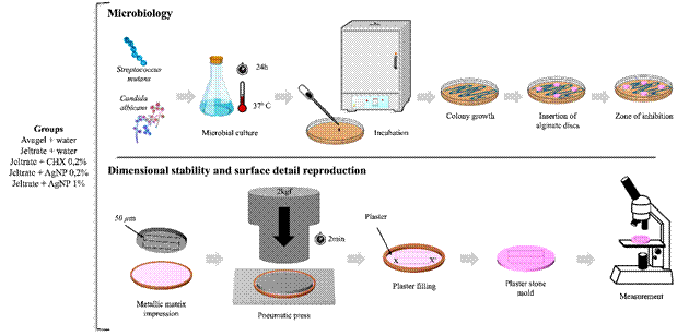
The methodological steps shown in Figure 1 were followed.

Biological synthesis of AgNPs
The fungus Trichoderma reesei was grown on Petri dishes containing MEX-agar medium and incubated in an oven at 28 °C until fungal growth and sporulation occurred within 7 days. The spores were then collected, inoculated into 100 mL of potato dextrose agar, and incubated at 30 °C under low-oxygen conditions in an oven for 7 days. Trichoderma reesei produced enzymes capable of generating AgNPs under low oxygen conditions. Silver was biologically synthesized in an amber-colored Erlenmeyer flask by mixing the culture supernatant with aqueous silver nitrate (AgNO3) at a concentration of 5 mM (range of 1 to 10 mM). After mixing, the pH was adjusted to 8.5. The mixture was stored in the dark at 40 °C under rotation at 200 rpm for ten days 21.
Microorganisms
Candida albicans CBS 562 and Streptococcus mutans UA159 were selected based on the microbial composition of oral biofilms and the microorganisms found on dental molds from patients.
Dimensional stability and surface detail reproduction
Dimensional stability and surface detail reproduction were assessed in accordance with ISO 1563. Five plaster stone models (n = 5) were manufactured for each group according to Guiraldo et al. 22. Molds were prepared on a matrix (outer diameter: 38 mm and inner diameter: 29.97 mm) containing three parallel lines measuring 20, 50, and 75 μm in width and 25 mm in length, spaced 2.5 mm apart. Two additional lines, X and X′, were used to determine dimensional stability and surface detail reproduction along the 50 μm line.
Before molding, the matrix was ultrasonically cleaned and dried with compressed air. The irreversible hydrocolloid alginate-based molding materials - Jeltrate Dustless (type II - regular setting) and Avagel, both from Dentsply Sirona, Milford, DE, USA, the latter of which contains CHX, were prepared according to the manufacturer’s instructions.
A perforated metal tray (inner diameter 31 mm, height 5 mm) was placed on a glass plate and filled with the molding material. The tray was joined to the matrix, and a 2 kgf pneumatic press was used to apply pressure and simulate the molding process, allowing the excess material to overflow. The molds were removed 2 min after the material lost its sticky consistency. The molds were rinsed with 150 mL of distilled water and dried afterward.
During the preparation of the alginate powder mixture according to the manufacturer’s instructions, the liquid component was completely replaced by disinfectant solutions. Distilled water was substituted with either 0.2% CHX, prepared at Proderma (Piracicaba, SP, Brazil), or with previously synthesized AgNPs solutions at concentrations of 0.2% and 1.0%. Biogenic AgNPs produced by Trichoderma reesei are typically reported to present particle sizes in the nanometric range (approximately 5-50 nm) 21. The manufacturer's recommended powder-to-liquid ratio was maintained for all groups.
A single operator manually stirred the mixture for 45 seconds using a spatula and a plastic bowl. The samples were then split into five groups based on the type of disinfection and the disinfectant solution used:
JW: Jeltrate Dustless + distilled water
JCHX: Jeltrate Dustless + 0.2% CHX solution
JAgNP_0.2%: Jeltrate Dustless + 0.2% AgNP solution
JAgNP_1%: Jeltrate Dustless + 1% AgNP solution
AW: Avagel + distilled water
The alginate molds were subsequently rinsed with 150 mL of distilled water, dried, and immediately filled with plaster (Durone IV; Dentsply Caulk), as per the manufacturer's instructions. The plaster stone models were removed from the trays containing the alginate mold 1 hour after the plaster was mixed.
Light microscopy
Surface detail reproduction measurements were conducted under a light microscope (SZM; Bel Engenharia, MI, Italy) on the plaster stone models at 4x to 12x magnifications to check whether the 50 μm-wide line was accurately replicated along the 25 mm length between the intersection of reference lines (X and X′), in compliance with ISO 1563 standard.
Dimensional stability measurements were conducted on the plaster stone models under a light microscope (STM; Olympus Optical Co Ltd, Japan) with a precision of 0.0005 mm, expressed as a percentage (L) and calculated in accordance with ISO 1563 standard using the equation:
L1 represents the distance between the lines in the matrix, while L2 denotes the distance between the lines in the plaster stone models.
Preparation of alginate discs
The powder-to-water ratio in the alginate mold material was adjusted according to the manufacturer’s instructions. The mixture was stirred by a single operator for 45 seconds using a spatula and a plastic bowl. The mixture was then poured into a cylinder (6 mm in diameter and 1 mm in thickness). Excess material was removed by compression with a sterile glass blade. Three discs were prepared for each group and bacterial strain 23.
Antimicrobial activity
Antimicrobial activity was assessed by the disc diffusion method on an agar medium. The discs were placed with sterile tweezers onto a Petri dish containing Mueller-Hinton (MH) agar, which had been previously inoculated with the microorganisms to be tested. The control sample was autoclaved with distilled water. Following incubation, clear zones, or zones of inhibition, are expected to form around the specimens. The following antibiotics were tested: 0.2% CHX and 0.2% and 1% AgNPs. Two microorganisms were used: S. mutans UA 159 and Candida albicans CBS 562.
The microbial plates and discs were incubated at 35-37 °C for bacteria for 24-48 h and at 25-27 °C for fungi for 48-72 h. The diameter of the inhibition zones was measured directly on the agar surface by a single operator using a millimeter ruler, following the shortest-path criterion 20. For each group and microorganism, the mean inhibition zone (in mm) was calculated from the three discs prepared for each condition.
Statistical analysis
The data were tested for normality and homogeneity. Subsequently, a one-way analysis of variance (ANOVA) was conducted, followed by Tukey’s post hoc test (α = 0.05). The significance level was set at 95%.
Results
Dimensional stability
The highest mean dimensional accuracy and the highest statistically significant level of dimensional accuracy were observed with Jeltrate + 0.2% CHX (Table 1).
Surface detail reproduction
All evaluated groups successfully reproduced the 50 μm line, regardless of the solution used for irreversible hydrocolloid manipulation.
Microbiological analysis
Only the JCHX and AW exhibited zones of inhibition for both tested microorganisms (Figures 2 and 3). The largest zone of inhibition was observed with JCHX against S. mutans, and the smallest was observed with AW against C. albicans. Table 2 shows the presence of an inhibition zone and mean inhibition zone diameters (mm) in groups JCHX (3.4 mm to S. mutans and 0.87 mm to C. albicans) and AW (0.87 mm to S. mutans and 0.15 mm to C. albicans). In contrast, no inhibition zones were observed in the JW (commercial negative control), AgNP_0.2%, and AgNP_1% groups.

Zone of inhibition (black arrow) formed against S. mutans. JW - Jeltrate + distilled water; JCHX - Jeltrate + 0.2% chlorhexidine digluconate; JAgNP_0.2% - Jeltrate + 0.2% silver nanoparticles; JAgNP_1% - Jeltrate + 1% silver nanoparticles; and AW - Avagel + distilled water.

Zone of inhibition (black arrow) formed against Candida albicans. JW - Jeltrate + distilled water; JCHX - Jeltrate + 0.2% chlorhexidine digluconate; JAgNP_0.2% - Jeltrate + 0.2% silver nanoparticles; JAgNP_1% - Jeltrate + 1% silver nanoparticles; and AW - Avagel + distilled water.
Discussion
The results showed that adding an AgNP solution to the alginate at either concentration did not inhibit microbial growth. However, surface detail reproduction and dimensional stability of the molds remained unchanged in all tested solutions. Therefore, all hypotheses stated in this study were confirmed. All plaster models, regardless of the solutions used, reproduced the 50 µm line, and the maximum variation in dimensional stability was 0.8 % (Jeltrate + water), in compliance with ISO 4823 (Dentistry - Elastomeric impression materials).
Alginates are irreversible hydrocolloid materials primarily composed of calcium sulfate, potassium, sodium, or ammonium alginates, sodium phosphate, and filler particles. They form when water interacts with the alginate powder, releasing calcium ions from calcium sulfate hemihydrate and forming cross-linking points. Alginates are often used in clinical practice because they are cost-effective and easy to handle compared to other impression materials. The success of clinical procedures involving molding is closely related to the material's ability to reproduce surface details and maintain dimensional stability, resulting in a plaster model that closely resembles the original structure 24.
Cross-contamination is frequently observed in dental clinical practice and must be mitigated to prevent the spread of microorganisms. Thus, decontamination of impression materials is crucial for controlling cross-infection. Oral microorganisms can easily adhere to impression materials after they set, and disinfection procedures should then be performed 6. Immersion or spraying are commonly used for disinfecting alginate molds, but these methods can only clean the surface, leading to dimensional changes in the mold 18. One of the key concerns with alginate models includes poor tear resistance, dimensional instability, especially after a delay in casting the stone model, and a weak ability to reproduce surface details. Therefore, the mold's precision can be directly affected by imbibition or syneresis, which can be easily prevented by immediately filling the mold with plaster 25. An alternative to spraying or immersion in disinfectant solution is the use of self-disinfecting materials. This approach provides better dimensional stability than the previous ones, reducing disinfection time 26.
The findings of this study demonstrate that mixing the material with a disinfectant solution does not compromise the dimensional stability of the molds, as it remains within the acceptable limits set by the standard (1.5 µm - ISO 4823, 2015); however, only 0.2% CHX yielded significantly larger models. In one of the tested groups, a disinfectant was present, but it was part of the Avagel alginate composition. Jeltrate + 0.2% CHX and Avagel + water were the only groups demonstrating antimicrobial activity, unlike the Jeltrate + water and the AgNP groups. Chlorhexidine is a broad-spectrum disinfectant considered the gold standard 27 and is known to be a safe and effective antimicrobial agent 26. It has bacteriostatic properties at low concentrations and bactericidal effects at higher concentrations. CHX uptake induces quick cell death by disrupting the bacterial cell wall 27. However, to prevent changes in mold dimensions, other antimicrobial agents, such as AgNP, can be a good alternative.
AgNPs are currently employed in medical products and equipment because they have been reported to exhibit broad-spectrum antimicrobial effects (against bacteria, fungi, protozoa, and some viruses) and low toxicity 28. AgNPs target multiple bacterial targets, reducing the likelihood of resistance. In contact with bacterial DNA and RNA, AgNPs bind to sulfur and phosphorus, thereby degrading associated nucleic acids 27. Studies have shown that AgNPs may be more effective than CHX under certain conditions. A previous study (27 demonstrated significantly stronger bacteriostatic and bactericidal effects against bacteria (Streptococcus mutans and oralis, Lactobacillus acidophilus and fermentum) and the fungus Candida albicans when compared to CHX, particularly when used as a solution. Another study (28 showed that disinfection effectiveness increased with higher AgNP concentrations (0.25, 0.50, and 1%), higher powder-to-water ratios, and longer handling times. In their study, AgNPs were incorporated into the powder rather than into the mixing solution, as done in the present study. Research (29 also found that incorporating AgNPs into alginate powder was more effective than disinfection with 2% glutaraldehyde, using alginate mold discs in a diffusion medium, as in the present study. Both the mode of incorporation of AgNPs into alginate and particle size appear to influence antibacterial activity. Ginjupalli et al. (18 assessed antimicrobial activity using AgNPs with different particle sizes (80-100, 50-80, 30-50, and 10-20 nm) incorporated into an irreversible hydrocolloid powder. They demonstrated that 80-100 nm AgNPs had the highest dose-dependent antimicrobial activity. While previous studies identified a maximal antibacterial effect for AgNPs in the ~80-100 nm size range 18, biosynthesis via Trichoderma reesei is typically associated with much smaller particles (e.g., ~5-50 nm) 30. When added as a solution rather than incorporated into powder and embedded in an alginate matrix, these smaller particles likely had reduced availability and diffusion. In the present study, AgNPs were synthesized biologically using Trichoderma reesei, a method known to produce particles with heterogeneous sizes and surface characteristics 30. They were added as a mixing solution rather than incorporated into the alginate powder. These factors, combined with the limited diffusion of nanoparticles through the set hydrocolloid matrix, likely reduced their availability for microbial interaction and may explain the absence of antimicrobial activity observed in the present study, despite the well-documented efficacy of AgNPs in other models.
Surface detail reproduction, as recommended by the standards, varies by impression material type. In general, the higher the precision of the material, the better its ability to reproduce surface details. Elastomeric impression materials exhibit surface detail reproduction ranging from 20 to 75 µm, depending on the specific type (ISO 4823, 2015), whereas alginates typically reproduce details of up to 50 µm (ISO 1563, 1990). Regarding dimensional stability, the standard allows variations of up to 1.5 µm (ISO 4823, 2015). Accordingly, mixing alginate with a disinfectant solution, whether or not AgNPs are present, does not appear to interfere with the material’s properties.
Some limitations of this study are noteworthy. The biogenically synthesized AgNPs were incorporated into the mixing solution without characterization of their size distribution, morphology, stability, or ion-release behavior, all of which can influence their antimicrobial performance. Additionally, no aging or storage protocols were applied, and the diffusion-based antimicrobial assay may have been limited by nanoparticle entrapment within the hydrocolloid matrix. The mode of incorporation - mixing AgNPs into the liquid phase rather than embedding them in the powder - may also have limited their interaction with microorganisms. Future studies should therefore include detailed nanoparticle characterization, ion-release analysis, aging protocols, and direct-contact antimicrobial models, as well as the evaluation of alternative incorporation strategies, to better elucidate the underlying mechanisms and optimize the antimicrobial potential of biogenically synthesized AgNPs in alginate impression materials.
In summary, Jeltrate mixed with CHX exhibited the highest dimensional stability. All groups reproduced the 50 µm line in the plaster models, regardless of the material or solution used. Jeltrate mixed with CHX and Avagel mixed with water inhibited bacterial growth. AgNPs incorporated into alginate did not alter the dimensional stability of alginate molds.
Acknowledgments
This study was supported by Conselho Nacional de Desenvolvimento Científico e Tecnológico - CNPq (#307434/2025-7) and PIC-FHO/2024.
References
-
1 Hardan L, Bourgi R, Cuevas-Suárez CE, Lukomska-Szymanska M, Cornejo-Ríos E, Tosco V, et al. Disinfection procedures and their effect on microorganism colonization of dental impression materials: a systematic review and meta-analysis of in vitro studies. Bioengineering (Basel) 2022;9:123. doi:10.3390/bioengineering9030123.
» https://doi.org/10.3390/bioengineering9030123 -
2 Niyazi AA, Alabrash MA, Elzouhiry MS, AlFarra RM, Althubyani NM, Huraib WM, et al. Comparative analysis of alginate dimensional stability with varied pouring intervals. J Pharm Bioallied Sci 2024;16(Suppl 5):S4599-S4603. doi:10.4103/jpbs.jpbs_762_24.
» https://doi.org/10.4103/jpbs.jpbs_762_24 -
3 Cervino G, Fiorillo L, Herford AS, Laino L, Troiano G, Amoroso G, et al. Alginate materials and dental impression technique: a current state of the art and application to dental practice. Mar Drugs 2019;17:18. doi:10.3390/md17010018.
» https://doi.org/10.3390/md17010018 -
4 Punj A, Bompolaki D, Garaicoa J. Dental impression materials and techniques. Dent Clin North Am 2017;61:779-796. doi:10.1016/j.cden.2017.06.004.
» https://doi.org/10.1016/j.cden.2017.06.004 -
5 Bendary IM, Omar AA, Goda RM, Ali AA, Lotfy KA, Shohayeb MM. Evaluation of two different self-disinfection alginate impression materials. BDJ Open 2024;10:84. doi:10.1038/s41405-024-00269-6.
» https://doi.org/10.1038/s41405-024-00269-6 -
6 Masih A, Gandagule RR, Tiwari A, Elangovan E, Choukse V, Saini N. Evaluating the influence of spray and immersion disinfection on the dimensional accuracy of impression materials: a comprehensive review. Cureus 2025;17:e88921. doi:10.7759/cureus.88921.
» https://doi.org/10.7759/cureus.88921 -
6 AlZain S. Effect of chemical, microwave irradiation, steam autoclave, ultraviolet light radiation, ozone and electrolyzed oxidizing water disinfection on properties of impression materials: a systematic review and meta-analysis study. Saudi Dent J 2020;32:161-170. doi:10.1016/j.sdentj.2019.12.003.
» https://doi.org/10.1016/j.sdentj.2019.12.003 -
8 Savabi O, Nejatidanesh F, Bagheri KP, Karimi L, Savabi G. Prevention of cross-contamination risk by disinfection of irreversible hydrocolloid impression materials with ozonated water. Int J Prev Med 2018;9:37. doi:10.4103/ijpvm.IJPVM_143_16.
» https://doi.org/10.4103/ijpvm.IJPVM_143_16 -
9 Ahmad A, Haneef M, Ahmad N, Kamal A, Jaswani S, Khan F. Biological synthesis of silver nanoparticles and their medical applications: a review. World Acad Sci J 2024;6:22. doi:10.3892/wasj.2024.237.
» https://doi.org/10.3892/wasj.2024.237 -
10 Hossain Z, Yasmeen F, Komatsu S. Nanoparticles: synthesis, morphophysiological effects, and proteomic responses of crop plants. Int J Mol Sci 2020;21:3056. doi:10.3390/ijms21093056.
» https://doi.org/10.3390/ijms21093056 -
11 Yin IX, Zhang J, Zhao IS, Mei ML, Li Q, Chu CH. The antibacterial mechanism of silver nanoparticles and its application in dentistry. Int J Nanomedicine 2020;15:2555-2562. doi:10.2147/IJN.S246764.
» https://doi.org/10.2147/IJN.S246764 -
12 Gemishev OTs, et al. Silver nanoparticles synthesis and characterization. IOP Conf Ser Mater Sci Eng 2021;1117:012007. doi:10.1088/1757-899X/1117/1/012007.
» https://doi.org/10.1088/1757-899X/1117/1/012007 -
13 Noronha VT, Paula AJ, Durán G, Galembeck A, Cogo-Müller K, Franz-Montan M, et al. Silver nanoparticles in dentistry. Dent Mater 2017;33:1110-1126. doi:10.1016/j.dental.2017.07.002.
» https://doi.org/10.1016/j.dental.2017.07.002 -
14 Bapat RA, Chaubal TV, Joshi CP, Bapat PR, Choudhury H, Pandey M, et al. An overview of application of silver nanoparticles for biomaterials in dentistry. Mater Sci Eng C Mater Biol Appl 2018;91:881-898. doi:10.1016/j.msec.2018.05.069.
» https://doi.org/10.1016/j.msec.2018.05.069 -
15 Beuter L, Bourauel C, Singer L. Assessing the impact of an environmentally friendly approach on irreversible dental hydrocolloid performance. Sci Rep 2024;14:30516. doi:10.1038/s41598-024-83035-w.
» https://doi.org/10.1038/s41598-024-83035-w -
16 Gurunathan T, Mohanty S, Nayak SK. A review of the recent developments in biocomposites based on natural fibres and their application perspectives. Compos Part A Appl Sci Manuf 2017;77:1-25. doi:10.1016/j.compositesa.2015.06.007.
» https://doi.org/10.1016/j.compositesa.2015.06.007 -
17 Zhang XF, Liu ZG, Shen W, Gurunathan S. Silver nanoparticles: synthesis, characterization, properties, applications, and therapeutic approaches. Int J Mol Sci 2016;17:1534. doi:10.3390/ijms17091534.
» https://doi.org/10.3390/ijms17091534 -
18 Ginjupalli K, Shaw T, Tellapragada C, Alla R, Gupta L, Perampalli NU. Does the size matter? Evaluation of effect of incorporation of silver nanoparticles of varying particle size on the antimicrobial activity and properties of irreversible hydrocolloid impression material. Dent Mater 2018;34:e158-e165. doi:10.1016/j.dental.2018.03.016.
» https://doi.org/10.1016/j.dental.2018.03.016 -
19 Ginjupalli K, Alla RK, Shaw T, Tellapragada C, Upadhya N. Comparative evaluation of efficacy of zinc oxide and copper oxide nanoparticles as antimicrobial additives in alginate impression materials. Mater Today Proc 2018;5:16258-16266. doi:10.1016/j.matpr.2018.05.117.
» https://doi.org/10.1016/j.matpr.2018.05.117 - 20 Nia AF, Ataei M, Zeighami H. A comparative study on the antimicrobial activity of irreversible hydrocolloid mixed with silver nanoparticles and chlorhexidine. Dent Res J (Isfahan) 2020;17:120-125.
-
21 Vahabi K, Mansoori GA, Karimi S. Biosynthesis of silver nanoparticles by fungus Trichoderma reesei. Insciences J 2011;1:65-79. doi:10.5640/insc.010165.
» https://doi.org/10.5640/insc.010165 -
22 Guiraldo RD, Moreti AF, Martinelli J, Berger SB, Meneghel LL, Caixeta RV, et al. Influence of alginate impression materials and storage time on the reproduction of surface detail and the dimensional accuracy of stone models. Acta Odontol Latinoam 2015;28:156-161. doi:10.1590/S1852-48342015000200010.
» https://doi.org/10.1590/S1852-48342015000200010 -
23 Benakatti VB, Patil AP, Sajjanar J, Shetye SS, Amasi UN, Patil R. Evaluation of antibacterial effect and dimensional stability of self-disinfecting irreversible hydrocolloid: an in vitro study. J Contemp Dent Pract 2017;18:887-892. doi:10.5005/jp-journals-10024-2144.
» https://doi.org/10.5005/jp-journals-10024-2144 - 24 Hussain MW, Chaturvedi S, Naqash TA, Ahmed AR, Das G, Rana MH, et al. Influence of time, temperature and humidity on the accuracy of alginate impressions. J Ayub Med Coll Abbottabad 2020;32:S659-S667.
-
25 Qiu Y, Xu J, Xu Y, Shi Z, Wang Y, Zhang L, et al. Disinfection efficacy of sodium hypochlorite and glutaraldehyde and their effects on the dimensional stability and surface properties of dental impressions: a systematic review. PeerJ 2023;11:e14868. doi:10.7717/peerj.14868.
» https://doi.org/10.7717/peerj.14868 -
26 Alqarni H, Jamleh A, Chamber MS. Chlorhexidine as a disinfectant in prosthodontic practice: a comprehensive review. Cureus 2022;14:e30566. doi:10.7759/cureus.30566.
» https://doi.org/10.7759/cureus.30566 -
27 Panpaliya NP, Dahake PT, Kale YJ, Dadpe MV, Kendre SB, Siddiqi AG, et al. In vitro evaluation of antimicrobial property of silver nanoparticles and chlorhexidine against five different oral pathogenic bacteria. Saudi Dent J 2019;31:76-83. doi:10.1016/j.sdentj.2018.10.004.
» https://doi.org/10.1016/j.sdentj.2018.10.004 -
28 Jafari A, Fard RMN, Shahabi S, Abbasi F, Shahedin GJ, Bakhtiari R. Optimization of antimicrobial efficiency of silver nanoparticles against three oral microorganisms in irreversible hydrocolloid impressions. Iran J Microbiol 2021;13:862-870. doi:10.18502/ijm.v13i6.8091.
» https://doi.org/10.18502/ijm.v13i6.8091 -
29 Rajendran V, Suma K, Ali SA, Karthigeyan R, Kalarani G. Antimicrobial efficacy of irreversible hydrocolloid impression impregnated with silver nanoparticles compared to surface disinfected impressions: an in vivo study. J Pharm Bioallied Sci 2021;13:S532-S536. doi:10.4103/jpbs.JPBS_565_20.
» https://doi.org/10.4103/jpbs.JPBS_565_20 -
30 Amaral MVMV, Carraro CB, Antoniêto ACC, Costa MN, Fraga-Silva TFC, Cipriano UG, et al. Biogenic silver nanoparticles produced by Trichoderma reesei inhibit SARS-CoV-2 infection, reduce lung viral load and ameliorate acute pulmonary inflammation. Curr Res Biotechnol 2025;9:100277. doi:10.1016/j.crbiot.2025.100277.
» https://doi.org/10.1016/j.crbiot.2025.100277
The research data are available upon request.