 Identification of Neuroregenerative Protein Networks
Abstract
One of the main limitations of Regenerative Medicine was growth factor selection for stem cell culture to obtain optimal differentiation, resulting in functional tissue. For neural regeneration tissue engineering techniques, this choice became even harder due to tissue complexity. This work aimed to use a systems biology approach allied to bioinformatics to identify growth factors for nervous cell regeneration in protein-protein interaction (PPI) networks, potentially resulting in more effective therapy design. Articles were curated using the PubMed database, and protein coding genes related to neuroregeneration and nervous tissue engineering were extracted for PPI network construction, in which enrichment analysis of Gene Ontology, clustering, and protein centrality were applied. The work generated a main network with 64 nodes, 440 edges, and 3 clusters: one with 21 nodes and 187 edges, another with 6 nodes and 12 edges, and a third one with 3 nodes and 3 edges. Some of the proteins identified, such as NGF, BDNF, GDNF and NTFs, had known functions in neuroregeneration, validating our method efficiency. Protein-coding genes with ambiguous functions or less described in the literature were also found, such as IL6, IL1B, and CXCL12, suggesting more studies to clarify their roles in neuroregeneration.
Keywords:
systems biology; neuroregeneration; proteomics; bioinformatics; regenerative medicine
HIGHLIGHTS
Some genes encode proteins with ambiguous or less known functions in nervous system regenerative processes, like IL6, IL1B, and CXCL12.
PPI networks were effectively used to analyze nervous system regeneration, providing insights into centrality and protein connections.
Cytoscape is a platform for working with PPI networks.
INTRODUCTION
Regenerative Medicine’s goal is to restore lost cellular functions of damaged organs via cellular regeneration or transplantation using a healthy equivalent [1]. Regenerative Medicine has predominantly used graft implants or tissue restoration, but there is a race to develop therapeutic processes that reduce or eliminate the long wait for an organ donor and decrease the chances of organ or tissue rejection [2].
In vitro tissue engineering needs Pluripotent Stem Cells (iPSC), and a scaffold made of biopolymers that can be integrated or degraded by the organism in the presence of biomolecular inducers of tissue growth and differentiation [3, 4]. Despite the promising results of Regenerative Medicine in epithelial and connective tissues [5, 6], there is still a lot to advance regarding cost reduction and its use for complex tissue regeneration, such as muscular and nervous tissue.
Nervous tissue regeneration is very limited, especially the central nervous system [7, 8], which results in many people having irreversible sequels resulting from traumas, accidents, and degenerative diseases. One of the main hurdles for Regenerative Medicine is to control stem cell fate and phenotype [9, 10], which will be greatly improved upon a better understanding of signaling pathways involved in the desired differentiation [11, 12].Given the complexity of protein-protein interaction networks, its analysis requires robust bioinformatics tools, such as Cytoscape [13, 14].
Nerve tissue regeneration needs better scaffold matrixes and better selection of stem cell lineages [15-19]. To our knowledge, there is no in silico study of nervous system growth factor interaction applied to tissue engineering. Here we seek to find relevant proteins involved in nervous tissue regeneration that could be better evaluated in vitro and in vivo, for their potential therapeutic application.
MATERIAL AND METHODS
Manual Curation
The assembly of protein-protein interaction networks, to carry out in silico analyses of growth factors involved in nervous system regeneration, started by using a PubMed search tool to find articles about proteins with neural regeneration potential. This step was divided into two secondary procedures: a search for articles and data analysis, directly applying the methodology for building protein-protein interaction networks developed by our research group [20, 21].
Keywords used for the search were: “((regeneration AND nervous system) OR neuro regeneration) AND tissue engineering AND growth factors”; filters applied in the search were: Species “Human” | Text Availability: “Free full text”. We aimed to obtain articles about nervous tissue regeneration and human tissue engineering, resulting in growth factors and proteins that could be applied to in vitro studies.
The construction of protein-protein interaction networks utilized the "STRING App," an extension of the "Cytoscape 3.9.1" software. Proteins were added with the data source adjusted as "protein query" and the species configuration set as "Homo sapiens" to ensure compatibility. Considering the large number of proteins, the interaction reliability cutoff value was raised to 50% to increase accuracy. Maximum number of additional interactors was set to 0.
To identify the most relevant proteins within this cluster, two measures of centrality were used: the betweenness centrality (BC), which calculates how central a protein is in the network based on the number of shortest paths through any given vertices that pass through a node; and Closeness Centrality (CC), which takes into account the sum of the shortest paths between a node and all other network nodes.
Subsequently, the "MCODE App" was used to calculate the weight of certain nodes and the clustering coefficient of correlated molecules within the network [22]. To increase specificity and associate PPI network regions with biological processes and molecular functions, pathway enrichment analysis was conducted. The "STRING enrichment" tool was utilized to display different divisions within GO databases, KEGG Pathways, and WikiPathways metabolic pathways databases, providing insights into biological processes, molecular functions, metabolic pathways, tissue specificity, and cellular localization of protein activity.
Furthermore, the significance of the correlation between interacting proteins and categories was assessed using False Discovery Rate (FDR) value calculated by the STRING App. Lower FDR values indicated lower chances of false positives, enabling result ranking based on significance, with the most important associations listed first.
RESULTS AND DISCUSSION
We used 77 articles to extract 64 genes encoding proteins associated with neuroregenerative processes. The list of protein-coding genes is shown in the table below.
Proteins obtained after curation of PubMed articles. The table lists a series of proteins associated with neural regeneration and tissue repair. For each protein, three types of information are provided: the protein name, the associated code in the UNIPROT database, and the corresponding gene name. The listed proteins include growth factors, neurotrophins, interleukins, fibroblast growth factors, vascular endothelial growth factors, as well as other proteins and factors associated with neural regeneration and cell growth.
The result was a network with 64 nodes and 440 edges as shown in Figure 1.

Cytoscape PPI network resulting from curated proteins. In Red: Cluster 1; In Green: Cluster 2 and in Yellow: Cluster 3.
After assembling this network, the "MCODE App" was used to divide the network into three distinct groups, from which new networks were formed. The three networks were based on MCODE algorithms and analyzed by “STRING enrichment,” generating a Gene Ontology database that defines molecular function, called GO Molecular Function; a Gene Ontology database that defines biological processes, called GO Biological Process; and a WikiPathways and KEGG Pathways metabolic pathway database. The three clusters formed, their main proteins, their molecular functions, and the biological processes to which they are linked are described below.
Cluster 1:
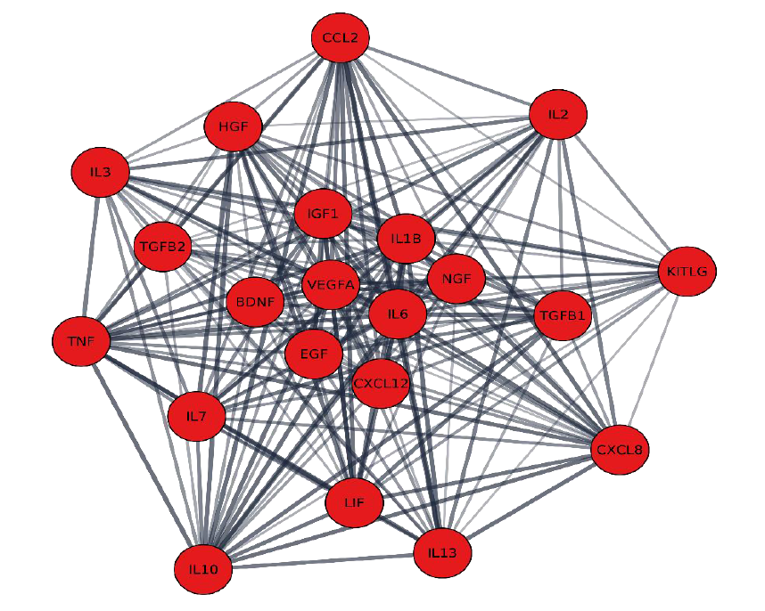
The largest cluster had 21 proteins (Table 2), forming a network of 21 nodes and 187 edges (Figure 2). Molecular function most correlated was that of receptor-ligand activity (GO: 0048018), with an FDR of 1,2E-30.
Binding to cytokine receptors (GO:0005126) presented a relevant FDR of 4.24E-28, added to the fact that 18 of 21 molecules are related to such activity and that these molecules are involved in cytokine metabolic pathways and inflammatory processes present on WikiPathways (WP530 FDR of 1.19E-17), suggests that such proteins may be involved in signaling pathways important to nervous system regeneration. [23-25]
Another important molecular function shared by 16 of the 21 proteins found is their activity as growth factors (GO:0008083), with an FDR of 1.25E-26. Understanding the differences between cytokines and growth factors and their mechanisms of action is relevant for the development of more effective therapies [26]. Cytokines can regulate inflammatory processes, which can lead to neurotoxic processes or even neural cell apoptosis [27]. They can also mobilize glial cell activity as a first response to damage and trauma [27, 28].
In betweenness centrality (BC) and closeness centrality (CC) analyses, the proteins considered most important within the cluster are those whose genes are listed in Table 3. This suggests that these proteins play crucial roles in the protein-protein interaction network , significantly influencing communication and interaction between cluster components [29]:
Vascular Endothelial Growth Factor A (VEGFA) is a proangiogenic molecule that induces vascularization, thus allowing the arrival of nutrients, oxygen and other molecules necessary for regeneration [30, 31]. Molecules from the VEGF family provide conditions for cell survival, explaining their high degree of centrality.
Epidermal Growth Factor (EGF) induces cell proliferation and differentiation with a well-known role in nervous system regeneration, being used in neural regeneration clinical trials [32-34].
Neural Growth Factor (NGF) is a member of the neurotrophin family, with more specific actions on nervous tissue cell proliferation, differentiation, and survival [35]. NGF increases the number of myelinated axons, matured nerves, myelin sheath thickness, fiber diameter, and population density. In the absence of NGF, apoptosis is likely to occur, supporting its role as an inhibitor of programmed cell death [36].
Insulin-Type I Growth Factor (IGF1) is a molecule produced in the liver and stimulated by growth hormones acting on cellular growth and development processes, as well as promoting neurogenesis [37].
Interleukin-6 (IL6) is an inflammatory cytokine that shows ambiguous effects on the nervous system. Despite appearing as a harmful cytokine in nervous development [38], there are studies that suggest a role in differentiation of oligodendrocytes and astrocytes, with potentially neurotrophic action [37].
Brain-Derived Neurotrophic Factor (BDNF), another growth factor of the neurotrophic family, has NGF like properties, sharing with it a genetic homology of 54% [39]. BNDF can help axon functional recovery, promoting survival and differentiation of human neural precursor cells (HNPC) [40].
Interleukin 1-beta (IL1B) is a pro-inflammatory cytokine that has neurodegenerative effects [41], however, in cases of trauma or neurological damage, IL1B can induce reactive astrocytes. Astrocyte reactivity, like the formation of oligodendrocytes and astrocytes induced by IL6, has ambiguous effects, and may promote neuroprotection, synaptic loss, or neurotoxicity [38, 42-44].
Stromal Cell-Derived Factor 1 (SDF-1) encoded by the CXCL12 gene is a small chemokine that acts on leukocyte migration (44), also showing nervous system angiogenic, neurogenic and anti-apoptotic properties. [45].
Interestingly, genes encoding proteins with ambiguous functions in the nervous system were centrally interacting cluster proteins, such as IL6, IL1 and CXCL12.
Cluster 2:
The second most relevant cluster is composed of 6 proteins, which form a network of 6 nodes and 12 edges, containing the proteins listed in Table 4. Based on STRING Enrichment data, biological processes that presented the lowest FDR value showed a negative regulation of neuronal apoptosis (GO:0043524) with an FDR of 2.09E-6 and a negative regulation of apoptotic processes (GO:0043066) with an FDR of 2.38E-5, compatible with pluripotent stem cell differentiation pathway in the WikiPathways database (WP2848) with an FDR of 2.2E-4.
Negative regulation of apoptosis can lead to neurodegenerative processes, which have a negative impact in the nervous system [46]. The protein with the highest degree of network centrality shown in Figure 3 was Glial Cell Lineage-Derived Neurotrophic Factor (GDNF), with a score of BC = 0.0707 and CC = 0.625. GDNF acts as a nerve growth factor, promoting axonal growth and neuron myelination [47, 48]. GDNF can induce neuroprotective effects by preventing astrogliosis harmful effects [49].

Nestin (NES) (BC = 0.0611 and CC = 0.588) can promote neurogenesis, survival, and renewal of neural progenitor cells [50].
The third protein with the highest degree of centrality was Sonic Hedgehog (SHH) with a score of BC = 0.0361 and CC = 0.576. SHH acts as a growth factor in spinal cord development, promoting neurites differentiation and inhibiting astrocyte lineages, preventing pathogenic inflammatory processes [51].
Neurotrophin-3 (NT-3), encoded by the NTF3 gene, belongs to the neurotrophin family (BC = 0.0121 and CC = 0.555). NT-3 has axonal regeneration and neuron myelination action, possibly promoting neuron protection [52, 53]. NT-3 has chemotaxis properties, guiding axon growth to form nerves [54]. Ciliary Neurotrophic Factor (CTNF, BC = 0.0080 and CC = 0.550) increases survival of ganglion neurons, preventing neuron degeneration after axotomy [55]. Furthermore, CTNF can promote axon regeneration by inducing the proliferation of Schwann cells [56]. The protein with the lowest centrality in the cluster was Transforming Growth Factor beta-3 (TGFB3, BC = 0.0016 and CC = 0.530), which has an effect on differentiation and proliferation of the nervous system [57].
Cluster 3:
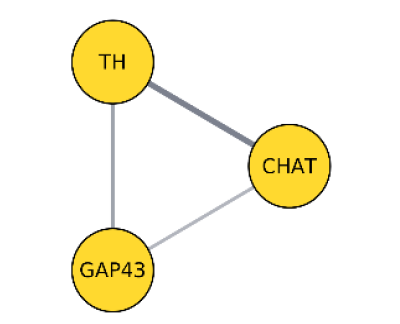
The third and smallest cluster resulted in a network of three nodes and three edges, with the proteins listed in Table 5. STRING Enrichment results indicated the category with the lowest FDR of WikiPathways metabolic pathway, biogenic amine synthesis (WP550), with an FDR of 0.0014 and biological processes from the GO database as being related to neurotransmitter biosynthetic processes (GO:0042136), with an FDR of 0.0308.
For this small network (Figure 4), with only three nodes, the centrality value of each one of them is of little relevance due to high error rates.

Although these are genes involved in neurotransmitter biosynthesis, their direct effect on nervous tissue regeneration is not clearly known. Some studies indicate that neurotransmitters can contribute to developmental processes as signals and regulators of nerve cell growth and differentiation [58-60].
In this work, the construction of protein-protein interaction networks (PPI) proved to be a valuable tool in the analysis nervous system regeneration data, providing centrality data and degree of connection within a complex system. In addition, enrichment tools added a layer of network functionality. Cytoscape demonstrated its effectiveness as a satisfactory platform, and with the availability of applications (Apps), created by other study groups, it may have unforeseen future use. Open Source tools in science is increasingly present and moves a considerable portion of scientific production.
Several network proteins, such as NGF, BDNF, GDNF and NTFs, are already known for their functions in neural regeneration, validating our results. In addition, we also found genes encoding proteins with ambiguous or less known functions in nervous system regenerative processes, such as IL6, IL1B and CXCL12, with a further need to better evaluate their importance in neuroregeneration.
This study proved possible to obtain useful information from noisy data and to interpret it for the benefit of medical treatments.
CONCLUSION
Protein-protein interaction networks (PPI) were used for the analysis of nervous system regeneration, providing information about centrality and protein connections. Cytoscape software and applications confirmed the importance of NGF, BDNF and GDNF in neural regeneration. Ambiguous proteins like IL6, IL1B and CXCL12 need further investigation.
Acknowledgments
Not applicable.
REFERENCES
- 1 Mao AS, Mooney DJ. Regenerative medicine: Current therapies and future directions. Proc Natl Acad Sci 2015; 112: 14452-9.
- 2 Edgar L, Pu T, Porter B, Aziz JM, La Pointe C, Asthana A, et al. Regenerative medicine, organ bioengineering and transplantation. Br J Surg 2020; 107: 793-800.
- 3 Mason C, Dunnill P. A brief definition of regenerative medicine. Regen Med 2008;3:1-5.
- 4 Ryan AR, Cleaver O. Plumbing our organs: Lessons from vascular development to instruct lab generated tissues. pp.165-94.
- 5 Fishman JM, Wiles K, Lowdell MW, De Coppi P, Elliott MJ, Atala A, et al. Airway tissue engineering: an update. Expert Opin Biol Ther 2014;14:1477-91.
- 6 Shin’oka T, Imai Y, Ikada Y. Transplantation of a Tissue-Engineered Pulmonary Artery. N Engl J Med 2001; 344:532-3.
- 7 Cajal SR y, May RM. Cajal’s Degeneration and Regeneration of the Nervous System. Oxford University Press. Epub ahead of print 14 November 1991.
- 8 Niu J. Neurodegeneration and regeneration. J Neurosci Res 2017;95:1525-7.
- 9 Fortin JM, Azari H, Zheng T, Darioosh RP, Schmoll ME, Vedam-Mai V, et al. Transplantation of Defined Populations of Differentiated Human Neural Stem Cell Progeny. Sci Rep 2016;6:23579.
- 10 Zakrzewski W, Dobrzyński M, Szymonowicz M, Rybak Z. Stem cells: past, present, and future. Stem Cell Res Ther 2019; 10: 68.
- 11 Lu P, Jones LL, Snyder EY, Tuszynski MH. Neural stem cells constitutively secrete neurotrophic factors and promote extensive host axonal growth after spinal cord injury. Exp Neurol 2003; 181: 115-29.
- 12 Alamán‐Díez P, García‐Gareta E, Arruebo M, Pérez MÁ. A bone‐on‐a‐chip collagen hydrogel‐based model using pre‐differentiated adipose‐derived stem cells for personalized bone tissue engineering. J Biomed Mater Res Part A 2023; 111: 88-105.
- 13 Shannon P, Markiel A, Ozier O, Baliga NS, Wang JT, Ramage D, et al. Cytoscape: A Software Environment for Integrated Models of Biomolecular Interaction Networks. Genome Res 2003; 13: 2498-504.
- 14 Utriainen M, Morris JH. clusterMaker2: a major update to clusterMaker, a multi-algorithm clustering app for Cytoscape. BMC Bioinformatics 2023;24:134.
- 15 Sensharma P, Madhumathi G, Jayant RD, Jaiswal AK. Biomaterials and cells for neural tissue engineering: Current choices. Mater Sci Eng C 2017;77:1302-15.
- 16 Boni R, Ali A, Shavandi A, Clarkson AN. Current and novel polymeric biomaterials for neural tissue engineering. J Biomed Sci 2018;25:90.
- 17 Hsu CC, Serio A, Amdursky N, Besnard C, Stevens MM. Fabrication of Hemin-Doped Serum Albumin-Based Fibrous Scaffolds for Neural Tissue Engineering Applications. ACS Appl Mater Interfaces 2018; 10: 5305-17.
- 18 Knowlton S, Anand S, Shah T, Tasoglu S. Bioprinting for Neural Tissue Engineering. Trends Neurosci 2018; 41:31-46.
- 19 Zhang X, Jiang W, Xie C, Wu X, Ren Q, Wang F, et al. Msx1+ stem cells recruited by bioactive tissue engineering graft for bone regeneration. Nat Commun 2022;13:5211.
-
20 Casotti MC, Meira DD, Aguiar C de, Duque D de A, Souza K de, Portella LO, et al. [Building protein-protein interaction networks by manual curation]. Braz de Bioinfo Mag, https://bioinfo.com.br/ building-protein-interaction-networks-by-manual-curation (2021, accessed 20 July 2023).
» https://bioinfo.com.br/ building-protein-interaction-networks-by-manual-curation - 21 Bastos LL, Puelles AA, Paixão V, Fioriniet G, Utsch E, Nogueira WG, et al. BIOINFO #02 - Braz J Bioinf Comput Biol. Alfahelix. Epub ahead of print July 2022.
- 22 Bader GD, Hogue CW V. An automated method for finding molecular complexes in large protein interaction networks. BMC Bioinformatics 2003; 4: 2.
- 23 Ochi M, Nakasa T, Kamei G, Usman MA, Mahmoud E. Regenerative medicine in orthopedics using cells, scaffold, and microRNA. J Orthop Sci 2014; 19: 521-8.
- 24 Lin XY, Lai BQ, Zeng X, Che MT, Ling EA, Wu W, et al. Cell Transplantation and Neuroengineering Approach for Spinal Cord Injury Treatment: A Summary of Current Laboratory Findings and Review of Literature. Cell Transplant 2016;25:1425-38.
- 25 Brick RM, Sun AX, Tuan RS. Neurotrophically Induced Mesenchymal Progenitor Cells Derived from Induced Pluripotent Stem Cells Enhance Neuritogenesis via Neurotrophin and Cytokine Production. Stem Cells Transl Med 2018;7:45-58.
- 26 Burgess AW. Growth Factors and Cytokines. In: Reviews in Cell Biology and Molecular Medicine, pp.104-26.
- 27 De Haas AH, van Weering HR, de Jong EK, Boddeke HW, Biber KP. Neuronal Chemokines: Versatile Messengers In Central Nervous System Cell Interaction. Mol Neurobiol 2007;36:137-51.
- 28 Filbin MT. How inflammation promotes regeneration. Nat Neurosci 2006;9:715-7.
- 29 Klein DJ. Centrality measure in graphs. J Math Chem 2010; 47: 1209-23.
- 30 Baffert F, Le T, Sennino B, Thurston G, Kuo CJ, Hu-Lowe D, et al. Cellular changes in normal blood capillaries undergoing regression after inhibition of VEGF signaling. Am J Physiol Circ Physiol 2006;290:H547-H559.
- 31 Yin X, Mead BE, Safaee H, Langer R, Karp JM, Levy O, et al. Engineering Stem Cell Organoids. Cell Stem Cell 2016;18:25-38.
- 32 Xian, Cory J. EGF family of growth factors: essential roles and functional redundancy in the nerve system. Front Biosci 2004;9:85.
- 33 Ninomiya M, Yamashita T, Araki N, Okano H, Sawamoto K. Enhanced neurogenesis in the ischemic striatum following EGF-induced expansion of transit-amplifying cells in the subventricular zone. Neurosci Lett 2006; 403:63-7.
- 34 Sun D, Bullock MR, Altememi N, Zhou Z, Hagood S, Rolfe A, et al. The Effect of Epidermal Growth Factor in the Injured Brain after Trauma in Rats. J Neurotrauma 2010;27:923-38.
- 35 Eddie Ma CH, Palmer A, Taylor JSH. Synergistic effects of osteonectin and NGF in promoting survival and neurite outgrowth of superior cervical ganglion neurons. Brain Res 2009; 1289: 1-13.
- 36 Freeman RS, Burch RL, Crowder RJ, Lomb DJ, Schoell MC, Straub JA, et al. NGF deprivation-induced gene expression: after ten years, where do we stand? pp. 111-126.
- 37 Islam O, Gong X, Rose-John S, Heese K. Interleukin-6 and Neural Stem Cells: More Than Gliogenesis. Mol Biol Cell 2009;20:188-99.
- 38 Teh DBL, Prasad A, Jiang W, Ariffin MZ, Khanna S, Belorkar A, et al. Transcriptome Analysis Reveals Neuroprotective aspects of Human Reactive Astrocytes induced by Interleukin 1β. Sci Rep 2017;7:13988.
- 39 Kingham PJ, Terenghi G. Bioengineered nerve regeneration and muscle reinnervation. J Anat 2006;209:511-26.
- 40 Jiao Y, Palmgren B, Novozhilova E, Englund Johansson U, Spieles-Engemann AL, Kale A, et al. BDNF Increases Survival and Neuronal Differentiation of Human Neural Precursor Cells Cotransplanted with a Nanofiber Gel to the Auditory Nerve in a Rat Model of Neuronal Damage. Biomed Res Int 2014; 2014: 1-11.
- 41 Kazantseva J, Ivanov R, Gasik M, Neuman T, Hussainova I. Graphene-augmented nanofiber scaffolds demonstrate new features in cells behaviour. Sci Rep 2016; 6: 30150.
- 42 Jiao Y, Palmgren B, Novozhilova E, Englund Johansson U, Spieles-Engemann AL, Kale A, et al. Interleukin‐1β induces angiogenesis and innervation in human intervertebral disc degeneration. J Orthop Res 2011; 29: 265-9.
- 43 Gomes FCA, Tortelli VP, Diniz L. Glia: from old concepts to the new functions of today and those to come. Adv Stud. 2013;27:61-84.
- 44 Pekny M, Wilhelmsson U, Pekna M. The dual role of astrocyte activation and reactive gliosis. Neurosci Lett 2014;565:30-8.
- 45 Janssens R, Struyf S, Proost P. The unique structural and functional features of CXCL12. Cell Mol Immunol 2018;15:299-311.
- 46 Yuan J, Yankner BA. Apoptosis in the nervous system. Nature 2000; 407: 802-9.
- 47 Li Q, Ping P, Jiang H, Liu K. Nerve conduit filled with GDNF gene-modified schwann cells enhances regeneration of the peripheral nerve. Microsurgery 2006; 26: 116-21.
- 48 Zhang L, Ma Z, Smith GM, Wen X, Pressman Y, Wood PM, et al. GDNF-enhanced axonal regeneration and myelination following spinal cord injury is mediated by primary effects on neurons. Glia 2009; 57: 1178-91.
- 49 Awad BI, Carmody MA, Steinmetz MP. Potential Role of Growth Factors in the Management of Spinal Cord Injury. World Neurosurg 2015; 83: 120-31.
- 50 Lagace DC, Whitman MC, Noonan MA, Ables JL, DeCarolis NA, Arguello AA, et al. Dynamic Contribution of Nestin-Expressing Stem Cells to Adult Neurogenesis. J Neurosci 2007;27:12623-9.
- 51 Lowry N, Goderie SK, Lederman P, Charniga C, Gooch MR, Gracey KD, et al. The effect of long-term release of Shh from implanted biodegradable microspheres on recovery from spinal cord injury in mice. Biomaterials 2012;33:2892-1.
- 52 Nectow AR, Marra KG, Kaplan DL. Biomaterials for the Development of Peripheral Nerve Guidance Conduits. Tissue Eng Part B Rev 2012;18:40-50.
- 53 Önger ME, Delibaş B, Türkmen AP, Erener E, Altunkaynak BZ, Kaplan S. The role of growth factors in nerve regeneration. Drug Discov Ther 2016;10:285-91.
- 54 Genç B, Ozdinler PH, Mendoza AE, Erzurumlu RS. A Chemoattractant Role for NT-3 in Proprioceptive Axon Guidance. PLoS Biol 2004;2:e403.
- 55 Pasquin S, Sharma M, Gauchat J-F. Ciliary neurotrophic factor (CNTF): New facets of an old molecule for treating neurodegenerative and metabolic syndrome pathologies. Cytokine Growth Factor Rev 2015;26:507-15.
- 56 Ma F, Wang H, Yang X, Wu Y, Liao C, Xie B, et al. Controlled release of ciliary neurotrophic factor from bioactive nerve grafts promotes nerve regeneration in rats with facial nerve injuries. J Biomed Mater Res Part A. 2022;110:788-96.
- 57 Luo SX, Huang EJ. Dopaminergic Neurons and Brain Reward Pathways. Am J Pathol 2016; 186: 478-88.
- 58 Herlenius E, Lagercrantz H. Neurotransmitters and neuromodulators during early human development. Early Hum Dev 2001;65:21-37.
- 59 Nguyen L, Rigo JM, Rocher V, Belachew S, Malgrange B, Rogister B, et al. Neurotransmitters as early signals for central nervous system development. Cell Tissue Res 2001;305:187-202.
- 60 Ruediger T, Bolz J. Neurotransmitters and the Development of Neuronal Circuits BT - Axon Growth and Guidance. In: Bagnard D (ed). New York, NY: Springer New York, pp. 104-14.