 Central Effects of Acute and Sub-Chronic Administration of L-theanine in Animal Model: Behavioral and Biochemical Aspects
Abstract
L-theanine, a naturally occurring amino acid found in green tea (Camellia sinensis), exhibits complex central mechanism of action. This study explored the effects of acute and subchronic oral Ltheanine administration on various behavioral and biochemical parameters in male Swiss mice. Animals received different doses of L-theanine and underwent diverse behavioral tests. Additionally, levels of reduced glutathione (GSH) and malondialdehyde (MDA) were evaluated. Acute L-theanine administration did not alter the locomotor activity but increased immobility time in the tail suspension test (1 mg/kg, p<0.05). L-theanine did not show anxiolytic effects but increased anxiety at high doses (60 mg/kg, p<0.01). L-theanine reduced sleep duration at a high dose (60 mg/kg, p<0.05) but did not affect aversive memory. Subchronic L-theanine administration (30 mg/kg) improved spatial memory (p<0.05) and reversed methylphenidate-induced appetite suppression (p<0.01). Subchronic L-theanine administration increased GSH levels (p<0.05), while reduced MDA in the hippocampus (p<0.01) and in the prefrontal cortex (p<0.05). These findings suggest that L-theanine possess diverse pharmacological properties, including the potential to modulate sleep, enhance cognitive function, and exert antioxidant effects in the brain. Further research is warranted to elucidate the underlying mechanisms of action and explore the therapeutic potential of L-theanine for various neurological conditions.
Keywords:
Camellia sinensis; L-theanine; Central Nervous System; Psychostimulant Activity; Behavior.
HIGHLIGHTS
L-theanine exerts modulator effects on the central nervous system.
L-theanine possesses antioxidant effects in the brain.
L-theanine enhances cognitive responses.
L-theanine does not affect the exploratory activity.
INTRODUCTION
L-theanine (N-ethyl-γ-L-glutamine) is a water-soluble, free, non-proteinogenic amino acid. It constitutes over 50% of the total free amino acids present in the leaves of green and black tea plants (Camellia sinensis) [1]. Green and black teas are widely consumed beverages with traditional uses for treating sleep disorders, depression, and anxiety [2]. The biosynthesis of L-theanine occurs in the roots of the tea plant and involves the enzymatic conversion of glutamic acid and ethylamine by the enzyme theanine synthetase [1].
L-theanine has excitatory and inhibitory functions in the central nervous system (CNS), but there are undefined doses and posology of treatment to achieve these effects [3]. This highlights the need for further research to determine optimal administration strategies. It is also shown that L-theanine increases the expression of GABAergic and serotoninergic receptors and the levels of dopamine, melatonin, serotonin, and antioxidants, improving cognitive functions by directly binding to α-amino-3-hydroxy-5-methyl-4- isoxazolpropionic acid (AMPA), kainate and N-methyl-D-aspartate (NMDA) receptors [4,5]. Additionally, Ltheanine administered orally is rapidly absorbed in the intestine, mainly via sodium-coupled cotransporters in the mucosa of the intestinal margin, and crosses the blood-brain barrier, acting on various tissues and organs, including the brain [1,3].
Despite encouraging preclinical findings, a significant knowledge gap exists regarding the precise behavioral and biochemical effects of L-theanine [1,4,5]. The current understanding of L-theanine's neuropharmacology suggests its potential as a cognitive enhancer and neuroprotective agent within the central nervous system (CNS). This emphasizes the need for further research to elucidate the mechanisms by which L-theanine exerts its effects in the CNS.
The growing need for therapeutic agents that enhance cognitive function and protect the central nervous system (CNS) from oxidative stress prompted this study. While the pharmacological properties of L-theanine are known, significant gaps persist regarding its precise behavioral and biochemical effects under different administration protocols. Our central hypothesis was that L-theanine would exert distinct effects within the CNS dependent on the treatment duration.
Accordingly, the present work aimed to comprehensively evaluate the effects of both acute and subchronic oral L-theanine administration on a wide range of behavioral and biochemical parameters in Swiss mice, including locomotor and exploratory activities, anxietyand depression-like behavior, appetite, aversive and spatial memory, and psychostimulant activity.
The novelty of this study lies in the direct comparison between the effects of acute and subchronic regimens, the extensive battery of behavioral tests and biochemical evaluations conducted, and the analysis of L-theanine's interaction with methylphenidate, providing clarification regarding its potential mechanisms of action which demonstrate doseand duration-dependency.
MATERIAL AND METHODS
Animals and ethical aspects
Swiss male mice (n = 212), body mass between 25 and 35 grams, were obtained from the bioterium of the Christus University (Unichristus) and housed in boxes (41 cm × 34 cm × 16 cm). The mice were maintained under adequate conditions (12/12-hour light/dark cycle at ± 26 ± 2 °C), receiving filtered water and food ad libitum before being evaluated.
The animals were handled according to the ethical principles of animal experimentation by the Ethics and Research Committee for the Use of Animals (CEUA) of Unichristus (protocol n° 017/21 and 010/23) in accordance with the Guide for the Care and Use of Laboratory Animals of the National Institute of Health (NIH). All experiments were also performed according to the Directive 2010/63/EU of the European Parliament and of the Council of 09/22/2010 on the protection of animals used for scientific purposes. Every effort was made to minimize the suffering of the animals.
Drugs
Diazepam and phenobarbital sodium were acquired from Cristália Laboratory (Brazil), imipramine from União Química (Brazil) and methylphenidate from Novartis (Brazil). All other chemicals and reagents were of analytical grade. L-theanine (Excipienta®, China, Lot nº. 20210510) was shown to be a white crystalline powder, easily dissolved in distilled water and insoluble in ethanol, possessing molecular weight of 174.1986 g/mol, density of 0.28 g/mL, molecular formula of C7H14N2O3, pH between 5.0 and 6.0, and high-performance liquid chromatography (HPLC) content of 100%. All drugs were solubilized in distilled water.
Treatment
Mice were divided into groups of 6 - 10 animals in a randomized and double-blinded manner and treated per oral (p.o.) with L-theanine (LT) at 1, 30, 60, 90, 120 mg/kg or associated with methylphenidate (2.5 mg/kg) by two different protocols: acute regimen (only one administration); or subchronic regimen (single daily doses for 14 days). The control group received distilled water, which was also used as a vehicle for L-theanine dilution. In addition, the animals received by intraperitoneal route (i.p) diazepam (1-10 mg/kg), imipramine (10 mg/kg) or methylphenidate (2.5 mg/kg) as reference drugs.
Behavioral tests were performed 60 min after the administration of L-theanine or the reference drugs. At the end of the tests, the brains were removed, and the prefrontal cortex (PFC) and hippocampus (HC) were evaluated for the oxidative stress markers: reduced glutathione (GSH) and malondialdehyde (MDA).
Behavioral evaluation
The acute treatment with LT was evaluated for the following behaviors: anxiety (hole board), depressor (tail suspension), hypnosis/sedation (phenobarbital sleep induction), exploratory (open field) and aversive memory (active avoidance). The subchronic treatment with LT was evaluated for the following behaviors: exploratory (open field), spatial memory (Y-maze) and psychostimulant (appetite suppression).
During the behavioral tests, the devices were sanitized with 5% alcohol solution to minimize the effects of the odors produced by the animals previously tested.
Assessment of anxiolytic and antidepressant activity
Hole board test
To assess the anxiety behavior, the animals were placed at a suspended plate (15 cm height, area of 20 × 20 cm containing 16 evenly spaced holes) for analysis of the following parameters: number of insertions of the animals head in the holes (headdips) and the time of permanence of the head in the holes during five minutes of observation, without environmentalization [6].
Tail suspension test
This test was performed to assess the depressive activity. For this, the animals were suspended and fastened by adhesive tape at approximately 1 cm from the tip of the tail on a platform positioned 50 cm above the bench for five minutes. Mice were tested individually for the immobility time (seconds), being considered immobile when they remained passively suspended [7].
Assessment of hypnotic-sedative activity by the phenobarbital sleep induction test
The animals received phenobarbital (50 mg/kg i.p.) as a sleep-inducing 60 minutes after the treatment and were individually observed for sleep latency and sleep time. Sleep latency corresponds to the interval between phenobarbital administration and loss of the righting reflex. Sleep time corresponds to the interval between the loss and recovery of the rightening reflex, for a period up to three hours. Loss of the righting reflex is the inability of the animal to return to its normal position when placed in the supine position. The criterion for reflex recovery was determined when the animal lives the position three times in a row [8].
Assessment of locomotor and exploratory activities by the open field test
The test was performed in a squared fenced arena (40 × 60 × 50 cm), made of acrylic, with a floor divided into nine equal quadrants for evaluation during five minutes, after one min adaptation of the parameters: number of crossings, rearings and groomings [9].
Assessment of aversive and spatial memories
Active avoidance test
To assess the aversive memory, the animals were placed in the apparatus, consisting of a chamber with two compartments composed of metals and, between these compartments, a statically opened door. For the animals adaptation, a training course was carried out 24 hours before the test. Initially, the animals received a sound stimulus for five seconds followed by constant shocks] at 0.2 mA on the paws. The sound stimulus and shocks ceased as the animal crossed from one compartment to another. Five cycles (with intervals of 60 seconds) of stimuli were applied. The results were expressed as escape latency, i.e., the time that the animals took to change compartments [10].
Y-maze test
To assess spatial memory (learning and occurrence of repetitive/restrictive behaviors), the animals were placed individually in the center of a Y-shaped labyrinth (composed by three gray and opaque "arms" of 10 × 50 cm, positioned at a 120° angle of separation between) for 8 minutes. The number of entries and the sequence of entries in each "arm" were recorded, and the number of sequences in hits and missed entries made by the animals were analyzed. An entry was considered when the four limbs of the animal were in the "arm" of the labyrinth [11].
Assessment of psychostimulant activity by the appetite suppression test
The animals were placed in individual cages and deprived of food for 24 hours. After 30 minutes of treatment, the animals received a previously weighed portion of food. At 60 and 300 minutes after receiving the food, the consumption was evaluated by reweighing the food. The difference between the first and second weights was determined as the amount of food consumed [12].
Evaluation of oxidative stress markers
Malondialdehyde
Lipid peroxidation in the PFC and HC was evaluated by spectrophotometry measuring the thiobarbituric reactive substances (TBA). The tissue homogenate was mixed with 2 mL (1:1) of TBA-TCAHCl reagent (0.37% TBA, 15% TCA and 0.25 N HCl) and incubated at 95 °C for 30 minutes. The reading was performed at 535 nm, and the values expressed as MDA/g of total tissue [13].
Reduced-Glutathione
Trichloroacetic acid 50% (TCA) were added to the homogenate samples (PFC and HC), stirred and centrifuged (5,000 rpm/15 min/4°C). Hydroxymethyl buffer (Tris), 0.4 M HCl (pH 8.9) and 0.01 M DTNB were added, and the absorbance was measured at 412 nm. The GSH concentration was expressed in μmol/mL [14]
Statistical analysis
Results are presented as mean ± standard error of the mean (SEM). Data were analyzed using the Shapiro-Wilk test for normality. Parametric data were then analyzed by ANOVA followed by Tukey's test, and nonparametric data were analyzed by ANOVA followed by Dunn's test. Values of p < 0.05 were considered significant. Statistical analyses were performed using GraphPad Prism version 8.0.1 software.
RESULTS AND DISCUSSION
Acute effect of L-theanine on anxiety and depressive behaviors
Analysis of anxiety-like behavior using the hole board test revealed that LT did not significantly alter the number of head dips at any dose tested compared to the control group (data not shown). However, LT administration did reduce the time spent with the head in the holes at the 1 mg/kg dose (15.5 ± 2.0 vs. control: 25.5 ± 2.5 s). While both 30 mg/kg (16.7 ± 2.4 s) and 60 mg/kg (17.0 ± 3.0 s) doses of LT further decreased this parameter, these reductions were not statistically significant compared to the control group. In contrast, diazepam at 1 mg/kg significantly increased the time spent with the head in the holes (46.1 ± 2.1 s vs. control) (Figure 1A).

Acute effect of L-theanine on the anxiety and depressive behaviors. (A) Time spent in the hole of the plate by the animal's head in the hole board test. (B) Immobility time in the tail suspension test. Control = distilled water; DZP = diazepam 1 mg/kg, i.p.; IMI = imipramine 10 mg/kg, i.p.; LT = L-theanine 1, 30 and 60 mg/kg p.o. The tests were performed 60 min after drugs administration. Mean ± SEM, n = 8 - 10. ANOVA followed by Tukey as post hoc test. *p<0.05 and ****p<0.0001 vs. control.
Analysis of depressive-like behavior in the tail suspension test revealed that LT) administration increased the immobility time at doses of 1 mg/kg (152.0 ± 10.6 s vs. control: 95.5 ± 12.5 s) and 60 mg/kg (137.4 ± 13.9 s vs. control). In contrast, imipramine (35.1 ± 9.0 s vs. control) significantly reduced immobility time (Figure 1B).
The evaluation of the effect of LT, administered in the acute regimen, in the tail suspension test demonstrated that LT (1 mg/kg, p.o.) increases the immobility time of the animals, indicating a depressant effect on the CNS. However, the literature shows that the treatment of rodents with LT (4 mg/kg, p.o./day for six weeks) has an antidepressant effect [15]. Another study, in which LT (1, 4 and 20 mg/kg, p.o.) was administered in a subchronic regimen for ten days, also revealed antidepressant effect [16]. These contradictory effects of LT may be explained by the differences in the doses administered, as well as by the duration of its use. In addition, the unpredictable chronic stress and tail suspension tests can be used to evaluate different aspects of the depressive behavior. Accordingly, the tail suspension test evaluates behaviors of despair and helplessness in a short period of time [7], while the test of chronic unpredictable stress evaluates continuous stressors over time related to chronic depression and underlying mechanisms [17]. However, the results obtained in our study in the acute treatment with LT, indicating lack of antidepressant effect, do not exclude the possibility of an antidepressant effect of LT in chronic depression, which may be explained by the antidepressant effects of LT in subchronic and chronic forms reported previously [15, 16].
Depression is also closely related to anxiety. It is common for depressed patients to have generalized anxiety and vice versa. This is due to the pathophysiological mechanisms of depression and anxiety are related; for example, a reduction in serotonin is among the causes of depression and anxiety [18]. In this context, the evaluation of LT in the anxiety test, demonstrated that LT (1, 30 and 60 mg/kg, p.o.) given in acute treatment regimen does not show anxiolytic effect; in contrast, it showed increased anxiety (1mg/kg). Unlike our results, using higher doses and chronic treatment, a clinical trial conducted in humans demonstrated reduced signs of anxiety after treatment with LT (200 mg/kg p.o., daily, for two months) [19].
The antidepressant and anxiolytic effects of LT can be assigned to its antagonism with glutamatergic receptors, including AMPA and NMDA [20, 3], although it is already known that LT is a partial antagonist of N-methyl-d-aspartate (NMDA) receptors in brain regions such as hippocampus [21, 22]. Thus, it is understandable that when LT is used at low doses in acute treatment regimen, as in our study, this amino acid exerts agonist effects on receptors, since partial agonist drugs present this duality; but at higher doses, they act as antagonists. These pleiotropic effects had been already observed for LT [23].
Acute effect of L-theanine on the phenobarbital sleep induction test
L-theanine (LT) administration in the acute regimen tended to increase sleep latency at the highest dose tested (60 mg/kg; 123.9 ± 14.8 vs. control: 71.6 ± 7.6 min). However, this increase was not statistically significant (p = 0.5602) (Figure 2A). In contrast, the reference drug diazepam significantly decreased sleep latency. On the other hand, L-theanine at 60 mg/kg significantly reduced sleep time compared to the control group (26.4 ± 7.3 vs. control: 85.2 ± 12.3 min) (Figure 2B).

Acute effect of L-theanine on hypnosis/sedation behavior in the phenobarbital sleep induction test. (A) Sleep latency and (B) sleep time. Control = distilled water; DZP = diazepam 10 mg/kg, i.p.; LT = L-theanine 1, 30 and 60 mg/kg p.o. The drugs were administered 60 minutes before sleep induction with phenobarbital sodium (50 mg/kg, i.p.). Mean ± SEM (n = 10). ANOVA followed by Dunn’s as post hoc test. *p<0.05 vs. control.
Our results also showed that the acute treatment regimen with LT (1, 30 and 60 mg/kg, p.o.) did not show hypnotic effect. However, at 60 mg/kg decreased the sleep duration of the animals. In a recent study in which it was analyzed the effect of LT alone and in combination with GABA, the administration of LT alone at 40 mg/kg did not increase either the onset time or the total sleep time induced by pentobarbital in mice. These data demonstrate the complex profile of this molecule, since in the same study at lower doses (20 mg/kg, p.o.), the authors observed decreased sleep latency and increased sleep time in the animals [24].
It is important to highlight that hypnotic mechanisms can be ascribed to the increase in α brain waves and the activation of the GABAergic system with consequent inhibitory effect on the CNS. In this line, it has been reported that the use of LT increased both the brain α waves and the expression of GABAergic receptors, being the hypnotic effect associated to this mechanism [4]. In view of this, we suggest that for the hypnotic effect to occur via GABA, continuous stimulation is necessary, i.e., the continuous use (chronic or subchronic) of LT. In fact, the literature shows that the chronic use of LT (200 mg/day, p.o. for four weeks) in humans reduces sleep latency and increases sleep time compared to placebo, indicating an improvement in the sleep quality [25].
Therefore, we suggest that LT has effects on the sleep quality depending on the duration of its use. It is important to highlight that the modulation of glutamatergic receptors is related to sleep quality, when activated, they mediate stimulating effects reducing sleep quality, and when inactivated, they mediate the improvement in the sleep quality. From this perspective, the distinct effects of LT on sleep, depending on the duration of its use, can be attributed to its chemical structure, which resembles glutamate. Thus, to explain this hypnotic effect of LT, we refer to the fact that this amino acid is a partial agonist, and in acute use, it can bind to and activate glutamate receptors and in chronic use, act as an antagonist inactivating these receptors [20, 3].
These variable outcomes across different doses and administration regimens highlight the complex pharmacological profile of L-theanine and necessitate cautious interpretation. As discussed, factors such as treatment duration, dose-dependent effects, and potential partial agonist activity at glutamatergic receptors may contribute to these differing behavioral responses.
Effect of L-theanine treatment on exploratory behavior
Acute administration of L-theanine (LT) per oral (1, 30, and 60 mg/kg) did not significantly alter the number of crossings in the open field test (Figure 3A). Similarly, no significant changes were observed in grooming behavior at any dose tested (Figure 3C). However, L-theanine at doses of 30 mg/kg (13.7 ± 2.9 vs. control: 34.3 ± 1.9) and 60 mg/kg (13.7 ± 2.5 vs. control) significantly increased the number of rearing events compared to the control group (Figure 3B). These findings suggest that acute L-theanine administration may enhance specific aspects of exploratory behavior in mice.

Effect of L-theanine on the exploratory behavior in the Open field test. (A-C) Acute regimen (LT 1, 30, 60 mg/kg): (A) crossing, (B) rearing, (C) grooming. Mean ± SEM (n = 10). ANOVA and Tukey’s test. *p<0.05, ***p<0.001, ****p<0.0001 vs. control. Control = distilled water; DZP = diazepam 2 mg/kg, i.p. (D-F) Chronic regimen (LT 30 mg/kg and LT 1 mg/kg + MPD 2,5 mg/kg p.o): (D) crossing, (E) rearing, (F) grooming. Mean ± SEM (n = 7). ANOVA and Dunn’s test. p<0.05. *vs. control; §vs. MPD and #vs. LT. MPD = methylphenidate 2.5 mg/kg, i.p. The drugs were administered 60 min before the tests.
L-theanine administered in a subchronic regimen (30 mg/kg) did not significantly alter any of the measured parameters in the open field test. Interestingly, co-administration of a low dose of L-theanine (1 mg/kg) with methylphenidate (2.5 mg/kg) significantly increased the number of crossings compared to the control group (81.4 ± 9.0 vs. control: 42.8 ± 5.4), the LT 30 mg/kg group (81.4 ± 9.0 vs. 39.4 ± 3.9), and the MPD group alone (81.4 ± 9.0 vs. 40.2 ± 3.4) (Figure 3D). Similarly, the combination of MPD and LT significantly increased the number of rearing events compared to the control group (17.5 ± 1.4 vs. 5.8 ± 1.1) and the LT 30 mg/kg group (17.5 ± 1.4 vs. 6.4 ± 2.0) (Figure 3E). No significant differences were observed in grooming behavior between any of the groups (Figure 3F). These results suggest that co-administration of L-theanine with methylphenidate may have synergistic effects on exploratory behavior in mice
A possible explanation may be the complementarity of the mechanism of action, methylphenidate acts primarily as a dopamine and noradrenaline reuptake inhibitor via the increase in availability of these neurotransmitters at the synapse. On the other hand, LT can modulate neurotransmission, increasing the release of dopamine and serotonin [26, 27].
Effect of L-theanine acute treatment on aversive memory
Administration of L-theanine (LT) per oral (1, 30, and 60 mg/kg) did not significantly alter escape latency in the active avoidance test compared to the control group. Escape latencies were 6.6 ± 1.6 s (1 mg/kg), 4.5 ± 1.0 s (30 mg/kg), and 4.3 ± 1.1 s (60 mg/kg) compared to the control group (4.4 ± 1.2 s). Diazepam, the reference drug, significantly increased escape latency (13.4 ± 3.0 s) compared to the control group (Figure 4A). These results suggest that L-theanine, at the doses tested, does not affect aversive memory in mice.

Effect of L-theanine on aversive and spatial memory. (A) Active avoidance test (escape latency) - Acute regimen. Control = distilled water; DZP = diazepam 2 mg/kg, i.p.; MPD = methylphenidate 2.5 mg/kg, i.p.; LT = L-theanine 1 - 60 mg/kg p.o. The tests were performed 60 min after a single dose of treatment (n = 10). (B) Y maze (number of hits) - subchronic regimen. LT (30 mg/kg, p.o); LT (1 mg/kg, p.o) + MPD (2.5mg/kg, i.p.). The tests were performed 60 min after the last dose of treatment. Mean ± SEM (n = 6). ANOVA and Tukey’s test. p<0.05. *vs. control. §vs. MPD.
Importantly, there are several types of memories, including the aversive memory, which consists of stimulus conditioning memory, and spatial memory, involving the encoding and retrieval of information on the environment and spatial orientation. These types of memories are crucial for the long-term preservation of information and are closely involved in the processes of memory consolidation. In our study, when aversive memory was assessed, we demonstrated that the acute treatment with LT (1, 30, or 60 mg/kg, p.o.) did not differ from the control, indicating that it does not improve aversive memory.
Effect of L-theanine subchronic treatment on spatial memory
L-theanine administered in a subchronic regimen (30 mg/kg) significantly increased the number of entries into the novel arm of the Y-maze test compared to the control group (20.3 ± 1.4 vs. 12.6 ± 1.1, respectively). Methylphenidate, used as a reference drug, did not significantly alter the number of novel arm entries (14.1 ± 1.3 vs. control). Interestingly, co-administration of MPD (1 mg/kg) with LT (1 mg/kg) further increased the number of novel arm entries compared to both the control group (21.6 ± 0.8 vs. 12.6) and the MPD group alone (21.6 ± 0.8 vs. 14.1 ± 1.3), suggesting a potential synergistic effect (Figure 4B).
The mechanisms involved in the beneficial action of LT on memory can be attributed to its ability to inactivate kinase/p38 and nuclear factor kappa-B and reduce redox imbalance. In addition, LT increases dopamine release [20, 28]. In this sense, dopaminergic elevation in the hippocampus results in activation of the cAMP/PKA pathway, which increases the activation of newly formed neuronal assemblies to allow memory consolidation. In addition, dopamine stimulated synthesis of brain-derived neurotrophic factor (BDNF), which is also related to memory consolidation [29].
Effect of L-theanine acute treatment on food intake
L-theanine (LT) administered in an acute regimen via oral gavage (1, 30, 60 mg/kg) significantly increased food intake at doses of 60 (1.7 ± 0.01 g), 90 (1.9 ± 0.02 g), and 120 mg/kg (2.0 ± 0.02 g) compared to the control group (1.2 ± 0.01 g). In contrast, methylphenidate (MPD), the reference drug, significantly reduced food intake to 0.7 ± 0.01 g compared to the control group (Figure 5A).

Effect of L-theanine on the appetite suppression test. (A) Acute regimen: MPD 2.5 mg/kg, i.p; LT 60 to 120 mg/kg, p.o. Control = distilled water, MPD = methylphenidate, LT = L-theanine. Mean ± SEM (n= 10). (B) Subchronic regimen: LT (30 mg/kg, p.o); LT (1 mg/kg, p.o.) + MPD (2.5 mg/kg, i.p.). Mean ± SEM (n= 6-7). The food intake was measured 300 min later the last treatment. ANOVA and Tukey’s test, p<0.05. *vs. control and §vs. MPD.
Effect of the association of L-theanine and MPD on appetite suppression
To investigate the potential synergistic effects of L-theanine and MPD, a low dose of L-theanine (1 mg/kg) was co-administered with MPD (2.5 mg/kg) and compared to L-theanine alone (30 mg/kg). The results demonstrated that both L-theanine (30 mg/kg) and the combination of MPD (2.5 mg/kg, i.p.) with L-theanine (1 mg/kg, p.o.) (LT+MPD) significantly increased food intake compared to MPD (2.5 mg/kg) (LT: 0.56 ± 0.14 g and LT+MPD: 0.52 ± 0.07 g vs. control: 0.87 ± 0.04 g) (Figure 5B). This suggests that even at low dose, L-theanine can modulate the appetite-suppressive effects of MPD.
As already mentioned, L-theanine can influence the dopaminergic system in the brain, which has implications in the mood, cognition, and potentially appetite. Studies have shown that L-theanine increases dopamine levels in specific brain regions, such as the striatum, which is involved in reward and motivation. This effect is significant, since dopamine plays a key role in the brain's reward system, influencing behaviors related to pleasure and motivation, including eating. Thus, L-theanine potentially may enhance feelings of well-being and reduce stress via modulation of dopamine levels, which might indirectly affect appetite. Increased dopamine can sometimes lead to an increased desire for food, particularly in situations where eating is associated with reward or stress relief [30, 31].
Effect of L-theanine acute treatment on oxidative stress markers: MDA and GSH
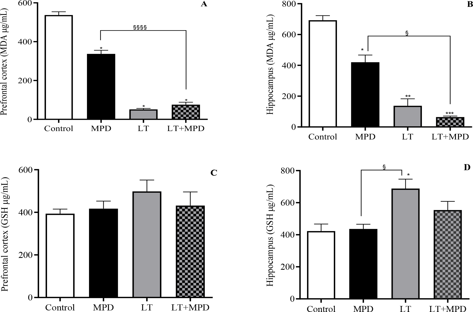
Administration of LT at 30 mg/kg did not alter MDA or GSH concentrations in the prefrontal cortex or hippocampus compared to the control group (data not shown). Subchronic regimen. MDA production was significantly decreased in the prefrontal cortex of the MPD group compared to the control group (337.3 ± 18.6 vs. 537.4 ± 17.1). However, the group receiving LT at 30 mg/kg exhibited a marked reduction in MDA even when compared to the MPD group (37.7 ± 4.9 vs. 337.3 ± 18.6). This effect was also observed when LT was used at a dose of 1 mg/kg in combination with MPD (45.7 ± 12.3 vs. 337.3 ± 18.6). (Figure 6A).

Effect of L-theanine on oxidative stress markers: malondialdehyde (MDA) and glutathione (GSH). Control = distilled water. MPD = methylphenidate (2.5 mg/kg, i.p.). LT = L-theanine (30 mg/kg, p.o.) or LT (1 mg/kg) when co-administered with MPD. (A/B) MDA: (A) Prefrontal cortex; (B) Hippocampus. (C/D) GSH: (C) Prefrontal cortex; (D) Hippocampus. ANOVA and Dunn’s or Tukey’s test. p<0.05. *vs. control, §vs. MPD.
The same pattern of effect for these drugs observed in the prefrontal cortex was also observed in the hippocampus, where the MPD-treated group had a significant reduction compared to the control (420.4 ± 46.3 vs. 692.9 ± 30.1). The LT 30 mg/kg-treated group still had a more pronounced reduction compared to MPD (137.7 ± 45.5 vs. 420.4 ± 46.3), and the group that received LT 1 mg/kg in combination with MPD maintained this low level of MDA, which was not different from the group that received LT 30 mg/kg. (Figure 6B).
With respect to GSH, LT 30 mg/kg increased GSH levels in the hippocampus (688.4 ± 58.3 vs. control: 422.7 ± 44.4 mg/ml) (Figure 6D). The combination of LT 1 mg/kg with MPD was not significantly different from the LT 30 mg/kg group, demonstrating that low doses of LT can still sustain higher GSH levels. In contrast, there was no difference in GSH levels in the prefrontal cortex among the groups evaluated (Figure 6C).
The oxidative stress is related to the initiation and development of a variety of CNS disorders, including changes in memory, appetite, depression, anxiety, and sleep. In this sense, the evaluation of LT administration on the levels of GSH and MDA, which are biomarkers of oxidative stress [31, 32], showed that the acute treatment with LT (1, 30 and 60 mg/kg, p.o.) did not alter MDA or GSH, indicating lack of antioxidant effect. However, the subchronic administration of LT (30 mg/kg, p.o.) increased GSH in the hippocampus, an antioxidant that reduces and activates glutathione peroxidase, allowing it to convert H2O2 into water [33].
In addition, the hippocampus has the main function to store recent memory, long-term memory formation, and spatial orientation. Thus, we suggest that subchronic treatment with LT (30 mg/kg, p.o.) has a neuroprotective effect, being related to memory improvement. In addition, LT (30 mg/kg, p.o.) given sub chronically reduced the concentration of MDA in the hippocampus and prefrontal cortex. MDA is an oxidizing agent and one of the end products of lipid peroxidation [32]. Therefore, we confirmed the antioxidant effect of LT and its relation with memory improvement. Similarly, one study demonstrated that the administration of LT (4 mg/kg/day for fourteen days) by subchronic regimen in male mice elevated GSH in astrocytes [3]. In addition, the literature describes the increase in GSH to the prevention of dopamine-induced neuronal cell death and the formation of quinoproteins, indicating that dopamine-quinone-induced dopaminergic neuronal cell damage can be prevented by GSH via quinone-inhibiting activity [3].
Our study, contributed to a better understanding of the effects of LT on the CNS, as demonstrated its beneficial effects, showing to be dependent on the dose and duration of treatment. Such contribution is in line with the absence of adverse effects after per oral administration at daily doses greater than 2,000 mg/kg, demonstrated previously in a clinical trial [33].
We emphasize that this research has some limitations, such as, the animal model used to study the effects of LT may not be entirely representative on how this substance works in humans, which may compromise our results. In addition, animal models exhibit varied responses based on genetic and environmental factors that may impact the reproducibility of results. Although diverse, the number of doses and duration of treatment do not yet cover all possible clinical therapeutic regimens. Despite the adequate number of behavioral tests applied in this study, it may have some bias, as the stress caused by the tests, as well the analysis of only two oxidative stress markers.
LIMITATIONS AND FUTURE PERSPECTIVES
Further research is necessary, being essential to carry out clinical studies to confirm the findings in animal models for better translation to medical practice. Studies assessing different doses and treatment duration could also clarify the efficacy of this substance for the treatment of specific CNS disorders, as well as the dose-response effect of this substance.
To fully characterize the behavioral profile of L-theanine and further investigate the doseand duration-dependent effects and observed inconsistencies, comprehensive dose-response studies and investigations employing extended observation periods are important considerations for future research.
A limitation of the present study is the absence of direct experimental measurements of the molecular pathways potentially involved in L-theanine's central effects. While our behavioral and biochemical findings, coupled with existing literature, suggest the involvement of GABAergic, glutamatergic, dopaminergic, and BDNF-related mechanisms, these pathways were not directly validated in this study. Consequently, the proposed involvement of these specific molecular targets remains putative and requires empirical confirmation through targeted molecular assays in future investigations. To provide a more complete mechanistic understanding, future research should extend beyond oxidative stress biomarkers to include a broader range of biochemical and molecular analyses.
While the present study focused on the fundamental preclinical characterization of L-theanine's central effects, the observed outcomes related to cognitive function, oxidative stress modulation, and behavioral aspects provide a foundation for future translational research. Exploring the potential therapeutic applications of L-theanine for specific neurological conditions, as well as investigating optimized formulation approaches or drug development pathways, represents a critical next step to enhance the clinical and technological relevance of these findings.
CONCLUSION
Acute treatment of L-theanine in mice decreased sleep duration and increased food intake, while subchronic treatment improved spatial memory and antioxidant activity in the hippocampus and prefrontal cortex, effects that were shown to be closely related to the dose and treatment duration. Notably, the association L-theanine and methylphenidate exerts modulator effect on appetite.
Funding:
Acknowledgments:
The authors acknowledge the Conselho Nacional de Desenvolvimento Científico e Tecnológico (CNPq), Fundação Cearense de Apoio ao Desenvolvimento Científico e Tecnológico (FUNCAP), and Christus University (Unichristus) for institutional support and for providing postgraduate fellowships to the students involved in this study. G.F.A (CNPq Research Productivity Fellowship, Process No. 305714/2022-8) and A.M.S.A (CNPq Research Productivity Fellowship, Process No. 308433/2017-3) are CNPq research productivity fellows.
Data Availability Statement:
Data are available on reasonable request for corresponding author.
Institutional Review Board Statement:REFERENCES
-
1 Chen S, Kang J, Zhu H, Wang K, Han Z, Wang L, et al. L-Theanine and immunity: a review. Molecules. 2023;28(9):3846. doi: 10.3390/molecules28093846.
» https://doi.org/10.3390/molecules28093846. -
2 Hayat K, Iqbal H, Malik U, Bilal U, Mushtaq S. Tea and its consumption: benefits and risks. Crit Rev Food Sci Nutr. 2015;55(7):939-54. doi: 10.1080/10408398.2012.678949.
» https://doi.org/10.1080/10408398.2012.678949. -
3 Takeshima M, Miyazaki I, Murakami S, Kita T, Asanuma M. L-theanine protects against excess dopamine-induced neurotoxicity in the presence of astrocytes. J Clin Biochem Nutr. 2016;59(2):93-9. doi: 10.3164/jcbn.16-15.
» https://doi.org/10.3164/jcbn.16-15. -
4 Dasdelen MF, Er S, Kaplan B, Celik S, Beker MC, Orhan C, et al. A novel theanine complex, Mg-L-theanine improves sleep quality via regulating brain electrochemical activity. Front Nutr. 2022;9:874254. doi: 10.3389/fnut.2022.874254.
» https://doi.org/10.3389/fnut.2022.874254. -
5 Ogawa S, Ota M, Ogura J, Kato K, Kunugi H. Effects of L-theanine on anxiety-like behavior, cerebrospinal fluid amino acid profile, and hippocampal activity in Wistar Kyoto rats. Psychopharmacology. 2018;235(1):37-45. doi: 10.1007/s00213-017-4743-1.
» https://doi.org/10.1007/s00213-017-4743-1. - 6 Clark G, Koster AG, Person DW. Principles of psychopharmacology. 2nd ed. New York: Academic Press; 1971. p. 169-71.
-
7 Steru L, Chermat R, Thierry B, Simon P. Tail suspension test: a new method for screening antidepressant in mice. Psychopharmacology. 1985;85(83):367-70. doi: 10.1007/BF00428203.
» https://doi.org/10.1007/BF00428203. - 8 Carlini EA. Practical pharmacology without equipment. São Paulo: Sarvier; 1973. p. 198.
-
9 Archer J. Tests for emotionality in rats and mice: a review. Anim Behav. 1973;21(2):205-35. doi: 10.1016/s0003-3472(73)80065-x.
» https://doi.org/10.1016/s0003-3472(73)80065-x. -
10 Banasr M, Chowdhury GM, Terwilliger R, Newton SS, Duman RS, Behar KL, et al. Glial pathology in an animal model of depression: reversal of stress-induced cellular, metabolic and behavioral deficits by the glutamate-modulating drug riluzole. Mol Psychiatry. 2010;15(5):501-11. doi: 10.1038/mp.2008.106.
» https://doi.org/10.1038/mp.2008.106. -
11 Li B, Arime Y, Hall FS, Uhl GR, Sora I. Impaired spatial working memory and decreased frontal cortex BDNF protein level in dopamine transporter knockout mice. Eur J Pharmacol. 2010;628(1-3):104-7. doi: 10.1016/j.ejphar.2009.11.036.
» https://doi.org/10.1016/j.ejphar.2009.11.036. -
12 Zelger JL, Carlini EA. Anorexigenic effects of two amines obtained Catha edulis Forsk. (Khat) in rats. Pharmacol Biochem Behav. 1980;12(5):701-5. doi: 10.1016/0091-3057(80)90152-5.
» https://doi.org/10.1016/0091-3057(80)90152-5. -
13 Nagababu E, Rifkind JM, Sesikeran B, Lakshmaiah N. Assessment of antioxidant activities of eugenol by in vitro and in vivo. Methods Mol Biol. 2011;610:165-80. doi: 10.1007/978-1-60327-029-8_10.
» https://doi.org/10.1007/978-1-60327-029-8_10. -
14 Sedlak J, Lindsay RH. Estimation of total, protein-bound, and nonprotein sulfhydryl groups in tissue with Ellman’s reagent. Anal Biochem. 1968;25(1):192-205. doi: 10.1016/0003-2697(68)90092-4.
» https://doi.org/10.1016/0003-2697(68)90092-4. -
15 Takeda A, Sakamoto K, Tamano H, Fukura K, Inui N, Suh SW, et al. Facilitated neurogenesis in the developing hippocampus after intake of theanine, an amino acid in tea leaves, and object recognition memory. Cell Mol Neurobiol. 2011;31(7):1079-88. doi: 10.1007/s10571-011-9707-0.
» https://doi.org/10.1007/s10571-011-9707-0. -
16 Yin C, Gou L, Liu Y, Yin X, Zhang L, Jia G, et al. Antidepressant-like effects of L-theanine in the forced swim and tail suspension tests in mice. Phytother Res. 2011;25(11):1636-9. doi: 10.1002/ptr.3456
» https://doi.org/10.1002/ptr.3456 -
17 Levinstein MR, Samuels BA. Mechanisms underlying the antidepressant response and treatment resistance. Front Behav Neurosci. 2014;8:208. doi: 10.3389/fnbeh.2014.00208.
» https://doi.org/10.3389/fnbeh.2014.00208. -
18 Lima CLS, Lira SM, Holanda MO. Physiological and pharmacological bases of anxiety disorder. RSD. 2020; 9(9):e808997780. doi: 10.33448/rsd-v9i9.7780.
» https://doi.org/10.33448/rsd-v9i9.7780. -
19 Rizzo R, Prato A, Scerbo M, Saia F, Barone R, Curatolo P. Use of nutritional supplements based on L-theanine and vitamin B6 in children with Tourette syndrome, with anxiety disorders: a pilot study. Nutrients. 2022;14(4):852. doi: 10.3390/nu14040852.
» https://doi.org/10.3390/nu14040852. - 20 Nathan PJ, Lu K, Gray M, Oliver C. The neuropharmacology of L-theanine (N-ethyl-L-glutamine): a possible neuroprotective and cognitive enhancing agent. J Herb Pharmacother. 2006;6(2):21-30. PMID: 17182482
-
21 Sebih F, Rousset M, Bellahouel S, Rolland M, de Ferreira MCJ, Guiramand J, et al. Characterization of L-theanine excitatory actions on hippocampal neurons: toward the generation of novel N-methyl-D-aspartate receptor modulators based on its backbone. ACS Chem Neurosci. 2017;8:1724-34. doi: 10.1021/acschemneuro.7b00036
» https://doi.org/10.1021/acschemneuro.7b00036 -
22 Wakabayashi C, Numakawa T, Ninomiya M, Chiba S, Kunugi H. Behavioral and molecular evidence for psychotropic effects in L-theanine. Psychopharmacology. 2012;219:1099-109. doi: 10.1007/s00213-011-2440-z.
» https://doi.org/10.1007/s00213-011-2440-z. - 23 Calvey N, Williams N. Drug action. Principles and practice of pharmacology for anaesthetists. 5th ed. Oxford: Wiley-Blackwell; 2008. p. 62. ISBN 978-1-405-19484-6.
-
24 Kim S, Jo K, Hong KB, Han SH, Suh HJ. GABA and L-theanine mixture decreases sleep latency and improves NREM sleep. Pharm Biol. 2019;57(1):65-73. doi: 10.1080/13880209.2018.1557698.
» https://doi.org/10.1080/13880209.2018.1557698. -
25 Hidese S, Ogawa S, Ota M, Ishida I, Yasukawa Z, Ozeki M, et al. Effects of l-theanine administration on stress-related symptoms and cognitive functions in healthy adults: a randomized controlled trial. Nutrients. 2019;11(10):2362. doi: 10.3390/nu11102362.
» https://doi.org/10.3390/nu11102362. -
26 Kakuda T. Neuroprotective effects of theanine and its preventive effects on cognitive dysfunction. Pharmacol Res. 2011;64(2):162-68. doi: 10.1016/j.phrs.2011.03.010.
» https://doi.org/10.1016/j.phrs.2011.03.010. -
27 Volkow ND, Wang GJ, Fowler JS, Telang F, Maynard L, Logan J, et al. Evidence that methylphenidate enhances the saliency of a mathematical task by increasing dopamine in the human brain. Am J Psychiatry. 2004;161(7):1173-80. doi: 10.1176/appi.ajp.161.7.1173.
» https://doi.org/10.1176/appi.ajp.161.7.1173. -
28 Saeed M, Khan MS, Kamboh AA, Alagawany M, Khafaga AF, Noreldin AE, et al. L-theanine: an astounding sui generis amino acid in poultry nutrition. Poult Sci. 2020;99(1):5625-36. doi: 10.1016/j.psj.2020.07.016.
» https://doi.org/10.1016/j.psj.2020.07.016. -
29 Zhu BT. Mechanistic explanation for the unique pharmacologic properties of receptor partial agonists. Biomed Pharmacother. 2005;59(3):76-89. doi: 10.1016/j.biopha.2005.01.010.
» https://doi.org/10.1016/j.biopha.2005.01.010. -
30 Yamada T, Terashima T, Kawano S, Furuno R, Okubo T, Juneja LR, et al. Theanine, gamma-glutamylethylamide, a unique amino acid in tea leaves, modulates neurotransmitter concentrations in the brain striatum interstitium in conscious rats. Amino Acids. 2009;36(1):21-7. doi: 10.1007/s00726-007-0020-7.
» https://doi.org/10.1007/s00726-007-0020-7. -
31 Delgobo M, Agnes JP, Santos VW, Parisotto EB, Zamoner A, Filho AZ. N-acetylcysteine and alpha-lipoic acid improve antioxidant defenses and decrease oxidative stress, inflammation and serum lipid levels in ovariectomized rats via estrogen-independent mechanisms. J Nutr Biochem. 2019;67:190-200. doi: 10.1016/j.jnutbio.2019.02.012.
» https://doi.org/10.1016/j.jnutbio.2019.02.012. -
32 Qi JH, Dong FX. The relevant targets of anti-oxidative stress: a review. J Drug Target. 2021;29(7):677-86. doi: 10.1080/1061186X.2020.1870987.
» https://doi.org/10.1080/1061186X.2020.1870987. -
33 Rao TP, Ozeki M, Juneja LR. In search of a safe natural sleep aid. J Am Coll Nutr. 2015;34(5):436-47. doi: 10.1080/07315724.2014.926153
» https://doi.org/10.1080/07315724.2014.926153