Cryopreservation toxicity and morphological outcomes in Piaractus brachypomus oocytes and embryos
Abstract
This study evaluated the toxicity of cryoprotectants and the performance of low-rate freezing protocols for oocytes and embryos of Piaractus brachypomus, a Neotropical fish of increasing relevant for Brazilian aquaculture and genetic conservation. Eight cryoprotectant solutions based on methanol (MeOH) or dimethyl sulfoxide (Me2SO), combined with 0.25 M sucrose in L-15 or HBSS media, were tested for oocytes toxicity at 28 °C and post-freezing viability. In spite of histological and scanning electron microscopy (SEM) analyses indicated preservation of gross morphological features, none of the cryopreserved oocytes supported embryonic development, indicating loss of functional viability following cryoprotectant exposure and freezing. For embryos, two low-rate freezing protocols were evaluated: Protocol 1 (P1E), based on gradual cooling to −13 °C, and Protocol 2 (P2E), involving linear cooling to −60 °C followed by storage in liquid nitrogen. In P1E, eight treatments using 3.1 M MeOH combined with different concentrations of polyvinylpyrrolidone (PVP) or sucrose were tested. The highest proportion of morphologically viable embryos (15.3%) was obtained with MeOT + 0.45 M sucrose (SC5), which different significantly from the other treatments. In contrast, no morphologically viable embryos were recovered after P2E, likely due to inadequate dehydration and intracellular ice formation. Overall, high concentrations of permeant cryoprotectants and prolonged equilibration times were detrimental to oocyte and embryo viability, while sucrose showed better cryoprotective performance than PVP. Even though protocols tested were insufficient to ensure consistent post-thaw viability, the partial success observed in P1E under MeOH and sucrose combinations provides a relevant experimental basis for future refinement of conservation strategies and contributes to development of ex situ germoplasm conservation approaches for P. brachypomus and other Neotropical species.
Keywords:
embryo chilling sensitivity; cryopreservation toxicity; Low-rate freezzig; neotropical freshwater fish
Introduction
Cryopreservation of fish gametes and embryos is a strategic tool for breeding programs and the conservation of genetic resources, particularly for species at risk of extinction. However, embryo cryopreservation in fish remains a significant challenge due to the large size, high water content, low membrane permeability, and extreme sensitivity to low temperatures exhibited by these cells (Zhang et al. 2006; Cabrita et al., 2006; Diwan et al., 2020). While significant challenges persist in fish embryo cryopreservation, emerging techniques have shown potential progress, exemplified by the partial success in zebrafish (Danio rerio) embryo cryopreservation combined with laser nanowarming (Khosla et al., 2020). Building on these advancements, oocyte cryopreservation has gained attention as a promising complementary approach, due to distinct cellular characteristics that may facilitate better preservation outcomes.
In this context, oocyte cryopreservation has been regarded as a promising alternative since the early 2000s for the preservation of the maternal genome, as oocytes present smaller cell volume and higher permeability compared to embryos (Isayeva et al., 2004). Despite the full viability of this technique still faces practical challenges, especially in non-model species, important progress has been made over the past decades, consolidating this approach as a relevant line of research in the fields of genetic conservation and aquaculture.
Studies using zebrafish have been conducted to advance the technique, with relevant outcomes reported for ovarian follicles (Tsai et al., 2009; Marques et al., 2015), ovarian tissue (Marques et al., 2019; Freitas et al., 2023), and oocytes at different developmental stages (Zampolla et al., 2009; Silva et al., 2018).
In neotropical species, most research has focused on Piaractus mesopotamicus, emphasizing embryo cryopreservation (Streit et al., 2007; Fornari et al., 2012, 2014; Neves et al., 2014; Lopes et al., 2011, 2014). On the other hand, Marques et al. (2018, 2021a) studied ovarian structures, in P. mesopotamicus and Brycon orbignyanus, respectively. Studies on oocyte cryopreservation in native South American species are scarce and, apart from a few works on Steindachneridion parahybae (Lopes et al., 2015, 2018, 2019), significant gaps remain regarding the application of the technique in roundfish species.
Piaractus brachypomus (pirapitinga) is a freshwater fish native to the Amazon and Orinoco River basins, with increasing economic importance in fish farming in Brazil and in several Asian countries (Guimarães and Martins, 2015; Kumar et al., 2018). Despite its productive and genetic relevance, no validated protocols currently exist for the cryopreservation of embryos or oocytes of this species.
To advance the development of such protocols, it is essential to understand the toxicity of cryoprotectants and the chilling sensitivity of oocytes and embryos. Compounds such as methanol (MeOT) and dimethyl sulfoxide (Me2SO) have been reported as effective due to their high permeability and low toxicity (Zhang et al., 2003, 2005; Seki et al., 2007, 2011). Additionally, the inclusion of non-permeating cryoprotectants like sucrose has shown protective effects (Xiao et al., 2008; Godoy et al., 2013; Marques et al., 2015).
Therefore, this study aimed to evaluate the toxic effects of cryoprotectants (MeOH and Me2SO combined with sucrose) on P. brachypomus oocytes and to test different cryopreservation protocols for embryos, with the goal of contributing to the ex situ genetic conservation of neotropical fish species.
Methods
Experiment side and animals
The study was conducted in a fish farm located in Rondônia, Brazil (11°41’46 0.95”S and 61°13’47 0.50”O). The broodstock fish used in the research were maintained in two ponds 800 m2 each, with a stocking density of 228 g of fish/m2. Five males and five females were used to obtain gametes, the reproduction method was induced spawning using carp pituitary extract (CPE), the average weight of male broodstock fish was 2.7 ± 1.2 and of female broodstock fish was 4.2 ± 0.9 kg. Fish were fed once daily 1% of body weight) a commercial diet with 32% crude protein and 3100 kcal digestible energy. Water quality parameters, mean temperature (29 ± 1 °C) and dissolved oxygen (5 mg/L ± 1.1), were measured daily.
Ethics statement
All experimental procedures used in this study follow the rules edited by the “National Council for Animal Experimentation Control” of the Brazilian Ministry of Science, Technology and Innovation. Project approved by the local “Ethics Committee on Animal Use” provided by Brazilian legislation (number 172/2012).
Oocyte collection
Four females (two for experiment 1 and two for experiment 2) were induced according to the recommendations of Vásquez-Torres (2005) to obtain oocytes. After 10 h of the second hormonal dose (290-310 Accumulated Thermal Unit - ATU) at 28 ± 2 °C, the females were manipulated and the oocytes were obtained through abdominal massage. Immediately after obtaining the oocytes, a pool was produced to carry out the tests described below.
Design experiment 1. Toxicity and cryopreservation of P. brachypomus oocytes
Toxicity test of cryoprotectants on oocytes at room temperature (28 ± 2 °C)
One thousand and eighty oocytes (with eccentric nucleus, suitable for fertilization) were randomly separated (Figure 1 - steps 1 and 2). The number of oocytes per treatment was defined based on logistical constraints related to the induced spawning condition in the field and the evaluation of a significant number of samples per treatment to ensure homogeneity of the developmental stage. Oocytes were randomly distributed into nine groups, which were eight treatments and one control. The control treatment was composed of oocytes not exposed to any medium and cryoprotectants. Only oocytes that presented macroscopic characteristics compatible with fertilization (eccentric nucleus) were selected. The experiment was repeated three times and 90 oocytes were used in each treatment (Table 1; Figure 1 – step 3a).
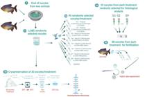
Infographic summarizing the development of the design of cryoprotectant toxicity tests and cryopreservation of Piaractus brachypomus oocytes.
Cryoprotective solutions for Piaractus brachypomus oocytes for the toxicity test. Methanol (MeOH), Dimethyl sulfoxide (Me2SO), Sucrose (SUC), Leibovitz medium (L-15) and Hanks balanced salt solution (HBSS).
To test the toxicity of cryoprotectant solutions, we used the equilibration time of 20 min (28 ± 2 °C) adapted from Plachinta et al. (2004), for embryos of native species (P. mesopotamicus) according to Streit (2005).
From the initial pool of 90 oocytes per treatment, 10 oocytes per each treatment were allocated for histological analysis (Figure 1 – step 4a). The remaining 80 oocytes from each treatment were fertilized with 30 μL of fresh milt obtained from a single male (Figure 1 – step 4b).
The hormonal induction protocol for semen collection followed the recommendations of Vásquez-Torres (2005). A volume of 0.1 mL of milt was collected for subjective evaluation of sperm motility and concentration. After checking for contamination according to Marques et al. (2021b), a sample of activated spermatozoa (2 μL) was obtained by diluting 20 μL of semen in 400 μL of distilled water and placing it on a slide for optical microscopy. It was then covered with a coverslip and immediately evaluated for sperm motility under optical microscopy at 400x magnification, with motility classified from 0 to 100%. To estimate sperm concentration, the samples were fixed in 10% buffered formaldehyde solution at a 1:1000 dilution (1 μL of semen: 999 μL of formaldehyde solution). An aliquot (10 μL) of the diluted semen was pipetted into each counting field of a Neubauer chamber covered with a coverslip, and allowed 15 min for cell stabilization. Using a microscope with 400x magnification and a manual counter, gametes were quantified by counting 10 squares. After cell counting, sperm concentration was calculated using the following equation (Sanches et al., 2011):
where: Spermatozoa mL−1: Number of spermatozoa per milliliter of sperm; ΣSPTZ: Total number of spermatozoa counted; 10 s.c: Squares counted; 25 t.s: Total squares; Dilution: Factor of dilution of the sperm by the fixative; Chamber depth: Normality 0.1 mm.
Based on the estimated motility (96%) and sperm concentration (17X109 sperm/mL), a ratio of 120,000 sperm/oocytes, the selected oocytes were fertilized with 2.9 μL milt. The eggs were placed in 5 L conical incubator (28 ± 0.2 °C) for embryonic development until the stage selected for cryopreservation tests with embryos. Sperm motility was subjectively assessed with values of 96% and 17 × 109 spermatozoa/mL, respectively. To ensure oocyte fertilization, a sperm:oocyte ratio of 120,000:1 was used according to Leite et al. (2013). In vitro incubation lasted 16 ± 0.5 h.
Slow rate freezing of oocytes
Thirty oocytes were selected based on macroscopic characteristics consistent with the eccentric nucleus stage, as previously reported from each treatment were exposed for 20 min to the cryoprotectant solutions (Table 1) and then placed into 0.5 mL straws (minitube for semen) (Figure 1 – step 3b). The cryopreservation curve used in the assays followed the protocol by Zhang et al. (1993). Samples, initially at room temperature of 29 ± 1 °C, were cooled at 1 °C/min using a controlled rate freezer (Dominium K, Biocom, Brazil) down to −60 °C, enucleation (moment that passes from liquid to solid state, ice) was performed at −5 at −7 °C, and then transferred to liquid nitrogen. After one month of storage in liquid nitrogen, the straws were heated in a water bath at 30 °C for 15 s, and the samples were then sent for scanning electron microscopy (SEM) analysis.
The following experiments are summarized in Figure 1.
Design experiment 2. Cryopreservation of P. brachypomus embryos
In the second experiment, from the two selected females induced for reproduction, as described in Oocyte collection, a total of 5000 oocytes were fertilized using semen from a second male specimen. The semen collection methodology, as well as the evaluation of motility and obtaining the sperm concentration, followed the methodology described in Experiment 1 - Cryoprotectant toxicity test in oocytes at room temperature (28 ± 2 °C). However, for this male, the motility obtained was 94% with a concentration of 18x109 sperm/mL, using the same insemination dose of 120,000:1 sperm:oocyte. After the fertilization process, the eggs were distributed in a 5 L conical incubator (28 ± 0.2 °C) for embryonic development until the stage selected for cryopreservation tests.
Slow rate freezing Protocol 1 for embryos (P1E)
Following incubation of 6 h at 28 ± 0.2 °C, 1,800 P. brachypomus viable embryos at 90% epiboly were selected by stereomicroscope (Table 2). Slow rate freezing of embryos was performed according to the cooling curve suggested by Lopes et al. (2011). Embryos were cooled from 28 ± 0.2 °C to 0 °C, keeping the temperature constant for 10 min every 10 °C drop. When the temperature reached 0 °C, the embryos were placed in a freezer at −13 °C for 8 h (Figure 2 – step 1). After the freezer storage period, the embryos were reheated, following the reverse of the cooling curve applied to the embryos, to 28 °C. Thus, the embryos, still at the 90% epiboly stage, were incubated for 8 ± 1 h until the larvae hatched at a temperature of 28 ± 0.2 °C in a 5 L conical incubator. Embryonic viability and morphological evaluation were performed at the end of organogenesis, pre-hatching of embryos, as morphologically described for P. brachypomus by Díaz-Olarte et al. (2010). Different embryonic stages (other than a differentiated pre-hatching embryo) have been classified as non-viable (Figure 2 – step 2a). From the 90% epiboly embryo samples that underwent the previously described heating curve, twenty embryos were sent for histological analysis (Figure 3 – step 2b). The experiment was repeated three times, and 200 embryos were used in each treatment, using 0.5 mL straws for filling and subsequent cooling.
Cryoprotective solutions used in cryopreservation tests of Piaractus brachypomus embryos. Methanol (MeOH), polivinilpirrolidona (PVP), sucrose (SUC) and Hanks balanced salt solution (HBSS).

Infographic summarizing the development of the design of cryopreservation of Piaractus brachypomus embryos.
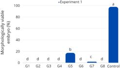
Rate morphologically viable embryo after hatching during 16 ± 0.5 hpf in Experiment 1 - Piaractus brachypomus oocytes exposed to the cryoprotectant solutions at room temperature (28 ± 2 °C). Bars with different superscript letters present statistical difference in significance level of 5% (P < 0.05).
Slow rate freezing Protocol 2 for embryos (P2E)
P. brachypomus viable embryos, in tailbud stage (bud), were selected using the stereomicroscope and distributed into five treatments, which were four treatments and one control (Table 3). Embryos in experimental treatments were exposed for 20 min to the cryoprotectant solutions. Control treatment, embryos not exposed to cryoprotectants, was kept in HBSS. Afterward, embryos were loaded into 0.5 mL straws. Straws were placed into a controlled rate freezer (Dominium K, Biocom, Brazil) and cooled at 1 °C/min down to −60 °C, seeding was performed at −7 °C, and then immersed in liquid nitrogen. Samples were stored for approximately 30 days in liquid nitrogen (Figure 2 – step 3a). All treatments were cultured in a incubator at 28 ± 0.2 °C until hatching. Eight embryos from each treatment were selected and heated in a water bath at 30 °C for 15 s, followed by histological processing of the samples (Figure 2 – step 3b). The experiment was repeated three times, testing a total of 24 embryos in each treatment.
Composition of cryoprotectant solutions for Piaractus brachypomus embryos used in Protocol 2 (P2E). Methanol (MeOH), sucrose (SUC) and Hanks balanced salt solution (HBSS).
The following experiments are summarized in Figure 2.
Histological analysis
The selected samples in both experiments (1 and 2) were fixed with glutaraldehyde (2.5% solution in 0.1 M sodium phosphate buffer, pH 7.2). For processing, the samples were dehydrated in a series of alcohol solutions (70 to 100%) and embedded in resin blocks. The blocks were cut into 2 µm thick sections using a rotary microtome (Leica RM2245, Germany), and the slides were stained with hematoxylin-phloxine (Tolosa et al., 2003). Images were captured using a light microscope (Olympus BX 41) coupled to a high-resolution camera using the Q Capture Pro 51 software.
Scanning Electron Microscopy (SEM)
For SEM analysis, samples containing 10 oocytes/treatment were fixed with glutaraldehyde (2.5% solution in 0.1 M sodium phosphate buffer, pH 7.2). Histological processing was the same as described above. For SEM analysis, samples were washed and post-fixed in osmium tetroxide 1% for 4 h. Then, samples were rewashed in buffer and subjected to dehydration in an ascending ethanol series. Drying was done with liquid CO2 followed by a coating of gold-palladium. Oocytes were examined and electronically micrographed by Scanning Electron Microscope JSM - 5410 (JEOL, USA).
Statistical analysis
When working on oocytes and freezing embryos at −13 °C, the statistical differences between treatments, using model methodology generalized linear. To this end, estimating the probabilities of embryos developed for each treatment and comparing them, through the chi-square implemented in SAS Proc GENMOD. In the estimation process, the logit link function was used considering that the residues presented binomial distribution.
In the work on freezing embryos in liquid nitrogen, it was estimated that relative frequency of embryos preserved after thawing.
Results
Experiment 1. Toxicity and cryopreservation of P. brachypomus oocytes
Toxicity test of cryoprotectants on oocytes at room temperature (28 ± 2 °C)
The cryoprotectant solutions were toxic when the oocytes are exposed at 28 ± 2 °C. Embryo hatching rate were null in most treatments, except in G5 and G7, which showed survival rates of 17.3% and 1.8% respectively, after 16 ± 0.5 h of incubation (hpf). However, both were significantly lower compared to the control (97.7%) (P < 0.05; Figure 3).
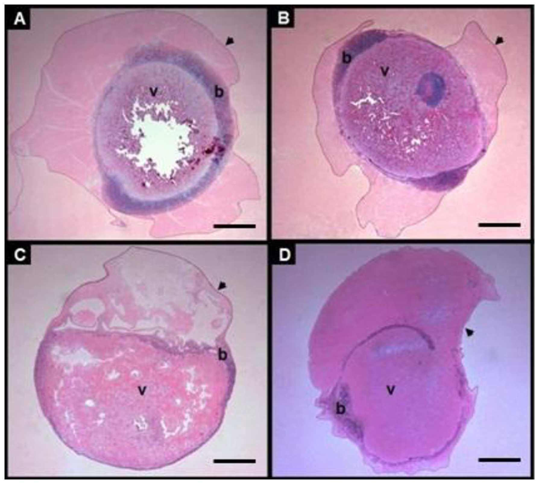
After the toxicity test, histological sections at low magnification suggested preservation of gross, without allowing detailed assessment of subcellular organization (Figure 4A). Figure 4B shows a morphologically developed embryo (pre-hatching), whereas Figure 4C presents a non-viable embryo (90% stage of epiboly), obtained after exposure of oocytes to cryoprotective solutions at room temperature (28 ± 2 °C). Histological observations revealed qualitative morphological alterations in non-viable embryos revealed blastoderm disorganization, irregular yolk morphology and alterations in chorionic integrity.

Histological analysis (hematoxylin-phloxine) of Piaractus brachypomus oocytes after toxicity test, 6 hpf (A). Developed morphologically embryo pre-hatching the final stage of the segmentation period with a free tail in 15-16 hpf (B) and non-viable embryo (90% stage of epiboly) (C). All the figures correspond following fertilization of Piaractus brachypomus oocytes exposed to the cryoprotectant solutions at room temperature (28 ± 2 °C). Yolk (v), zona radiate (zr), embryo (e), perivitelline space (ep), chorion (c) and blastoderm (b). This figure is in color in the electronic version.
Slow rate freezing of oocytes
Histological (Figures 55B) and scanning electron microscopy (SEM; Figure 5C), these observations allowed for a qualitative assessment of the overall surface morphology of the oocytes. However, they did not provide sufficient resolution to support detailed ultrastructural interpretations.

Histology of Piaractus brachypomus oocytes cryopreserved with 1.6M MeOT + 0.25M sucrose + Hank's solution (T5), cords with hematoxylinfloxin in (A, B) and in SEM (C). Yolk (v) and zona radiata (zr).
Experiment 2. Cryopreservation of P. brachypomus embryos
Slow rate freezing Protocol 1 (P1E) for embryos
Among the treatments, the SC5 result in the highest morphologically viable embryos (15.3%; P < 0.05; Figure 6) after 15-16 hpf. Nonetheless, this rate was significantly lower than the the control (98.7%). There was no difference among treatments SC3 (1.5%), SC6 (4.6%), SC7 (1.8%), and SC8 (1.9%). In treatments SC1, SC2 and SC4, all embryos were non-viable (90% stage of epiboly).

Morphologically viable Piaractus brachypomus larvae rate after 8 h in freezing −13 °C (P1E) from Experiment 2. Bars with different superscript letters present statistical difference in significance level of 5% (P < 0.05).
For histological analysis of embryos at the 90% epiboly stage (the end of gastrulation) after 8 h at −13 °C in Experiment 2, treatments SC4, SC5 and SC8 were selected for histological analysis, along with the control. These treatments were selected to histologically characterize the embryos when subjected to incubation at a temperature of 28 °C to complete the embryonic cycle until larval hatching. Therefore, treatment SC4 represented non-viable embryos; SC5 had the highest viability; SC8 had low viability. Control embryos showed mostly intact morphology, though some exhibited disorganized blastoderm (Figures 77B). In SC5, embryos displayed intact chorion, well defined blastoderm involving the yolk and no visible cell damage (Figurse 7C and 7D). In contrast, SC6 showed disrupted blastoderm and chorion rupture with yolk extrusion (Figures 77F). Regarding the embryos treated as SC4, after warming, they exhibited morphological alterations and did not progress beyond the gastrulation stage, with no evidence of developmental advancement toward organogenesis. (Figures 77H).

Photomicrograph of P. brachypomus embryos in 90% epiboly after 8 h in freezing −13 °C (P1E) from Experiment 2 (A and B) and experimental SC5 (C and D), SC7 (E and F) and SC4 (G and H). Blastoderm (b), yolk (v) and chorion (arrow head). Scale bars: Figures A, C, E, G = 100µm; Figures B, D, F, H = 50µm. This figure is in color in the electronic version.
Slow rate freezing Protocol 2 (P2E) for embryos
There was no morphologically viable embryo after cryopreservation using P2E, wuile in the control group, 98% of the larvae hatched at 16 ± 0.5 hpf. Embryos classified as exhibiting apparent structural integrity immediately after warming were characterized by a well defined blastoderm, intact chorion and uniform and enclosed uniform yolk (Figures 88B). Treatments SCT1 and SCT4 showed a higher proportion of embryos with this apparent structural condition, while no embryos meeting these criteria were observed in treatments SCT2 and SCT3. Embryos from SCT2 and SCT3 exhibited pronounced morphological alterations, including irregularity shape, disorganized blastoderm and fusion of yolk granules (Figures 88D). Although most embryos from all treatments presented an intact chorion, this observation was limited to macroscopic morphological characteristics visible a low magnification. This apparent structural integrity was transient and did not result in progression to subsequent developmental stages.

Photomicrograph of Piaractus brachypomus embryos after one month cryopreservation (P2E) from Experiment 2, illustrating general morphological characteristics observed at low magnification. Images A and B show embryos with apparent structural integrity immediately after thawing, characterized by a well defined blastoderm (b), intact chorion (arrow head) and uniform yolk (v). Images C and D show embryos with pronounced morphological alterations, including blastoderm disorganization and yolk granule fusion. Scale bars: 200µm. This figure is in color in the electronic version.
Discussion
In the present study, cryopreservation was applied at the end of the gastrulation phase and no morphologically viable embryos were observed after thawing in any of the treatment tested. In spite of treatments SCT1 and SCT4 showed a higher proportion of embryos with apparent structural integrity immediately after thawing, this condition did not translate into functional viability or progression to subsequent development stages. In contrast, embryos from treatments SCT2 and SCT3 consistently showed pronounced morphological alterations, including blastoderm disorganization and yolk granule fusion. These observations indicate that macroscopic morphological appearance immediately after thawing is not a guarantee of embryonic developmental competence.
Although apparent morphological integrity of P. brachypomus oocytes was observed after cryoprotectant in some treatments, preservation of structure alone does not necessarily reflect normal hydration dynamics, membrane permeability or osmotic functionality. In the presente study, the hydration capacity of the oocytes was not directly measured, since the experimental design focused on evaluating the toxicity of the cryoprotectant and structural integrity and not on osmotic functionality. Similar response patterns have been reported for other Neotropical species, in which embryos maintained apparent morphological integrity after cooling or freezing, but failed to develop or hatch. For example, in Prochilodus lineatus, moderate cooling (−8 °C to −10 °C) resulted in hatching rates of up to 25-30%, while deep freezing resulted in complete loss of hatching (Ninhaus-Silveira et al., 2009; Machado et al., 2019). Similarly, Piaractus mesopotamicus embryos of showed partial hatching success (≈64%) after 12 h of controlled cooling at −8 °C (Fornari et al., 2012), while more advanced developmental stages suffered irreversible damage from cryopreservation (Lopes et al., 2011). These findings reinforce the idea that morphological preservation does not necessarily indicate biological functionality and highlight that post-thaw hatching are more reliable predictive indicator.
Histological and scanning electron microscopy analyses provided qualitative information on the overall morphological condition of oocytes and embryos after cryopreservation. At the magnification used, these techniques allowed visualization of general structural characteristics, such as blastoderm organization, chorion continuity and yolk morphology, but did not allow a detailed assessment of cellular or subcellular integrity. Therefore, the observations obtained from histological sections and SEM images in this study should be interpreted as illustrative of general morphological outcomes and not as evidence of preservation of membrane functionality or ultrastructural stability.
Even though cell morphology appeared intact, the cryoprotective solutions used, based exclusively on MeOH or Me2SO as permeable cryoprotective agents, proved inadequate to support oocyte development. Notably, treatments containing high concentrations of MeOH (3.1 M) or Me2SO (1.3 M) did not result in morphologically viable embryos, suggesting cytotoxic effects at these levels, as reported in previous studies. The combination of high cryoprotectant concentration with prolonged equilibration time, as applied in this study, appears particularly detrimental to oocytes physiology. A similar finding was reported by Zampolla et al. (2009), who observed that MeOH concentrations between 3 and 4 mol/L led to reduced mitochondrial distribution in zebrafish oocytes. Similarly, Me2SO at concentrations of 2-4 mol/L caused immediate ATP depletion and impaired mitochondrial DNA maintenance, reinforcing the observed loss of viability.
Previous studies with zebrafish ovarian follicles showed that medium containing 50% L-15 was more effective than HBSS or KCl buffer in preserving cellular integrity after exposure to subzero temperatures (Zampolla et al., 2008; Tsai et al., 2009). In contrast, in the present study, the HBSS medium proved more suitable than the medium containing 50% L-15 for maintaining the viability of P. brachypomus oocytes. A possible explanation for this difference lies in the osmotic characteristics of the media.
Despite the trials were conducted independently, the current findings are consistent with those reported by Ahammad et al. (2003), who observed that embryos at more advanced developmental stages are more sensitive to cryopreservation than those at intermediate stages. In the present study, embryos subjected to Protocol 2 (P2E) were rendered nonviable after warming, likely due to insufficient dehydration and intracellular ice formation.
This result may be attributed, at least in part, to insufficient dehydration associated with the faster cooling rate applied in P2E, in compounded by the cryoprotectant solution used. Effective cryopreservation depends on the ability of the cell to lose water before intracellular freezing occurs. Optimal cooling rates are strongly influenced by cellular properties, as described by Mazur (1984) and Fuller (2004), including cell volume, membrane surface area and permeability to water and cryoprotectants. Fish embryos, characterized by large size, substantial yolk content and inherently low membrane permeability, are particularly susceptible to incomplete dehydration during rapid cooling. Under these conditions intracellular ice formation, is more likely to occur, representing one of the main causes of cryoinjury and post-thaw viability loss (Zhang and Rawson, 1995; Diwan et al., 2020).
The gradual cooling approach employed in Protocol 1 (P1E), with a 10-min equilibration period for each 10 °C reduction in temperature, was designed to facilitate the diffusion of the cryoprotectants into the cells and promote intracellular water efflux, thus reducing the risk of intracellular ice formation. However, the proportion of morphologically viable embryos after thawing remained low. This limited success may be related to the exclusive use of MeOH as a cryoprotectant, which appears insufficient to provide adequate protection under the tested conditions, a results comparable to the low viability observed in oocyte cryopreservation protocols based solely on MeOH.
Among the treatments tested in P1E, the SC5 cryoprotectant solution was the most effective among those evaluated, yielding 15.3% morphologically viable embryos after thawing. Notably, all solutions containing sucrose resulted in some degree of embryonic survival, whereas those incorporating polyvinylpyrrolidone (PVP) failed to produce any viable embryos. This observation align with previous findings by Dumoulin et al. (1994), who reported that PVP is less effective in preventing cryoinjury compared to other macromolecules. Although polymer-based cryoprotectants can enhance cell survival in certain contexts, PVP specifically has been linked to reduced post-thaw viability. In contrast, the combination of MeOH and sucrose has shown favorable results in other fish species, including Cyprinus carpio (Ahammad et al., 2003), zebrafish (Kopeika et al., 2006) and Prochilodus lineatus (Machado et al., 2019).
Sucrose contributes to cryoprotection by promoting osmotic dehydration and cell shrinkage, thereby minimizing the formation of intracellular ice crystals (Diwan et al., 2020). Methanol, due to its low molecular weight (32 g/mol), exhibits high membrane permeability. Because membrane transport and osmoregulation are central factors in cryodamage in embryonic cells, the combined use of MeOH and sucrose has been proposed as a suitable cryoprotective strategy for fish embryos, which are characterized by multiple compartments, high yolk content and inherently low membrane permeability.
Cell survival during cryopreservation is closely associated with membrane integrity, as the plasma membrane acts as a semipermeable barrier between the cytoplasm and the external environment. In the present study, frozen embryos may have experienced membrane-related damage resulting from the osmotic stress and uncontrolled water influx during the cooling process, as described for other biological system (Muldrew and Mcgann, 1994). However, it is important to emphasize that such damage could no be directly assessed by the histological and scanning electron microscopy (SEM) analyses performed. Membrane susceptibility to cryoinjury is also influenced by lipid composition, which affects key biochemical and biophysical properties, including resistance to thermal stress (Hayward et al., 2014). The presence of lipids, both as components of membrane architecture and as intracellular lipid droplets, has been identified as a major limiting factor for cryopreservation efficiency in several species, including fish (Isayeva et al., 2004). Even though, SEM images in the present study revealed structural disorganization in the cryopreserved embryos, particularly in the blastoderm region, these observations should be interpreted as qualitative indicators of morphological alteration rather than confirmation of specific molecular or cytoskeletal damage.
Intracytoplasmic lipid droplets are know to interact with intermediate filaments from the cytoskeleton (Okada et al., 2014) and such interaction have been associated with the maintenance of cell morphology (Bodin et al., 2005). Previous studies suggest that structural alterations in these domains can modify the physicochemical properties of the intracellular environment and disruption cytoskeleton organization (Isachenko et al., 2001), potentially leading to chromosomal abnormalities and defective cytokinesis, (Eroglu et al., 1998). In addition to structural injury, cryoprotectants themselves may alter gene expression patterns in embryos and larvae, as reported by (Zhang et al., 2021). These mechanisms were not directly evaluated in the present study and are discussed here as plausible explanations based on established cryobiological literature.
Notwithstanding previous studies have reported that P. brachypomus embryos can be preserved under cold storage condictions, such as at −10 °C for 6 h, 8 or 13 h post-fertilization (Pessoa et al., 2015). These protocols involved substantially less severe thermal exposure than the freezing conditions evaluated in the present study. Similar autcomes under moderate cooling have also reported for Rhinelepis aspera, P. mesopotamicus and Salminus brasiliensis using comparable cryoprotectants (Fornari et al., 2012, 2013, 2014). In contrast, the present study focused on more rigorous cryopreservation protocols intended for long-term storage, which expose embryos to greater osmotic and thermal stress. This methodological gap highlights the technical and biological challenges associated with transitioning from short-term cooling to effective cryogenic preservation, particularly in Neotropical fish species.
Conclusion
The cryopreservation protocols tested for P. brachypomus oocytes and embryos demonstrated limited efficacy, as no successful post-thaw fertilization was achieved and embryo survival rates remained low. Among the treatments evaluated, the protocol combining MeOH and sucrose (SC5), when applied to embryos, yielded the highest proportion of morphologically viable embryos (15.3%), suggesting potential for further refinement. The data highlight the cytotoxic effects of high concentrations of cryoprotectants and the structural challenges imposed by fish embryos. Despite these limitations, the partial success observed provides a valuable foundation for refining cryopreservation strategies and advancing germplasm conservation efforts in Neotropical fish species.
Acknowledgments
The authors would like to thank José Mário Mendez from Piscicultura Buriti and Miyuki Hyashida from Aquicultura São Paulo Farm for providing the facilities to carry out the study. The authors would like to thank Brazilian National Council for Scientific and Technological Development (CNPq – MCTI, Projetos em cooperação, Grant 403157/2024-2), FAPERGS (Programa de Pesquisa e Desenvolvimento voltado a Desastres Climáticos - Grant: 24/2551-0002156-6). J.A. Povh and D.P. Streit Jr. are research fellows of CNPq, Grants 312072/2021-0 and 305387/2022-7 respectively
Data availability statement
Research data is only available upon request.
-
Financial support.
This study was financed in part by the Universidade Federal de Mato Grosso do Sul – UFMS/MEC – Brasil - Finance Code 001. This study was also support of Coordenação de Aperfeiçoamento de Pessoal de Nível Superior (CAPES) [grant number 23038.011255/2021-17 and 88887.877624/2023-00].
-
How to cite:
Digmayer M, Fornari DC, Marques LS, Povh JA, Ribeiro RP, Nicoleti ET, Alencar LQ, Spica LN, Zhang T, Streit DP Jr. Cryopreservation toxicity and morphological outcomes in Piaractus brachypomus oocytes and embryos. Anim Reprod. 2026;23(2):e20250080. https://doi.org/10.1590/1984-3143-AR2025-0080
References
-
Ahammad MM, Bhattacharyya D, Jana BB. Hatching of common carp (Cyprinus carpio L.) embryos stored at 4 and −2 °C in different concentrations of methanol and sucrose. Theriogenology. 2003;60(8):1409-22. https://doi.org/10.1016/S0093-691X(03)00118-3 PMid:14519463.
» https://doi.org/10.1016/S0093-691X(03)00118-3 -
Bodin S, Soulet C, Tronchère H, Sié P, Gachet C, Plantavid M, Payrastre B. Integrin-dependent interaction of lipid rafts with the actin cytoskeleton in activated human platelets. J Cell Sci. 2005;118(Pt 4):759-69. https://doi.org/10.1242/jcs.01648 PMid:15671066.
» https://doi.org/10.1242/jcs.01648 -
Cabrita E, Robles V, Wallace JC, Sarasquete MC, Herráez MP. Preliminary studies on the cryopreservation of gilthead seabream (Sparus aurata) embryos. Aquaculture. 2006;251(2-4):245-55. https://doi.org/10.1016/j.aquaculture.2005.04.077
» https://doi.org/10.1016/j.aquaculture.2005.04.077 -
Díaz-Olarte J, Marciales-Caro LJ, Cristancho Vásquez F, Cruz-Casallas PE. Comparación del desarrollo embrionario de Piaractus brachypomus (Serrasalmidae) y Pseudoplatystoma sp. (Pimelodidae). Int J Morphol. 2010;28(4):1193-204. https://doi.org/10.4067/S0717-95022010000400033
» https://doi.org/10.4067/S0717-95022010000400033 -
Diwan AD, Harke SN, Gopalkrishna, Panche AN. Cryobanking of fish and shellfish egg, embryos and larvae: an overview. Front Mar Sci. 2020;7:251. https://doi.org/10.3389/fmars.2020.00251
» https://doi.org/10.3389/fmars.2020.00251 -
Dumoulin JC, Bergers-Janssen JM, Pieters MH, Enginsu ME, Geraedts JP, Evers JL. The protective effects of polymers in the cryopreservation of human and mouse zonae pellucidae and embryos. Fertil Steril. 1994;62(4):793-8. https://doi.org/10.1016/S0015-0282(16)57006-X PMid:7926089.
» https://doi.org/10.1016/S0015-0282(16)57006-X -
Eroglu A, Toth TL, Toner M. Alterations of the cytoskeleton and polyploidy induced by cryopreservation of metaphase II mouse oocytes. Fertil Steril. 1998;69(5):944-57. https://doi.org/10.1016/S0015-0282(98)00030-2 PMid:9591507.
» https://doi.org/10.1016/S0015-0282(98)00030-2 - Fornari DC, Ribeiro RP, Streit DP Jr, Vargas L, Godoy LC, Oliveira CAL, Digmayer M, Galo JM, Neves PR. Increasing storage capability of pacu (Piaractus mesopotamicus) embryos by chilling: development of a useful methodology for hatcheries management. Cryo Letters. 2012;33(2):126-33. PMid:22576116.
-
Fornari DC, Streit DP Jr, Galo JM, Guerreiro LJ, Povh JA, Barrero-Lopera NM, Oliveira D, Ribeiro RP. Viability of dourado embryos cooled in different cryoprotectant solutions. Pesqui Agropecu Bras. 2013;48(8):1-8. https://doi.org/10.1590/S0100-204X2013000800057
» https://doi.org/10.1590/S0100-204X2013000800057 -
Fornari DC, Ribeiro RP, Streit DP Jr, Godoy LC, Neves PR, Oliveira D, Sirol RN. Effect of cryoprotectants on the survival of cascudo preto (Rhinelepis aspera) embryos stored at −8 °C. Zygote. 2014;22(1):58-63. https://doi.org/10.1017/S0967199411000517 PMid:21867599.
» https://doi.org/10.1017/S0967199411000517 -
Freitas TR, Galuppo AG, Marques LS, Rodrigues RB, Perez-Atehortúa M, França TS, Teixeira NS, dos Santos WV, Gomes IC, Sachett A, Tercya H, Siqueira-Silva DH, Gamba D, Zhang T, Streit DP Jr. Hydrogel encapsulation as a handling and vitrification tool for zebrafish ovarian tissue. Theriogenology. 2023;198:153-63. https://doi.org/10.1016/j.theriogenology.2022.12.019 PMid:36586353.
» https://doi.org/10.1016/j.theriogenology.2022.12.019 - Fuller BJ. Cryoprotectants: the essential antifreezes to protect life in the frozen state. Cryo Letters. 2004;25(6):375-88. PMid:15660165.
-
Godoy LC, Streit DP Jr, Zampolla T, Bos-Mikich A, Zhang T. A study on the vitrification of stage III zebrafish (Danio rerio) ovarian follicles. Cryobiology. 2013;67(3):347-54. https://doi.org/10.1016/j.cryobiol.2013.10.002 PMid:24513460.
» https://doi.org/10.1016/j.cryobiol.2013.10.002 -
Guimarães IG, Martins GP. Nutritional requirement of two Amazonian aquacultured fish species, Colossoma macropomum (Cuvier, 1816) and Piaractus brachypomus (Cuvier, 1818): a mini review. J Appl Ichthyology. 2015;31:57-66. https://doi.org/10.1111/jai.12976
» https://doi.org/10.1111/jai.12976 -
Hayward SA, Manso B, Cossins AR. Molecular basis of chill resistance adaptations in poikilothermic animals. J Exp Biol. 2014;217(Pt 1):6-15. https://doi.org/10.1242/jeb.096537 PMid:24353199.
» https://doi.org/10.1242/jeb.096537 -
Isachenko V, Isachenko E, Michelmann HW, Alabart JL, Vazquez I, Bezugly N, Nawroth F. Lipolysis and ultrastructural changes of intracellular lipid vesicles after cooling of bovine and porcine GV‐oocytes. Anat Histol Embryol. 2001;30(6):333-8. https://doi.org/10.1046/j.1439-0264.2001.00339.x PMid:11820401.
» https://doi.org/10.1046/j.1439-0264.2001.00339.x -
Isayeva A, Zhang T, Rawson DM. Studies on chilling sensitivity of zebrafish (Danio rerio) oocytes. Cryobiology. 2004;49(2):114-22. https://doi.org/10.1016/j.cryobiol.2004.05.005 PMid:15351683.
» https://doi.org/10.1016/j.cryobiol.2004.05.005 -
Khosla K, Kangas J, Liu Y, Zhan L, Daly J, Hagedorn M, Bischof J. Cryopreservation and laser nanowarming of zebrafish embryos followed by hatching and spawning. Adv Biosyst. 2020;4(11):e2000138. https://doi.org/10.1002/adbi.202000138 PMid:32996298.
» https://doi.org/10.1002/adbi.202000138 - Kopeika J, Zhang T, Rawson D. Zebrafish embryos (Danio rerio) using microinjection. Cryo Letters. 2006;27(5):319-28. PMid:17256065.
-
Kumar P, Zampolla T, Zhang T, Rawson DM. Effect of cryoprotectant toxicity on mitochondrial function and distribution in zebrafish (Danio rerio) embryos. Aquaculture. 2018;495:332-40. https://doi.org/10.1016/j.aquaculture.2018.02.052
» https://doi.org/10.1016/j.aquaculture.2018.02.052 -
Leite LV, Melo MAP, Oliveira FCE, Pinheiro JPS, Campello CC, Nunes JF, Salmito-Vanderley CSB. Determinação da dose inseminante e embriogênese na fertilização artificial de tambaqui (Colossoma macropomum). Arq Bras Med Vet Zootec. 2013;65(2):421-9. https://doi.org/10.1590/S0102-09352013000200018
» https://doi.org/10.1590/S0102-09352013000200018 - Lopes TS, Streit DP Jr, Ribeiro RP, Romagosa E. Post-cooling damage to Piaractus mesopotamicus embryos at different stages of development. Atas Saúde Ambient. 2014;2:1-11.
-
Lopes TS, Romagosa E, Streit DP Jr, Ribeiro RP, Digmayer M. Cooling of pacu (Piaractus mesopotamicus) embryos at various stages of development for 6 or 10 hours. Theriogenology. 2011;75(3):570-6. https://doi.org/10.1016/j.theriogenology.2010.09.030 PMid:21074840.
» https://doi.org/10.1016/j.theriogenology.2010.09.030 -
Lopes TS, Sanches EA, Okawara RY, Romagosa E. Chilling of Steindachneridion parahybae (Siluriformes: Pimelodidae) embryos. Theriogenology. 2015;84(4):538-44. https://doi.org/10.1016/j.theriogenology.2015.04.005 PMid:25989879.
» https://doi.org/10.1016/j.theriogenology.2015.04.005 -
Lopes TS, Sanches EA, Caneppele D, Romagosa E. Surubim-do-Paraíba oocytes viability after being exposed to different cryoprotectants. Cienc Rural. 2018;48(6):e20170667. https://doi.org/10.1590/0103-8478cr20170667
» https://doi.org/10.1590/0103-8478cr20170667 -
Lopes TS, Sanches EA, Caneppele D, Silveira MM, Romagosa E. Chilling sensitivity of Steindachneridion parahybae (Siluriformes: Pimelodidae) oocytes in different cryoprotectants. Vet Anim Sci. 2019;7:100046. https://doi.org/10.1016/j.vas.2019.100046 PMid:32734068.
» https://doi.org/10.1016/j.vas.2019.100046 -
Machado GJ, Murgas LDS, Palhares PC, Assis IL, Castro TFD. Cooling of curimba (Prochilodus lineatus) embryos using different concentrations of dimethyl sulphoxide and methanol. Rev Bras Zootec. 2019;48:e20160377. https://doi.org/10.1590/rbz4820160377
» https://doi.org/10.1590/rbz4820160377 -
Marques LS, Bos-Mikich A, Godoy LC, Silva LA, Maschio D, Zhang T, Streit DP Jr. Viability of zebrafish (Danio rerio) ovarian follicles after vitrification in a metal container. Cryobiology. 2015;71(3):367-73. https://doi.org/10.1016/j.cryobiol.2015.09.004 PMid:26408854.
» https://doi.org/10.1016/j.cryobiol.2015.09.004 -
Marques LS, Fossati AAN, Leal MS, Rodrigues RB, Bombardelli RA, Streit DP Jr. Viability assessment of primary growth oocytes following ovarian tissue vitrification of neotropical teleost pacu (Piaractus mesopotamicus). Cryobiology. 2018;82:118-23. https://doi.org/10.1016/j.cryobiol.2018.03.009 PMid:29596843.
» https://doi.org/10.1016/j.cryobiol.2018.03.009 -
Marques LS, Fossati AAN, Rodrigues RB, Rosa HT, Izaguirry AP, Ramalho JB, Moreira JCF, Santos FW, Zhang T, Streit DP Jr. Slow freezing versus vitrification for the cryopreservation of zebrafish (Danio rerio) ovarian tissue. Sci Rep. 2019;9(1):15353. https://doi.org/10.1038/s41598-019-51696-7 PMid:31653925.
» https://doi.org/10.1038/s41598-019-51696-7 -
Marques LS, Freitas TR, Rodrigues RB, Teixeira NS, Atehortúa MP, Silva HTR, Moreira JCF, Streit DP Jr. Vitrification protocol for immature Brycon orbignyanus ovarian tissue as an extinction escape strategy. Cryobiology. 2021a;103:116-22. https://doi.org/10.1016/j.cryobiol.2021.08.004 PMid:34464611.
» https://doi.org/10.1016/j.cryobiol.2021.08.004 -
Marques LS, Guerrero Gracia LF, Pérez Atehortúa M, Rodrigues RB, Teixeira NS, Oberst ER, Streit DP Jr. Urine, feces, and blood contamination of frozen and fresh tambaqui (Colossoma macropomum Cuvier, 1818) sperm. Cryobiology. 2021b;102:121-6. https://doi.org/10.1016/j.cryobiol.2021.07.003 PMid:34245723.
» https://doi.org/10.1016/j.cryobiol.2021.07.003 -
Mazur P. Freezing of living cells: mechanisms and implications. Am J Physiol. 1984;247(3):C125-42. https://doi.org/10.1152/ajpcell.1984.247.3.C125 PMid:6383068.
» https://doi.org/10.1152/ajpcell.1984.247.3.C125 -
Muldrew K, McGann LE. The osmotic rupture hypothesis of intracellular freezing injury. Biophys J. 1994;66(2 Pt 1):532-41. https://doi.org/10.1016/S0006-3495(94)80806-9 PMid:8161706.
» https://doi.org/10.1016/S0006-3495(94)80806-9 -
Neves PR, Ribeiro RP, Streit DP Jr, Natali MR, Fornari DC, Santos AI, Godoy LC. Injuries in pacu embryos (Piaractus mesopotamicus) after freezing and thawing. Zygote. 2014;22(1):25-31. https://doi.org/10.1017/S096719941200024X PMid:22784589.
» https://doi.org/10.1017/S096719941200024X -
Ninhaus-Silveira A, Foresti F, de Azevedo A, Agostinho CÂ, Veríssimo-Silveira R. Cryogenic preservation of embryos of Prochilodus lineatus (Valenciennes, 1836) (Characiformes; Prochilodontidae). Zygote. 2009;17(1):45-55. https://doi.org/10.1017/S0967199408004991 PMid:19021922.
» https://doi.org/10.1017/S0967199408004991 -
Okada Y, Nishikawa JI, Semma M, Ichikawa A. Role of lipid raft components and actin cytoskeleton in fibronectin-binding, surface expression, and de novo synthesis of integrin subunits in PGE2- or 8-Br-cAMP-stimulated mastocytoma P-815 cells. Biochem Pharmacol. 2014;88(3):364-71. https://doi.org/10.1016/j.bcp.2014.01.039 PMid:24518258.
» https://doi.org/10.1016/j.bcp.2014.01.039 -
Pessoa NO, Galvão JAS, Souza FGM Fo, Sousa MLNM, Sampaio CMS. Cooling of pirapitinga (Piaractus brachypomus) embryos stored at −10 °C. Zygote. 2015;23(3):453-9. https://doi.org/10.1017/S0967199414000057 PMid:24666580.
» https://doi.org/10.1017/S0967199414000057 - Plachinta M, Zhang T, Rawson DM. Studies on cryoprotectant toxicity to zebrafish (Danio rerio) oocytes. Cryo Letters. 2004;25(6):415-24. PMid:15660168.
-
Sanches EA, Marcos RM, Baggio DM, Tessaro L, Balen RE, Bombardelli RA. Estimativa da concentração espermática do sêmen de peixe pelo método de espermatócrito. Rev Bras Zootec. 2011;40(6):1163-7. https://doi.org/10.1590/S1516-35982011000600001
» https://doi.org/10.1590/S1516-35982011000600001 -
Seki S, Kouya T, Tsuchiya R, Valdez DM Jr, Jin B, Koshimoto C, Edashige K. Cryobiological properties of immature zebrafish oocytes assessed by their ability to be fertilized and develop into hatching embryos. Cryobiology. 2011;62(1):8-14. https://doi.org/10.1016/j.cryobiol.2010.11.003 PMid:21114971.
» https://doi.org/10.1016/j.cryobiol.2010.11.003 -
Seki S, Kouya T, Valdez DM Jr, Jin B, Hara T, Saida N, Kasai M, Edashige K. The permeability to water and cryoprotectants of immature and mature oocytes in the zebrafish (Danio rerio). Cryobiology. 2007;54(1):121-4. https://doi.org/10.1016/j.cryobiol.2006.11.005 PMid:17217943.
» https://doi.org/10.1016/j.cryobiol.2006.11.005 -
Silva LA, Marques LS, Zhang T, Rodrigues RB, Eloy LR, Streit DP Jr. Production of live larvae following in vitro maturation of zebrafish oocytes. Theriogenology. 2018;105:115-9. https://doi.org/10.1016/j.theriogenology.2017.09.021 PMid:28950168.
» https://doi.org/10.1016/j.theriogenology.2017.09.021 -
Streit DP Jr, Digmayer M, Ribeiro RP, Sirol RN, Moraes GV, Galo JM. Embriões de pacu submetidos a diferentes protocolos de resfriamento. Pesqui Agropecu Bras. 2007;42(8):1199-202. https://doi.org/10.1590/S0100-204X2007000800018
» https://doi.org/10.1590/S0100-204X2007000800018 - Streit DP Jr. Crioprotetores e resfriamento de embriões de pacu (Piaractus mesopotamicus) [tese]. Maringá: Universidade Estadual de Maringá; 2005.
- Tolosa EM, Rodrigues CJ, Behmer OA, Freitas-Neto AG. Manual de técnicas para histologia normal e patológica. 2. ed. São Paulo: Manole; 2003. 331 p.
-
Tsai S, Rawson DM, Zhang T. Development of cryopreservation protocols for early stage zebrafish (Danio rerio) ovarian follicles using controlled slow cooling. Theriogenology. 2009;71(8):1226-33. https://doi.org/10.1016/j.theriogenology.2009.01.014 PMid:19250661.
» https://doi.org/10.1016/j.theriogenology.2009.01.014 - Vásquez-Torres W. A pirapitinga, reprodução e cultivo. In: Baldisserotto B, Gomes LC, editors. Espécies nativas para piscicultura no Brasil. Santa Maria: Editora UFSM; 2005. p. 203-21.
-
Xiao ZZ, Zhang LL, Xu XZ, Liu QH, Li J, Ma DY, Xu SH, Xue YP, Xue QZ. Effect of cryoprotectants on hatching rate of red seabream (Pagrus major) embryos. Theriogenology. 2008;70(7):1086-92. https://doi.org/10.1016/j.theriogenology.2008.06.028 PMid:18703224.
» https://doi.org/10.1016/j.theriogenology.2008.06.028 -
Zampolla T, Spikings E, Zhang T, Rawson DM. Effect of methanol and Me2SO exposure on mitochondrial activity and distribution in stage III ovarian follicles of zebrafish (Danio rerio). Cryobiology. 2009;59(2):188-94. https://doi.org/10.1016/j.cryobiol.2009.07.002 PMid:19595995.
» https://doi.org/10.1016/j.cryobiol.2009.07.002 - Zampolla T, Zhang T, Rawson DM. Evaluation of zebrafish (Danio rerio) ovarian follicle viability by simultaneous staining with fluorescein diacetate and propidium iodide. Cryo Letters. 2008;29(6):463-75. PMid:19280050.
-
Zhang T, Rawson DM, Moris GJ. Cryopreservation of pre-hatch embryos of zebrafish (Brachydanio rerio). Aquat Living Resour. 1993;6(2):145-53. https://doi.org/10.1051/alr:1993014
» https://doi.org/10.1051/alr:1993014 -
Zhang T, Rawson DM. Studies on chilling sensitivity of zebrafish (Brachydanio rerio) embryos. Cryobiology. 1995;32(3):239-46. https://doi.org/10.1006/cryo.1995.1023
» https://doi.org/10.1006/cryo.1995.1023 -
Zhang T, Liu XH, Rawson DM. Effects of methanol and developmental arrest on chilling injury in zebrafish (Danio rerio) embryos. Theriogenology. 2003;59(7):1545-56. https://doi.org/10.1016/S0093-691X(02)01199-8 PMid:12559459.
» https://doi.org/10.1016/S0093-691X(02)01199-8 -
Zhang T, Isayeva A, Adams SL, Rawson DM. Studies on membrane permeability of zebrafish (Danio rerio) oocytes in the presence of different cryoprotectants. Cryobiology. 2005;50(3):285-93. https://doi.org/10.1016/j.cryobiol.2005.02.007 PMid:15925580.
» https://doi.org/10.1016/j.cryobiol.2005.02.007 -
Zhang T, Wang RY, Bao QY, Rawson DM. Development of a new rapid measurement technique for fish embryo membrane permeability studies using impedance spectroscopy. Theriogenology. 2006;66(4):982-8. https://doi.org/10.1016/j.theriogenology.2006.02.038 PMid:16580717.
» https://doi.org/10.1016/j.theriogenology.2006.02.038 -
Zhang J, Tian Y, Wang L, Li Z, Wu Y, Li Z, Ma W, Zai J. Comparative transcriptome analyses reveal changes of gene expression in larvae hatched from fresh and cryopreserved kelp grouper (Epinephelus moara) embryos. Aquaculture. 2021;547:737459. https://doi.org/10.1016/j.aquaculture.2021.737459
» https://doi.org/10.1016/j.aquaculture.2021.737459
Edited by
-
Editor-in-Chief:
Carlos Eduardo Ambrósio.