
 Unraveling the relationship between white matter lesions in MRI and migraine: a systematic review
Abstract
Background White matter hyperintensities (WMH) are commonly detected on brain magnetic resonance imaging (MRI) scans of migraine patients, but their clinical relevance and underlying mechanisms remain uncertain.
Objective To systematically review the relationship between WMH and migraine, focusing on prevalence, progression, and associations with clinical and demographic characteristics.
Methods We conducted a systematic review of observational studies published between 1990 and May 2025, including adult patients with migraine (with or without aura) who underwent brain MRIs with at least 1.5T scanners. Data extraction was performed by two independent reviewers, with disagreements resolved by a third. Study quality was assessed using the Newcastle-Ottawa scale for observational studies.
Results A total of 25 studies were included, comprising approximately 3,600 participants, of whom 1,725 had migraine. Most participants were women and reported age means or medians typically between 30 and 60 years. Frequently, WMHs were observed in migraine patients, particularly in those with aura, longer disease duration, and higher headache frequency. No consistent association was found between WMH and comorbidities. Significant heterogeneity in imaging protocols, lesion quantification methods, and study design limited data comparability and precluded meta-analysis.
Conclusion Migraine patients often present with WMHs, but their clinical significance remains unclear. Future studies should employ standardized MRI protocols, volumetric lesion quantification, and consistent migraine phenotyping to clarify its pathophysiological role in migraine and potential implications for diagnosis and management.
Keywords
Migraine Disorders; Magnetic Resonance Imaging; White Matter; Neuroimaging
INTRODUCTION
As fairly common as migraine can be in the general population, so are the challenges it proposes to the clinical practitioner and referred neurologist.1 The imaging diagnostic tools are ever improving, and it is tempting to address a disease through them, especially in areas where misdiagnosis can lead to permanent impairment.
Usually, when there is clinical evidence of migraine, as stated by the International Classification of Headache Disorders 3rd edition (ICHD-3),2 and no new features or neurologic impairment, there is no imperative need of a neuroimaging study to establish a diagnosis.3
In outpatient settings, patients often seek medical help for headaches accompanied by anxiety, prompting the use of imaging resources to rule out other conditions, even when the clinical diagnosis is clear. While this approach is understandable from a patient care perspective, it can lead to unnecessary expenses. As the American Headache Society guidelines4 note, neuroimaging is generally unnecessary in migraine cases with a normal neurological exam and no atypical features or red flags, as incidental findings like white matter lesions (WMLs) or atrophy typically do not alter the diagnosis or management.
Migraines with aura (MA) can be mistaken for stroke or neurodegenerative diseases. A study by Vijiaratnam et al.5 found that MA patients were more likely to undergo computed tomography (CT) and magnetic resonance imaging (MRI), with the latter revealing more prevalent white matter hyperintensities (WMHs) compared to patients without aura.
Therefore, the routine use of imaging can increase costs, patient anxiety, and lead to further unnecessary tests. It is crucial to balance its need with potential harms. Certain headache characteristics may still justify neuroimaging, such as rapid onset, changes in pattern, or a lack of therapeutic response.6,7
Commonly, WMH can be observed in migraine patients, but their clinical significance remains unclear. Current models are speculative, and further research is needed to establish causality.7-9 There is an urge to establish such causal correlation, and other studies have attempted to address it in previous revisions and meta-analysis,10-12 but none exclusively focused on the WMH and their anatomical correlations, or course and duration of the disease. This systematic review attempts to provide the required data in medical literature, to base such relation and prompt more investigation into this matter.
METHODS
This review was conducted and reported in accordance with the Preferred Reporting Items for Systematic Reviews and Meta-Analyzes (PRISMA) 2020 guidelines. A protocol was registered on the PROSPERO platform (CRD42021265265). We included observational and cohort studies published between 1990 and May 2025 in indexed journals, investigating WMHs in patients with migraine, both with and without aura, aged over 16 years and without other neurological conditions, who underwent at least one brain MRI. The scan had to be performed on equipment with a minimum field strength of 1.5T.
Articles were retrieved from the Medical Literature Analysis and Retrieval System Online (MEDLINE/PubMed), Excerpta Medica (Embase), and Latin American and Caribbean Literature on Health Sciences (LILACS) databases using the search terms listed in Supplementary Material 1 (available at https://www.arquivosdeneuropsiquiatria.org/wp-content/uploads/2025/09/ANP-2024.0351-Supplementary-Material-1.docx), with the last search being conducted in May 2025.
Two independent reviewers screened titles and abstracts using the Rayyan (Qatar Foundation) platform, with disagreements resolved by a third reviewer. Data extraction was performed and compared by two investigators, and disagreements were solved by a third reviewer. Missing or unclear data were documented and, when necessary, the corresponding authors were contacted for clarification.
The extracted data included the number of subjects, gender, mean age, number of patients with and without aura, the number of subjects with WMH in MRI, and lesion volume in milliliters. Additional data included disease duration, headache frequency, and follow-up information. Studies were evaluated using the Newcastle-Ottawa Scale (NOS) for observational studies (Supplementary Material 2, https://www.arquivosdeneuropsiquiatria.org/wp-content/uploads/2025/09/ANP-2024.0351-Supplementary-Material-2.docx), including an adapted version for cross-sectional designs.13 Due to the methodological and statistical variability across the studies, a meta-analysis was not feasible. As a result, the findings were synthesized qualitatively.
RESULTS
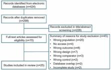
The search on the aforementioned platforms initially retrieved 424 studies. After manual exclusion of duplicates, 298 unique records remained. Two independent reviewers screened the titles and abstracts, excluding 228 records based on predefined criteria. Any discrepancies were resolved by a third reviewer. A total of 70 full-text articles were then assessed for eligibility, resulting in the exclusion of 45 with justification. Ultimately, 25 studies met the inclusion criteria and were part of the final analysis, with their characteristics presented in Table 1. The study selection process is detailed in Figure 1.
The strengths and weaknesses of each study can be found in Table 2 and will be further discussed. There was a considerable amount of missing data, and we tried to reach the authors of two studies but did not receive replies in 6 months. The study populations’ heterogeneity, the different machines and imaging methods, and mostly the difference in measuring outcomes (such as lesion volume) led to the realization that performing meta-analysis with this data was not the best practice.
Finally, most studies were performed with a small sample of participants, and for that reason their isolated results are not applicable for all populations. Only two studies had a large population-based design,14,15 but their data were also limited due to possible selection bias.
Participant characteristics
The included studies encompassed a total of approximately 3,600 participants, including 1,725 individuals with migraine and 1,878 control participants. The populations were predominantly composed of adults, with migraine subtypes including migraine without aura (MO), migraine with aura (MA), and chronic migraine (CM). Sample sizes varied widely, ranging from small, well-characterized clinical cohorts to large population-based samples.
Most studies reported a higher proportion of female participants, consistent with the epidemiology of migraine. Ages ranged from young to older adults, with reported means or medians typically between 30 and 60 years. Several studies provided age distributions stratified by presence of WMH, migraine subtype, or control status.
Approximately half of the studies included a control group, which varied in definition, from headache-free individuals to general population controls. Most studies applied standardized diagnostic criteria, such as those from the International Classification of Headache Disorders (ICHD). Some studies reported clinical features, including disease duration, attack frequency, comorbidities (e.g., cardiovascular risk factors), and aura status.
Several studies included in this review explored the relationship between comorbidities—particularly cardiovascular and metabolic risk factors—and WMHs in migraine patients. Trauninger et al.16 reported a possible association between it and elevated cholesterol, uric acid, and markers of endothelial dysfunction. Dinia et al.17 found WMH in 26 of 41 patients (63%) but did not identify significant associations with vascular risk factors, likely due to limited statistical power. Palm-Meinders et al.,18 in a Dutch cohort of 203 migraine patients and 83 controls, assessed multiple comorbidities, including hypertension, diabetes, and lipid levels, but found no consistent association with WMH burden. Hamedani et al.,19 using data from 93 migraine patients and 935 controls within the Atherosclerosis Risk in Communities (ARIC) study, emphasized the challenges of identifying robust associations given the multifactorial nature of WMH and the complexity of adjusting for confounders.
Other studies, including those by Dobrynina et al.20 (n = 92), Al-Hashel et al.21 (n = 60), Iyigundogdu et al.22 (n = 218), and Meilán et al.23 (n = 125), also collected data on cardiovascular risk factors, such as hypertension, diabetes, hyperlipidemia, obesity, and smoking. However, their findings regarding comorbidity associations were either not statistically conclusive or not the primary focus of analysis.
Overall, while comorbidities were frequently considered in study designs, the evidence remains heterogeneous and inconclusive. Larger, standardized, and statistically powered studies are needed to clarify the role of cardiovascular and systemic risk factors in the development of WMH among migraine patients.
Imaging protocols and analysis of WMHs
Most studies utilized accessible and widely available MRI protocols, primarily incorporating T2-weighted and FLAIR sequences, which allow for reliable WMH visualization. However, important limitations remain due to heterogeneity in image acquisition parameters, lesion definition, and quantification approaches. While many studies reported the presence or severity of WMH, only a subset performed volumetric measurements.
Studies such as Palm-Meinders et al.,18 Arkink et al.,24 Schramm et al.,25 and Lee et al.15 quantified WMH volume with consistent methodology and reported distribution across brain regions. Among these, only Palm-Meinders et al. and Arkink et al. included both control groups and follow-up volumetry, enhancing the validity of their longitudinal comparisons.
Several studies employed advanced segmentation strategies. Palm-Meinders et al.,18 Arkink et al.,24 and Lee et al.15 used automated or semi-automated methods for WMH detection, often combined with manual correction. Lee et al. notably introduced the deep white matter hyperintensity segmentation (DEWS) framework, a machine learning-based algorithm validated for high-sensitivity WMH detection in younger populations. Similarly, Ali et al.26 applied semi-automated segmentation with expert validation, enhancing reproducibility. Schramm et al.25 provided both volumetric and Fazekas scoring, offering robust multi-dimensional assessments of WMH burden.
On the other hand, several studies relied on manual visual assessment without volumetric quantification, including Dinia et al.,17 Erdélyi-Bótor et al.,14 Antony et al.,27 and Iyigundogdu et al.,22 which may introduce interobserver variability and limit comparability across studies. Visual scales, such as Fazekas and Scheltens, were applied in studies like Honningsvåg et al.,28 while Silva et al.29 employed voxel-based morphometry (VBM), and Sun et al.30 utilized a high-resolution 7T multimodal MRI, combining structural, diffusion tensor imaging (DTI), and fMRI data to characterize WMH microstructure.
Despite methodological advancements, challenges persist. Many studies lacked control groups, and few stratified findings by migraine subtype or reported interrater reliability. The diversity of approaches—ranging from visual inspection to artificial intelligence-based segmentation—reflects both innovation and a need for standardization. Future research would benefit from harmonized imaging protocols, consistent volumetric quantification, and systematic reporting of WMH distribution, to facilitate pooled analyses and improve comparability across studies.
Findings on migraine patients’ white matter
The main findings across the included studies were heterogeneous, although most shared the goal of evaluating the burden, distribution, and progression of WMHs in individuals with migraine. Most studies reported WMHs as supratentorial, small, and located predominantly in deep frontal white matter, with prevalence rates varying considerably across cohorts.
Trauninger et al.16 found no significant difference in WMH frequency between migraine with and without aura and observed that lesions were more frequent in patients with longer disease duration and higher attack frequency. Dinia et al.17 observed WMHs in 63.4% of migraine with aura patients at baseline, with 19.5% developing new lesions after a 33-month follow-up. Seneviratne et al.31 reported WMHs in 43.2% of patients and identified age, family history of migraine, and attack frequency as associated factors. In the community-based study by Palm-Meinders et al.,18 women with migraine, especially those without aura, had higher WMH volume and more lesion progression than controls, independently of comorbidities. Similarly, Hamedani et al.,19 using longitudinal data from the ARIC cohort, found that migraine without aura was associated with greater WMH burden, although this association was not significant after full adjustment for confounders.
Longitudinal imaging by Erdélyi-Bótor et al.14 showed that WMHs increased in number over three years, though lesion progression was not associated with clinical variables such as age or disease duration. The Nord-Trøndelag Health (HUNT) MRI study by Honningsvåg et al.28 did not find a significant association between migraine and WMHs but did identify one with tension-type headache.
Lee et al.15 found WMHs in nearly 70% of young migraine patients, although lesional burden was mild and only age correlated with volume. Arkink et al.,24 in a 9-year follow-up study, confirmed greater WMH volume progression in migraine patients compared with controls, particularly in those with aura.
Among more recent studies, Dobrynina et al.20 reported WMHs in 42.4% of migraine patients, and Al-Hashel et al.21 and Ahmed et al.32 found similar rates of 40 and 29%, respectively. Imai et al.,33 studying migraine in patients with reversible cerebral vasoconstriction syndrome (RCVS), identified WMHs in 46% of cases. Silva et al.29 reported WMHs in approximately 50% of migraine patients using VBM, while Antony et al.27 and Iyigundogdu et al.22 observed rates of 36 and 22%, respectively.
Meilán et al.23 found WMHs in 60.8% of patients, distributed similarly between episodic and chronic migraine, Schramm et al.25 observed a progressive increase in WMH volume in a large longitudinal cohort. However, migraine was not associated with higher volume or progression, as assessed by both volumetric analysis and Fazekas scoring.
Additional findings include Nanda et al.34 reporting WMHs in 36.7% of patients, Ali et al.26 identifying higher WMH volume in migraine with aura compared to controls, and Liu et al.35 and Abdalla et al.36 documenting WMHs in 22 and 46.1% of migraine patients, respectively. Finally, Sun et al.30 used 7T multimodal MRI to reveal microstructural white matter alterations even in patients without visible WMHs on conventional sequences, highlighting advanced imaging's potential to detect subtle abnormalities.
In summary, despite differences in design, imaging methodology, and patient characteristics, most studies confirm that WMHs are frequently observed in migraine patients. However, the extent, location, and clinical implications of these lesions remain variable, reinforcing the need for standardized imaging protocols and longitudinal studies with rigorous clinical phenotyping.
DISCUSSION
The objective of this systematic review was to analyze the relationship between WMH and migraine, focusing on their prevalence, progression, and potential associations with clinical and demographic factors. The key findings indicate that WMHs are frequently observed in migraine patients—particularly in those with MA, longer disease duration, and higher headache frequency. Prevalence rates among migraine patients varied across studies, ranging from 22 to over 60%, with some reporting small, deep, frontal WMHs as the most common pattern.20,23,26,29
Previous studies have often overlooked essential migraine characteristics and the influence of comorbidities. While many explored WMH prevalence, few addressed their progression or clear associations with clinical subtypes, such as aura or attack frequency. Our review bridges this gap by synthesizing studies assessing both baseline and longitudinal data, with an emphasis on disease chronicity and migraine phenotypes. Notably, studies like Dinia et al.17 and Erdélyi-Bótor et al.14 provided longitudinal follow-up and identified WMH progression in a subset of patients.
Our findings align with Zhang et al.,37 who performed a meta-analysis of 30 studies and found a pooled WMH prevalence of 44% among migraineurs, with a higher prevalence in patients with aura. While their study highlighted associations with age and comorbidities like hypertension and diabetes, our synthesis did not confirm consistent correlations with these conditions, suggesting potential differences in methodology, population characteristics, or WMH detection techniques. Indeed, several studies in our review, such as Trauninger et al.,16 Palm-Meinders et al.,18 and Erdélyi-Bótor et al.,14 found no statistically significant influence of comorbidities on WMH presence or progression.
Masson et al.38 added complementary evidence by examining structural brain changes beyond WMH, identifying alterations in both gray and white matter volume in migraine patients. Although our review specifically targeted WMH, their results support the hypothesis that migraine-related brain changes are multifaceted and may reflect broader anatomical or neurovascular dysfunctions.38
Designing robust WMH studies requires careful control of confounding factors. Participants’ mean age must be considered, as older individuals may naturally develop WMHs due to aging or vascular comorbidities. Using the latest ICHD criteria is essential for standardized migraine classification, and MRI acquisition protocols must be consistent across studies to ensure comparability.39,40
One persistent challenge is the absence of a validated quantitative or qualitative scale tailored for WMH assessment in migraine patients. This gap, along with its historical perception as a benign condition, may contribute to the scarcity of large, representative studies. However, given the impact of migraines on quality of life and productivity, advancing our understanding of WMH is critical. With growing insights into migraine pathophysiology, particularly involving calcitonin gene-related peptide (CGRP) signaling,41,42 a vascular component to WMH development appears plausible, although a direct mechanistic link remains speculative.
While Schramm et al.25 did not find a strong association between migraine and WMH progression, our analysis highlights the WMH burden in older patients and those with longer migraine history. For example, Meilán et al.23 observed WMHs in 60.8% of migraine patients, and Schramm et al.33 reported volumetric progression over time. Other studies using advanced imaging techniques, like Sun et al.30 with 7T MRI, demonstrated microstructural changes even in patients lacking visible WMH on conventional scans.
Interestingly, across multiple studies in our review,14,16-18 comorbidities such as hypertension or diabetes were not associated with WMH presence or progression. This contrasts with broader epidemiological findings where such comorbidities are established risk factors,43-45 indicating that WMH in migraine patients may follow distinct pathophysiological mechanisms.
This review underscores the need for future studies to investigate the cognitive and cerebrovascular implications of WMH in migraine. Although our focus was on lesion prevalence and progression, the long-term consequences of this condition remain poorly understood.
The strengths of our review include rigorous inclusion criteria and a comprehensive approach to both cross-sectional and longitudinal data. Limitations include heterogeneity in imaging methods, small sample sizes in some studies, and limited follow-up duration. Advancing knowledge on WMH in migraine will require coordinated efforts using standardized diagnostic criteria, quantitative imaging, and careful control of confounding variables.
In conclusion, across 25 studies (∼ 3,600 participants), WMHs are frequently observed in migraine, particularly in patients with aura, longer disease duration, and higher attack frequency. The strength of evidence is moderate and stems predominantly from cross-sectional designs, which consistently report higher WMH burden in migraine patients than in controls.
Evidence from prospective cohorts is inconsistent: some longitudinal studies show modest lesion accrual in subgroups, whereas others find no excess progression, so causality and prognostic significance remain uncertain. Findings from retrospective clinical samples generally echo the cross-sectional signal but are limited by referral bias, heterogeneous imaging protocols, and variable WMH definitions.
When present together, WMHs in migraine should be interpreted with caution: they are common incidental findings, typically small, supratentorial, and deep-frontal/parietal, and do not by themselves warrant changes in routine diagnostic workup or management in otherwise typical migraine presentations. Clinically, careful assessment is important to avoid overinvestigation and to assist in the differential diagnosis with demyelinating disease such as multiple sclerosis.46
To clarify pathophysiology and clinical relevance, well-designed prospective studies are needed. This includes use of standardized MRI acquisition, consistent volumetric and topographic WMH quantification, and rigorous migraine phenotyping (ICHD), with adjustment for vascular/metabolic comorbidities and evaluation of cognitive and cerebrovascular outcomes. These improvements will enhance comparability across studies and support evidence-based counselling for migraine patients who present with WMHs.
Data Availability Statement
Data will be available upon request to the corresponding author.
References
-
1 Özge A, Baykan B, Bıçakçı Ş, et al. Revolutionizing migraine management: advances and challenges in CGRP-targeted therapies and their clinical implications. Front Neurol 2024; 15:1402569. Doi: 10.3389/fneur.2024.1402569
» https://doi.org/10.3389/fneur.2024.1402569 -
2 Headache Classification Committee of the International Headache Society (IHS). The International Classification of Headache Disorders, 3rd edition. Cephalalgia 2018;38(01):1–211. Doi: 10.1177/0333102417738202
» https://doi.org/10.1177/0333102417738202 -
3 Detsky ME, McDonald DR, Baerlocher MO, Tomlinson GA, McCrory DC, Booth CM. Does this patient with headache have a migraine or need neuroimaging? JAMA 2006;296(10):1274-–1283. Doi: 10.1001/jama.296.10.1274
» https://doi.org/10.1001/jama.296.10.1274 -
4 Evans RW, Burch RC, Frishberg BM, et al. Neuroimaging for Migraine: The American Headache Society Systematic Review and Evidence-Based Guideline. Headache 2020;60(02):318–336. Doi: 10.1111/head.13720
» https://doi.org/10.1111/head.13720 -
5 Vijiaratnam N, Barber D, Lim KZ, et al. Migraine: Does aura require investigation? Clin Neurol Neurosurg 2016;148:110–114. Doi: 10.1016/j.clineuro.2016.07.006
» https://doi.org/10.1016/j.clineuro.2016.07.006 -
6 Holle D, Obermann M. The role of neuroimaging in the diagnosis of headache disorders. Ther Adv Neurol Disord 2013;6(06):369–374. Doi: 10.1177/1756285613489765
» https://doi.org/10.1177/1756285613489765 -
7 Guidelines from the American Academy of Neurology and US Headache Consortium. Neuroimaging in patients with nonacute headache [Internet]. Available from: http://tools.aan.com/professionals/practice/pdfs/gl0088.pdf
» http://tools.aan.com/professionals/practice/pdfs/gl0088.pdf -
8 Debette S, Markus HS. The clinical importance of white matter hyperintensities on brain magnetic resonance imaging: systematic review and meta-analysis. BMJ 2010;341:c3666. Doi: 10.1136/bmj.c3666
» https://doi.org/10.1136/bmj.c3666 -
9 Lipton RB, Pan J. Is migraine a progressive brain disease? JAMA 2004;291(04):493–494. Doi: 10.1001/jama.291.4.493
» https://doi.org/10.1001/jama.291.4.493 -
10 Bashir A, Lipton RB, Ashina S, Ashina M. Migraine and structural changes in the brain: a systematic review and meta-analysis. Neurology 2013;81(14):1260–1268. Doi: 10.1212/WNL.0b013e3182a6cb32
» https://doi.org/10.1212/WNL.0b013e3182a6cb32 -
11 Swartz RH, Kern RZ. Migraine is associated with magnetic resonance imaging white matter abnormalities: a meta-analysis. Arch Neurol 2004;61(09):1366–1368. Doi: 10.1001/archneur.61.9.1366
» https://doi.org/10.1001/archneur.61.9.1366 -
12 Moher D, Liberati A, Tetzlaff J, Altman DGPRISMA Group. Preferred reporting items for systematic reviews and meta-analyses: the PRISMA statement. PLoS Med 2009;6(07):e1000097. Doi: 10.1371/journal.pmed.1000097
» https://doi.org/10.1371/journal.pmed.1000097 -
13 Nayebirad S, Mohamadi A, Yousefi-Koma H, et al. Association of anti-Ro52 autoantibody with interstitial lung disease in autoimmune diseases: a systematic review and meta-analysis. BMJ Open Respir Res 2023;10(01):e002076. Doi: 10.1136/bmjresp-2023-002076
» https://doi.org/10.1136/bmjresp-2023-002076 -
14 Erdélyi-Bótor S, Aradi M, Kamson DO, et al. Changes of migraine-related white matter hyperintensities after 3 years: a longitudinal MRI study. Headache 2015;55(01):55–70. Doi: 10.1111/head.12459
» https://doi.org/10.1111/head.12459 -
15 Lee MJ, Park BY, Cho S, Park H, Chung CS. Cerebrovascular reactivity as a determinant of deep white matter hyperintensities in migraine. Neurology 2019;92(04):e342–e350. Doi: 10.1212/WNL.0000000000006822
» https://doi.org/10.1212/WNL.0000000000006822 -
16 Trauninger A, Leél-Ossy E, Kamson DO, et al. Risk factors of migraine-related brain white matter hyperintensities: an investigation of 186 patients. J Headache Pain 2011;12(01):97–103. Doi: 10.1007/s10194-011-0299-3
» https://doi.org/10.1007/s10194-011-0299-3 -
17 Dinia L, Bonzano L, Albano B, et al. White matter lesions progression in migraine with aura: a clinical and MRI longitudinal study. J Neuroimaging 2013;23(01):47–52. Doi: 10.1111/j.1552-6569.2011.00643.x
» https://doi.org/10.1111/j.1552-6569.2011.00643.x -
18 Palm-Meinders IH, Koppen H, Terwindt GM, et al. Structural brain changes in migraine. JAMA 2012;308(18):1889–1897. Doi: 10.1001/jama.2012.14276
» https://doi.org/10.1001/jama.2012.14276 -
19 Hamedani AG, Rose KM, Peterlin BL, et al. Migraine and white matter hyperintensities: the ARIC MRI study. Neurology 2013;81(15):1308–1313. Doi: 10.1212/WNL.0b013e3182a8235b
» https://doi.org/10.1212/WNL.0b013e3182a8235b -
20 Dobrynina LA, Suslina AD, Gubanova MV, et al. White matter hyperintensity in different migraine subtypes. Sci Rep 2021;11(01):10881. Doi: 10.1038/s41598-021-90341-0
» https://doi.org/10.1038/s41598-021-90341-0 -
21 Al-Hashel JY, Alroughani R, Gad K, Al-Sarraf L, Ahmed SF. Risk factors of white matter hyperintensities in migraine patients. BMC Neurol 2022;22(01):159. Doi: 10.1186/s12883-022-02680-8
» https://doi.org/10.1186/s12883-022-02680-8 -
22 Iyigundogdu I, Derle E. Do Mean Platelet Volume and Platelet Distribution Width Have An Association with White Matter Hyperintensities in Migraine Patients? Ann Indian Acad Neurol 2023;26(04):435–440. Doi: 10.4103/aian.aian_183_23
» https://doi.org/10.4103/aian.aian_183_23 -
23 Meilán A, Larrosa D, Ramón C, et al. No association between migraine frequency, white matter lesions and silent brain infarctions: a study in a series of women with chronic migraine. Eur J Neurol 2020;27(08):1689–1696. Doi: 10.1111/ene.14284
» https://doi.org/10.1111/ene.14284 -
24 Arkink EB, Palm-Meinders IH, Koppen H, et al. Microstructural white matter changes preceding white matter hyperintensities in migraine. Neurology 2019;93(07):e688–e694. Doi: 10.1212/WNL.0000000000007940
» https://doi.org/10.1212/WNL.0000000000007940 -
25 Schramm SH, Tenhagen I, Jokisch M, et al. Migraine or any headaches and white matter hyperintensities and their progression in women and men. J Headache Pain 2024;25(01):78. Doi: 10.1186/s10194-024-01782-7
» https://doi.org/10.1186/s10194-024-01782-7 -
26 Ali M, van der Weerd N, van Os HJA, et al.;CREW Consortium. Coagulation Factors and White Matter Hyperintensities in Middle-Aged Women With and Without Migraine and Ischemic Stroke. Eur J Neurol 2025;32(03):e70063. Doi: 10.1111/ene.70063
» https://doi.org/10.1111/ene.70063 -
27 Antony J, Namboothiri AD, Mathew L. Association between Migraine Patterns and White Matter Hyperintensities in MRI Brain: A Cross-sectional Analytical Study. J Clin Diagn Res 2023;17(10):TC01–TC06. Doi: 10.7860/JCDR/2023/62607.18539
» https://doi.org/10.7860/JCDR/2023/62607.18539 -
28 Honningsvåg LM, Håberg AK, Hagen K, Kvistad KA, Stovner LJ, Linde M. White matter hyperintensities and headache: A population-based imaging study (HUNT MRI). Cephalalgia 2018;38(13):1927–1939. Doi: 10.1177/0333102418764891
» https://doi.org/10.1177/0333102418764891 -
29 Silva N, Maciel NM, Nather JC Jr, et al. White Matter Lesions Identified by Magnetic Resonance in Women with Migraine: A Volumetric Analysis and Clinical Correlations. Diagnostics (Basel) 2023;13(04):799. Doi: 10.3390/diagnostics13040799
» https://doi.org/10.3390/diagnostics13040799 -
30 Sun Y, Ma L, Wang S, et al. Neuroimaging differences between chronic migraine with and without medication overuse headache: a 7. Tesla multimodal MRI study. J Headache Pain 2025;26(01):54. Doi: 10.1186/s10194-025-01988-3
» https://doi.org/10.1186/s10194-025-01988-3 -
31 Seneviratne U, Chong W, Billimoria PH. Brain white matter hyperintensities in migraine: clinical and radiological correlates. Clin Neurol Neurosurg 2013;115(07):1040–1043. Doi: 10.1016/j.clineuro.2012.10.033
» https://doi.org/10.1016/j.clineuro.2012.10.033 -
32 Ahmed SR, Mohamed AAM, Salem HH, Helmy S, Moustafa RR, Borham SMF. Association of white matter hyperintensities with migraine phenotypes and response to treatment. Acta Neurol Belg 2023;123(05):1725–1733. Doi: 10.1007/s13760-022-02015-x
» https://doi.org/10.1007/s13760-022-02015-x -
33 Imai M, Shimoda M, Oda S, et al. Reversible Cerebral Vasoconstriction Syndrome Patients with a History of Migraine: A Retrospective Case-control Study. Intern Med 2023;62(03):355–364. Doi: 10.2169/internalmedicine.9776-22
» https://doi.org/10.2169/internalmedicine.9776-22 -
34 Nanda C, Sachdev N. Assessment of White Matter Alterations in Patients of Migraine Using Diffusion Tensor Imaging. Neurol India 2024;72(06):1169–1173. Doi: 10.4103/ni.ni_894_22
» https://doi.org/10.4103/ni.ni_894_22 -
35 Liu ZZ, Yu HY, Li YH, et al. Comparison of Syn T2-FLAIR and Syn DIR with conventional T2-FLAIR in displaying white matter hyperintensities in migraine patients. Neuroradiology 2025;67(01):49–56. Doi: 10.1007/s00234-024-03477-x
» https://doi.org/10.1007/s00234-024-03477-x -
36 Abdalla K, Alawneh KZ, Al-Bdour M, Abu-Salih AQ. Migraine and MRI: uncovering potential associations. Head Face Med 2025;21(01):6. Doi: 10.1186/s13005-024-00478-2
» https://doi.org/10.1186/s13005-024-00478-2 -
37 Zhang W, Cheng Z, Fu F, Zhan Z. Prevalence and clinical characteristics of white matter hyperintensities in migraine: A meta-analysis. Neuroimage Clin 2023;37:103312. Doi: 10.1016/j.nicl.2023.103312. migraine with the headache characteristics and response to treatment. Revista Latinoamericana de Hipertension 2021;15(5):345–351 DOI: 10.5281/zenodo.4487139
» https://doi.org/10.1016/j.nicl.2023.103312 -
38 Masson R, Demarquay G, Meunier D, et al. Is Migraine Associated to Brain Anatomical Alterations? New Data and Coordinate-Based Meta-analysis. Brain Topogr 2021;34(03):384–401. Doi: 10.1007/s10548-021-00824-6
» https://doi.org/10.1007/s10548-021-00824-6 -
39 Scheltens P, Barkhof F, Leys D, et al. A semiquantative rating scale for the assessment of signal hyperintensities on magnetic resonance imaging. J Neurol Sci 1993;114(01):7–12. Doi: 10.1016/0022-510x(93)90041-v
» https://doi.org/10.1016/0022-510x(93)90041-v -
40 Park BY, Lee MJ, Lee SH, et al. DEWS (DEep White matter hyperintensity Segmentation framework): A fully automated pipeline for detecting small deep white matter hyperintensities in migraineurs. Neuroimage Clin 2018;18:638–647. Doi: 10.1016/j.nicl.2018.02.033
» https://doi.org/10.1016/j.nicl.2018.02.033 -
41 Wattiez AS, Sowers LP, Russo AF. Calcitonin gene-related peptide (CGRP): role in migraine pathophysiology and therapeutic targeting. Expert Opin Ther Targets 2020;24(02):91–100. Doi: 10.1080/14728222.2020.1724285
» https://doi.org/10.1080/14728222.2020.1724285 -
42 Fazekas F, Koch M, Schmidt R, et al. The prevalence of cerebral damage varies with migraine type: a MRI study. Headache 1992;32(06):287–291. Doi: 10.1111/j.1526-4610.1992.hed3206287.x
» https://doi.org/10.1111/j.1526-4610.1992.hed3206287.x -
43 Bachmann D, von Rickenbach B, Buchmann A, et al. White matter hyperintensity patterns: associations with comorbidities, amyloid, and cognition. Alzheimers Res Ther 2024;16(01):67. Doi: 10.1186/s13195-024-01435-6
» https://doi.org/10.1186/s13195-024-01435-6 -
44 Ashina M, Hansen JM, Do TP, Melo-Carrillo A, Burstein R, Moskowitz MA. Migraine and the trigeminovascular system-40 years and counting. Lancet Neurol 2019;18(08):795–804. Doi: 10.1016/S1474-4422(19)30185-1
» https://doi.org/10.1016/S1474-4422(19)30185-1 -
45 Eikermann-Haerter K, Huang SY. White Matter Lesions in Migraine. Am J Pathol 2021;191(11):1955–1962. Doi: 10.1016/j.ajpath.2021.02.007
» https://doi.org/10.1016/j.ajpath.2021.02.007 -
46 Barkhof F, Filippi M, Miller DH, et al. Comparison of MRI criteria at first presentation to predict conversion to clinically definite multiple sclerosis. Brain 1997;120(Pt 11):2059–2069. Doi: 10.1093/brain/120.11.2059
» https://doi.org/10.1093/brain/120.11.2059 - 47 Kocatürk M, Kocatürk Ö White matter hyperintensities and carotid intima media thickness in migraine without aura patients. Neurol Asia 2020;25(04):501–508
-
48 Alkhaffaf WH, Naif MM, Ahmed RN. The Association of MRI findings in migraine with the headache characteristics and response to treatment. Revista Latinoamericana de Hipertension 2021;15(05):345–351. https://doi.org/10.5281/zenodo.4487139
» https://doi.org/10.5281/zenodo.4487139
Edited by
-
Editor-in-Chief:
Hélio A. G. Teive. https://orcid.org/0000-0003-2305-1073.
-
Associate Editor:
Pedro André Kowacs. https://orcid.org/0000-0001-7770-7475.

