 A case of familial partial lipodystrophy type 2 masquerading as Cushing syndrome: Explaining an atypical phenotype by whole-exome sequencing
SUMMARY
Familial partial lipodystrophy type 2 is a rare disease, particularly when it is caused by nonclassical gene variants. A high index of suspicion is essential for a timely diagnosis. We present the case of a 32-year-old woman, referred to evaluation of a possible Cushing syndrome, which was clinically and biochemically ruled out. Yet, due to the finding of a rather abnormal fat distribution during physical examination, the diagnosis of lipodystrophy was cogitated. Whole-exome sequencing revealed a missense variant of exon 11 R582H of the gene encoding Laminin A (rs57830985,c.1745G>A, p.Arg582His). The patient presented some clinical and biochemical characteristics discordant with those previously reported in patients harboring other classical variants of this gene.
INTRODUCTION
Lipodystrophies are a group of heterogeneous, low prevalence diseases (one case per million) (1), characterized by total or partial loss of adipose tissue, which can be congenital or acquired. Although their most constant feature is adipose tissue hypotrophy, some subtypes present with a rather abnormal distribution of fat, which can accumulate in certain organs (1,2). Some lipodystrophies are accompanied by metabolic alterations, namely: insulin resistance; type 2 diabetes (T2D); hypertriglyceridemia; non-alcoholic fatty liver disease (NAFLD); hypoalphalipoproteinemia, and polycystic ovary syndrome (3).
Different subtypes of lipodystrophy include generalized congenital lipodystrophy, acquired generalized lipodystrophy, familial (congenital) partial lipodystrophy, and acquired partial lipodystrophy. Dunnigan’s syndrome is the most common type of familial partial lipodystrophies (FPLD2), holding an autosomal dominant inheritance, although a more severe phenotype has been described in patients harboring homozygous pathogenic variants (1-3).
FPLD2 is caused by a missense pathogenic variant of the LMNA gene. This gene, located at 1q21-22 and consisting of 12 exons, encodes laminin A/C (LMNA/C), which along with laminin B, forms a protein network found below the inner nuclear membrane. These proteins are crucial for the preservation of nuclear membrane mechanical stability, anchoring proteins in the nuclear pore complex, and organizing chromatin; they also have a fundamental role in the regulation of DNA replication and transcription (4). So-called laminopathies are a group of diseases caused by various pathogenic variants of this gene. The most common or “typical” FPLD2 pathogenic variant is found in exon 8, specifically in codon R482, while all other pathogenic variants are known as atypical (5). The classical phenotype usually occurs in post-pubertal women; it is characterized by the loss of adipose tissue in the legs, arms, buttocks, and trunk and an accumulation of fat in the neck, face, dorsocervical region, labia majora, and visceral area, frequently resulting in a cushingoid appearance (6). Limb muscle hypertrophy, especially in the legs, and vein enlargement are remarkable characteristics. Most individuals develop insulin resistance, which frequently evolves into type 2 diabetes (T2D). Other hormonal and metabolic abnormalities include menstrual disorders, moderate to severe hypertriglyceridemia, NAFLD, and hypoalphalipoproteinemia (3-6).
In patients with atypical FPLD2, particularly those with the exon 11 R582H variant, only laminin A is affected, since laminin C is the product of the alternative splicing encompassing exons 1 to 10 (7,8). These patients have a milder form of the condition, with less severe fat loss and metabolic symptoms. They may experience mild hypertriglyceridemia and absence of acanthosis nigricans or hirsutism (5-7,9). In this study, we present the case of a female patient with atypical FPLD2, due to a missense variant in exon 11 of the gene encoding Laminin A (rs57830985,c.1745G>A, p.Arg582His).
CASE REPORT
A 32-year-old woman, presented with a history of oligomenorrhea, hirsutism, increased adipose tissue on the face, neck, and abdomen, as well as marked muscular hypertrophy of the lower limbs since puberty. She was initially diagnosed with polycystic ovary syndrome (PCOS) for which she was treated with combined oral contraceptives, with no clinical improvement. Given her physical appearance - including moon facies and apparent central obesity - we speculated Cushing syndrome diagnosis, which was unequivocally ruled out twice by a normal 24-hour urinary free cortisol and a low-dose dexamethasone suppression test. For the past four years her menstrual abnormalities and hirsutism had significantly worsened and she had been recently diagnosed with type 2 diabetes.
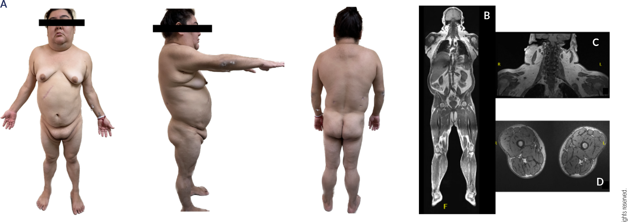
On the physical examination the following measurements were taken: blood pressure of 130/80 mm Hg; pulse 86 beats/min, weight 93 kg, 1.65 m of height, body mass index of 34 kg/m2, and 108 cm of waist circumference. The patients presented a striking cushingoid appearance, round, and plethoric face. Severe acanthosis nigricans in the posterior neck, and hirsutism (Ferriman- Gallwey Score 24), as well as a bizarre and abnormal adipose tissue distribution, with fat accumulation in the neck, face, dorsocervical region, and labia majora, accompanied by a total absence of adipose tissue in the lower limbs, and significant hypertrophy of thighs and calves (Figure 1A).
(A) Frontal, lateral and posterior view of the patient. Abnormal fat distribution is evident with accumulation of fat in the neck, face, dorsocervical region, and labia majora. Also, a lack of fat in the lower limbs; significant hypertrophy of thighs and calves. (B, C) Increased accumulation of subcutaneous adipose tissue in the face, neck, superior chest and back. Hepatomegaly. (D) Decreased accumulation of subcutaneous adipose tissue in the lower limbs.
Laboratory evaluation (Table 1) revealed severe hypertriglyceridemia, moderate hypercholesterolemia, and uncontrolled diabetes (glycated hemoglobin 9.1%), with a Homeostatic Model Assessment for Insulin Resistance (HOMA-IR) of 23. Prolactin levels, total testosterone, androstenedione, and dehydroepiandrosterone sulfate (DHEA-S) were within normal ranges. Additionally, an adrenocorticotropic hormone (ACTH) stimulation test was performed to rule out nonclassical congenital adrenal hyperplasia, with negative results. Therefore, the diagnosis of polycystic ovary syndrome was concluded (Table 1). A total body MRI confirmed abnormal fat distribution and revealed the presence of a lipoma adjacent to the right trapezius muscle, as well as mild hepatomegaly with grade 1 steatosis (Figure 1B-D). Echocardiography ruled out cardiac structural abnormalities. She was started on high-dose statins and fibrates as well as metformin, sitagliptin, dapagliflozin, insulin glargine, and oral contraceptives.
MATERIALS AND METHODS
DNA purification
Total DNA was extracted from peripheral blood mononuclear cells. After lysis with proteinase K solution, 300 μL of 5M ammonium acetate were added to precipitate proteins and cellular components. The aqueous phase was transferred to a fresh tube, 600 μL of isopropanol was added and the mixture was incubated overnight at -20 °C, and then centrifuged at 14,000 rpm for 30 min. The resulting DNA pellet was washed with 1 mL 75% ethanol and centrifuged at 10,000 rpm for 5 min; the pellet was air-dried, and DNA resuspended in nuclease free water (10).
Whole-exome sequencing (WES)
The genomic DNA (gDNA) was shipped to the Genomics Core Lab of the Instituto Tecnológico y de Estudios Superiores de Monterrey for exome sequencing. Then, gDNA was quantified using Qubit dsDNA BR Assay Kit (Invitrogen, Carlsbad, CA, USA). Quality was determined spectrophotometrically using a Nanodrop One spectrophotometer (Thermo Fisher Scientific, Waltham MA, USA). WES libraries were prepared using Illumina DNA Prep with Exome 1.0 Enrichment (Illumina, San Diego CA, United States). All libraries were quantified with the Qubit dsDNA BR Assay Kit (Invitrogen, Carlsbad, CA, USA), libraries sizes were analyzed in S2 Standard DNA Cartridge for Sep 400 (BiOptic, New Taipei City, Taiwan), and sequencing was performed in a NovaSeq 6000 sequencer (Illumina, San Diego CA, United States) in a 150 bp pair-end configuration.
FastQC and preprocessing
The quality assessment of the exome libraries was performed with FastQC (Babraham Bioinformatics) to determine the quality of the sequencing. All raw sequences passed the initial quality filter. Adapters were removed and a quality and length filter were performed with Trimmomatic 0.40 (11).
Computational WES analysis
Preprocessed sequences were aligned to the human reference sequence (GRCh38) using the Illumina-Dragen Enrichment pipeline (llumina, San Diego CA, United States). This pipeline was set to produce copy number variants (-enable-cnv true). The BAM files resulting from the enrichment were removed from PCR duplicates using Picard tools (http://broadinstitute.github.io/picard). Each BAM file was used to obtain somatic variants with the GATK pipeline (https://github.com/broadinstitute/gatk/ releases), and variants were annotated by ANNOVAR using the following databases: Clinvar, gnomAD, refGene, cytoBand, exac03, avsnp147, dbnsfp30a (12). Genomics were uploaded into the cloud using Docker, GATK, and WDL in Terra. O’Reilly Media. Somatic variants were then transformed to Maf using Funkotator from GATK. Additionally, converted annotated variant files were analyzed with the Maftools package from R programming language to visualize the landscape of critical pathogenic variants.
The pathogenic variants analysis was carried out using Maftools, the variations were filtered using subsetMaf with the parameter “Variant_Classification == Missense_Mutations”. Graphs of pathogenic variants in genes of interest were constructed using lollipop plot to observe variants in general (13).
WES RESULTS
The analysis identified a heterozygous missense variant of the LMNA gene (rs57830985 c. G1745A, p. R582H), a pathogenic variant associated with familial partial lipodystrophy type 2 (Figure 2).

In addition, we searched the whole exome for gene variants linked to glucose metabolism, insulin resistance, adipogenesis, lipid metabolism, and steroidogenesis. Table 2 presents the aforementioned findings.
DISCUSSION
FPLD2 patients with atypical (and generally heterozygous) pathogenic variants in the LMNA gene encoding have different phenotypes and metabolic manifestations than those harboring more typical pathogenic variants (p.Arg482Trp; p.Arg482Gln; p.Arg482Leu; p.Lys486Asn; p.Lys486Asn) (5). Garg and cols. reported three patients with the atypical p.R582H pathogenic LMNA variant. These patients had lower levels of triglycerides and a milder limb fat loss than the patients with the classical R482 and K486 pathogenic variants (5). Furthermore, heterozygous patients for the atypical p.R582H pathogenic variant have a less severe phenotype and clinical course than homozygous patients, who behave clinically like subjects with typical pathogenic variants (14). Neither acanthosis nigricans nor hirsutism are present in patients harboring atypical LMNA pathogenic variants, particularly when heterozygous (5,7). Although our patient was found to have one of these atypical heterozygous LMNA pathogenic variants, she displayed a rather severe and aggressive phenotype characterized by a younger age at diagnosis, higher BMI, severe hypertriglyceridemia, marked insulin resistance, hirsutism, and menstrual abnormalities (Table 3). Having sequenced her whole exome, we looked for the concomitant occurrence of pathological variants in genes associated with glucose metabolism, insulin resistance, lipid metabolism, and steroidogenesis that could be associated with the clinical and biochemical characteristics of our patient. In doing so, we found polymorphisms in several genes that participate in different metabolic pathways, including the vitamin D receptor (VDR), the regulatory subunit of phosphaytidylinositol 3-kinase (PIK3R1), the glucokinase regulatory protein (GCKR), the 17β-hydroxysteroid dehydrogenase type 5 (HSD17B5), the LH and chorionic gonadotropin hormone receptor (LHCGR), and the 21-hydroxilase enzyme, among others.
Comparison of clinical and biochemical characteristics in typical FPLD2, atypical FPLD2 and our patient
The VDR is ubiquitously expressed in various tissues, including muscle, bone, visceral and subcutaneous adipose tissue, liver, and pancreas, and thus participates in several biological processes beyond calcium metabolism (15). Our patient harbored the VDR gene missense rs2228570 (c.2T>C, p.M1) variant, which modifies the translation initiation codon and results in a shorter protein product that could potentially have a higher binding affinity for 1,25-dihydroxyvitamin D3 (16,17). Although its clinical significance remains controversial (18), patients harboring this VDR gene variant have been reported to have an increased BMI, waist circumference, total body fat, and sum of skinfold thickness (15-17, 19-21). The association of the rs2228570 variant with the development of T2D has been confirmed by several meta-analyses, particularly in Asian populations (22-24). Even when the exact molecular mechanism is unknown, there is evidence that the VDR regulates insulin synthesis and secretion, and it may have a role in beta cell growth and differentiation, and a protective effect against cytokine mediated inflammation, which contributes to beta cell dysfunction and insulin resistance (25,26).
The PIK3R1 gene encodes for the p85 regulatory subunit of the enzyme phosphaytidylinositol 3-kinase (PIK3). This subunit stabilizes the p110 catalytic subunit of the enzyme and determines its biological activity (27). WES of our patient revealed the variant rs3730089 (c.978G>A, p.Met326Ile) of the PIK3R1 gene. This variant leads to a constitutive activation of PI3K, as well as to downstream alterations in protein kinase B (AKT), a signaling cascade that regulates growth, differentiation, survival, and cellular glucose uptake. This variant has been associated with an increased risk of type 2 diabetes, as well as obesity and dyslipidemia (27-29).
GCKR is an allosteric regulator that modulates the activity and mobilization of glucokinase, favoring a rapid response to changes in glucose concentrations. Our patient had the GCKR gene variant rs1260326 (c.1337T>C, p.Leu446Pro), which generates a functional alternative of the protein that promotes glucose uptake and a consequent increment in VLDL and triglyceride synthesis (30-32). The same variant has been associated with non-alcoholic fatty liver disease and steatohepatitis in Danish, Swedish, Japanese, and Han Chinese populations (33-36). This variant could contribute to the severe hypertriglyceridemia presented by our patient, in contrast with the mild-moderate hypertriglyceridemia reported in other atypical cases of FPLD2 (4-7).
The LHCGR is expressed in the ovary (theca and granulosa cells, stromal cells, and luteinized cells) as well as in adipose tissue (37,38). Its function is essential for sexual differentiation in fetuses and reproductive function in adult life (38,39). The LHCGR polymorphism (rs2293275, c.935A > G, p. Asn312Ser) found in our patient is a missense single-nucleotide variant, identified as a risk factor for the development of PCOS (39-41), although the evidence is controversial (42). This variant is associated with an abnormal glycosylation of the LHCGR, which may affect its trafficking, stability, and sensitivity (43). These alterations have been associated with abnormal LH signaling, which seems to have a key role in ovarian androgen production, follicular development, and ovulation (43,44).
The patient also has a variant in the HSD17B5 gene. This enzyme is a member of the aldo-keto reductase (AKR) superfamily. It catalyzes the synthesis of potent androgens in peripheral tissues (theca cells and adrenal glands), converting androstenedione to testosterone, activating the androgen receptor, and acting as a substrate for aromatase (45,46). The rs12529 (c.15C>G, p.His5Gln) is a non-synonymous single nucleotide variant in exon 1 of the HSD17B5 gene that involves a buried charge change and thus results in a more unstable protein product (47,48). The clinical impact of rs12529 in women is controversial. Some studies report its association with increased levels of testosterone and insulin resistance (49,50).
Finally, our patient also harbored a variant of the CYP21A2 gene (rs6471, c.844G>T, p.Val282Leu), which encodes the steroidogenic enzyme 21-hydroxilase. This polymorphism reduces the enzymatic activity by 50%-80% and is associated with non-classic congenital adrenal hyperplasia (51-53). The heterozygous condition of this variant implies that a single normal allele in the patient was enough to preclude the appearance of the associated phenotype as reported by Inácio and cols. (54). Adrenal hyperplasia was excluded with an ACTH stimulation test during evaluation for hirsutism and amenorrhea.
Our case suggests that the atypical manifestations of FPLD2 are associated with pathogenic variants in the laminin genes, but they also may be related to variations in other genes. Several genes are required to regulate different metabolic pathways. The presence of other polymorphisms and pathogenic variants may change the phenotype of atypical cases. Other case studies could help clarify if the alterations detected in our patient are common in cases with aggressive metabolic manifestations. This case is an example of the differential diagnostic algorithm in patients with severe metabolic conditions in which other neuroendocrine causes have been suspected and excluded.
An important limitation of this study is that the exome analysis was not performed in relatives of the patient. An evaluation of family members with and without manifestations of metabolic disease and lipodystrophy would be ideal for calculating the weight of the exome variations in the phenotype of the patient.
It is important to understand the benefits and limitations of exome sequencing in order to interpret the clinical relevance of genomic variants. In our case, we tried to identify clinical and biochemical aspects that did not coincide with what has been reported in the literature, and we looked for genetic variants that could be associated with these characteristics. The clinical significance of the variants found in our patient is still subject to debate, since their association with metabolic alterations has not been consistent in all studies.
In conclusion, the clinical and biochemical characteristics of patients with heterozygous FPLD2 harboring an atypical variant (rs57830985, c.1745G>A, p.Arg582His) are rare. Exome sequencing can contribute to the understanding of unusual or aggressive phenotypes. The true contribution of other genetic variants detected in the exomes of such patients requires validation in larger cohorts of patients and genetically similar populations.
-
Funding statement:
this work did not receive any financial support.
Data availability statement:
whole-exome sequencing data is available by contacting Etual Espinosa-Cardenas (espinosaetual@gmail.com) upon reasonable request.
-
Disclosure:
no potential conflict of interest relevant to this article was reported.
REFERENCES
-
1 Lightbourne M, Brown RJ. Genetics of lipodystrophy. Endocrinol Metab Clin North Am. 2017;46(2):539-54. doi: 10.1016/j.ecl.2017.01.012.
» https://doi.org/10.1016/j.ecl.2017.01.012. -
2 Garg A. Clinical review#: Lipodystrophies: Genetic and acquired body fat disorders. J Clin Endocrinol Metab. 2011;96 (11):3313-25. doi: 10.1210/jc.2011-1159.
» https://doi.org/10.1210/jc.2011-1159. -
3 Hussain I, Garg A. Lipodystrophy syndromes. Endocrinol Metab Clin North Am. 2016;45(4):783-97. doi: 10.1016/j.ecl.2016.06.012.
» https://doi.org/10.1016/j.ecl.2016.06.012. - 4 Martinez B. Bases Moleculares de las Lipodistrofias Familiares. [thesis]. Spain: Universidad de Santiago de Compostela; 2012.
-
5 Garg A, Vinaitheerthan M, Weatherall PT, Bowcock AM. Phenotypic heterogeneity in patients with familial partial lipodystrophy (Dunnigan variety) related to the site of missense mutations in lamin A/C gene. J Clin Endocrinol Metab. 2001;86(1):59-65. doi: 10.1210/jcem.86.1.7121.
» https://doi.org/10.1210/jcem.86.1.7121. -
6 Cecchetti C, D’Apice MR, Morini E, Novelli G, Pizzi C, Pagotto U, et al. Case report: An atypical form of familial partial lipodystrophy type 2 due to mutation in the rod domain of lamin A/C. Front Endocrinol (Lausanne). 2021:12:675096. doi: 10.3389/fendo.2021.675096.
» https://doi.org/10.3389/fendo.2021.675096. -
7 Fernandez-Pombo A, Diaz-Lopez EJ, Castro AI, Sanchez-Iglesias S, Cobelo-Gomez S, Prado-Moraña T, et al. Clinical spectrum of LMNA-associated type 2 familial partial lipodystrophy: A systematic review. Cells. 2023;12(5):725. doi: 10.3390/cells12050725.
» https://doi.org/10.3390/cells12050725. -
8 Speckman RA, Garg A, Du F, Bennett L, Veile R, Arioglu E, et al. Mutational and haplotype analyses of families with familial partial lipodystrophy (dunnigan variety) reveal recurrent missense mutations in the globular C-terminal domain of lamin A/C. Am J Hum Genet. 2000;66(4):1192-8. doi: 10.1086/302836.
» https://doi.org/10.1086/302836. -
9 Decaudain A, Vantyghem MC, Guerci B, Hécart AC, Auclair M, Reznik Y, et al. New metabolic phenotypes in laminopathies: LMNA mutations in patients with severe metabolic syndrome. J Clin Endocrinol Metab. 2007;92(12):4835-44. doi: 10.1210/jc.2007-0654
» https://doi.org/10.1210/jc.2007-0654 -
10 Taniguchi-Ponciano K, Andonegui-Elguera S, Peña-Martínez E, Silva-Román G, Vela-Patiño S, Gomez-Apo E, et al. Transcriptome and methylome analysis reveals three cellular origins of pituitary tumors. Sci Rep. 2020;10(1):19373. doi: 10.1038/s41598-020-76555-8.
» https://doi.org/10.1038/s41598-020-76555-8. -
11 Bolger AM, Lohse M, Usadel B. Trimmomatic: a flexible trimmer for Illumina sequence data. Bioinformatics. 2014;30(15):2114-20. doi: 10.1093/bioinformatics/btu170.
» https://doi.org/10.1093/bioinformatics/btu170. -
12 Wang K, Li M, Hakonarson H. ANNOVAR: functional annotation of genetic variants from high-throughput sequencing data. Nucleic Acids Res. 2010;38(16):e164 doi: 10.1093/nar/gkq603.
» https://doi.org/10.1093/nar/gkq603. -
13 Mayakonda A, Lin DC, Assenov Y, Plass C, Koeffler HP. Maftools: efficient and comprehensive analysis of somatic variants in cancer. Genome Res. 2018;28(11):1747-56. doi: 10.1101/gr.239244.118.
» https://doi.org/10.1101/gr.239244.118. -
14 Soyaltin UE, Simsir IY, Akinci B, Altay C, Adiyaman SC, Lee K, et al. Homozygous LMNA p.R582H pathogenic variant reveals increasing effect on the severity of fat loss in Lipodystrophy. Clin Diabetes Endocrinol. 2020:6:13. doi: 10.1186/s40842-020-00100-9.
» https://doi.org/10.1186/s40842-020-00100-9. -
15 Reis A, Hauache O, Velho G. Vitamin D endocrine system and the genetic susceptibility to diabetes, obesity and vascular disease. A review of evidence. Diabetes Metab. 2005;31(4):318-25. doi: 10.1016/s1262-3636(07)70200-8.
» https://doi.org/10.1016/s1262-3636(07)70200-8. -
16 Arai H, Miyamoto K, Taketani Y, Yamamoto H, Lemori Y, Morita K, et al. A vitamin D receptor gene polymorphism in the translation initiation codon: Effect on protein activity and relation to bone mineral density in Japanese women. J Bone Miner Res. 1997;12(6):915-21. doi: 10.1359/jbmr.1997.12.6.915.
» https://doi.org/10.1359/jbmr.1997.12.6.915. -
17 Gross C, Krishnan AV, Malloy PJ, Eccleshall TR, Zhao X, Feldman D. The vitamin D receptor gene start codon polymorphism: A functional analysis of FokI variants. J Bone Miner Res. 1998;13(11):1691-9. doi: 10.1359/jbmr.1998.13.11.1691.
» https://doi.org/10.1359/jbmr.1998.13.11.1691. -
18 Alathari BE, Sabta AA, Kalpana CA, Vimaleswaran KS. Vitamin D pathway-related gene polymorphisms and their association with metabolic diseases: A literature review. J Diabetes Metab Disord. 2020;19(2):1701-29. doi: 10.1007/s40200-020-00561-w.
» https://doi.org/10.1007/s40200-020-00561-w. -
19 Bienertová-Vašků J, Zlámal F, Pohořalá A, Mikeš O, Goldbergová-Pávková M, Novák J, et al. Allelic variants in vitamin D receptor gene are associated with adiposity measures in the Central-European population. BMC Med Genet. 2017 Aug 22;18(1):90. doi: 10.1186/s12881-017-0454-z.
» https://doi.org/10.1186/s12881-017-0454-z. -
20 Shen F, Wang Y, Sun H, Zhang D, Yu F, Yu S, et al. Vitamin D receptor gene polymorphisms are associated with triceps skin fold thickness and body fat percentage but not with body mass index or waist circumference in Han Chinese. Lipids in Health and Disease. Lipids Health Dis. 2019;18(1):97. doi: 10.1186/s12944-019-1027-2.
» https://doi.org/10.1186/s12944-019-1027-2. -
21 Roth SM, Zmuda JM, Cauley JA, Shea PR, Ferrell RE. Vitamin D receptor genotype is associated with fat-free mass and sarcopenia in elderly men. J Gerontol A Biol Sci Med Sci. 2004;59(1):10-5. doi: 10.1093/gerona/59.1.b10
» https://doi.org/10.1093/gerona/59.1.b10 -
22 Aravindhan S, Almasoody MF, Selman NA, Andreevna AN, Ravali S, Mohammadi P, et al. Vitamin D receptor gene polymorphisms and susceptibility to type 2 diabetes: Evidence from a meta-regression and meta-analysis based on 47 studies. J Diabetes Metab Disord. 2021;20(1):845-67. doi: 10.1007/s40200-020-00704-z.
» https://doi.org/10.1007/s40200-020-00704-z. -
23 Li L, Wu B, Liu JY, Yang LB. Vitamin D receptor gene polymorphisms and type 2 diabetes: A meta-analysis. Arch Med Res. 2013;44(3):235-41. doi: 10.1016/j.arcmed.2013.02.002.
» https://doi.org/10.1016/j.arcmed.2013.02.002. -
24 Yu F, Cui LL, Li X, Wang CJ, Ba Y, Wang L, et al. The genetic polymorphisms in vitamin D receptor and the risk of type 2 diabetes mellitus: an updated meta-analysis. Asia Pac J Clin Nutr. 2016;25(3):614-24. doi: 10.6133/apjcn.092015.12.
» https://doi.org/10.6133/apjcn.092015.12. -
25 Pilz S, Kienreich K, Rutters F, de Jongh R, van Ballegooijen AJ, Grübler M, et al. Role of vitamin D in the development of insulin resistance and type 2 diabetes. Curr Diabetes Rep. 2012;13(2):261-70. doi: 10.1007/s11892-012-0358-4.
» https://doi.org/10.1007/s11892-012-0358-4. -
26 Van Belle TL, Gysemans C, Mathieu C. Vitamin D and diabetes: The odd couple. Trends Endocrinol Metab. 2013;24(11):561-8. doi: 10.1016/j.tem.2013.07.002.
» https://doi.org/10.1016/j.tem.2013.07.002. -
27 Karadogan A, Arikoglu H, Gokturk F, Iscioglu F, Ipekci S. PIK3R1 gene polymorphisms are associated with type 2 diabetes and related features in the Turkish population. Adv Clin Exp Med. 2018;27(7):921-7. doi: 10.17219/acem/68985.
» https://doi.org/10.17219/acem/68985. -
28 Mir R, Elfaki I, Duhier FM, Alotaibi MA, AlAlawy AI, Barnawi J, et al. Molecular determination of mirrna-126 RS4636297, phosphoinositide-3-kinase regulatory subunit 1-gene variability RS7713645, RS706713 (tyr73tyr), rs3730089 (met326ile) and their association with susceptibility to T2D. J Pers Med. 2021;11(9):861. doi: 10.3390/jpm11090861.
» https://doi.org/10.3390/jpm11090861. -
29 Xue A, Wu Y, Zhu Z, Zhang F, Kemper KE, Zheng Z, et al. Genome-wide association analyses identify 143 risk variants and putative regulatory mechanisms for type 2 diabetes. Nat Commun. 2018;9(1):2941. doi: 10.1038/s41467-018-04951-w.
» https://doi.org/10.1038/s41467-018-04951-w. -
30 Orho-Melander M, Melander O, Guiducci C, Perez-Martinez P, Corella D, Roos C, et al. Common missense variant in the glucokinase regulatory protein gene is associated with increased plasma triglyceride and C-reactive protein but lower fasting glucose concentrations. Diabetes. 2008;57(11):3112-21. doi: 10.2337/db08-0516.
» https://doi.org/10.2337/db08-0516. -
31 Rees MG, Raimondo A, Wang J, Ban MR, Davis MI, Barrett A, et al. Inheritance of rare functional GCKR variants and their contribution to triglyceride levels in families. Hum Mol Genet. 2014;23(20):5570-8. doi: 10.1093/hmg/ddu269.
» https://doi.org/10.1093/hmg/ddu269. -
32 De Castro-Orós I, Cenarro A, Tejedor MT, Baila-Rueda L, Mateo-Gallego R, Lamiquiz-Moneo I, et al. Common genetic variants contribute to primary hypertriglyceridemia without differences between familial combined hyperlipidemia and isolated hypertriglyceridemia. Circ Cardiovasc Genet. 2014;7(6):814-21. doi: 10.1161/circgenetics.114.000522.
» https://doi.org/10.1161/circgenetics.114.000522. -
33 Zain SM, Mohamed Z, Mohamed R. A common variant in the glucokinase regulatory gene RS780094 and risk of nonalcoholic fatty liver disease: A meta-analysis. J Gastroenterol Hepatol. 2015;30(1):21-7. doi: 10.1111/jgh.12714.
» https://doi.org/10.1111/jgh.12714. -
34 Mohammadi S, Farajnia S, Shadmand M, Mohseni F, Baghban R. Association of RS780094 polymorphism of glucokinase regulatory protein with non-alcoholic fatty liver disease. BMC Res Notes. 2020;13(1):26. doi: 10.1186/s13104-020-4891-y.
» https://doi.org/10.1186/s13104-020-4891-y. -
35 Sparsø T, Andersen G, Nielsen T, Burgdorf KS, Gjesing AP, Nielsen AL, et al. The GCKR rs780094 polymorphism is associated with elevated fasting serum triacylglycerol, reduced fasting and OGTT-related insulinaemia, and reduced risk of type 2 diabetes. Diabetologia. 2007;51(1):70-5. doi: 10.1007/s00125-007-0865-z.
» https://doi.org/10.1007/s00125-007-0865-z. -
36 Yuan F, Gu Z, Bi Y, Yuan R, Niu W, Ren D, et al. The association between rs1260326 with the risk of NAFLD and the mediation effect of triglyceride on NAFLD in the elderly Chinese Han population. Aging. 2022;14(6):2736-47. doi: 10.18632/aging.203970.
» https://doi.org/10.18632/aging.203970. -
37 Lidaka L, Bekere L, Rota A, Isakova J, Lazdane G, Kivite-Urtane A, et al. Role of single nucleotide variants in FSHR, GNRHR, ESR2 and LHCGR genes in adolescents with polycystic ovary syndrome. Diagnostics. 2021;11(12):2327. doi: 10.3390/diagnostics11122327.
» https://doi.org/10.3390/diagnostics11122327. -
38 Narayan P. Genetic models for the study of luteinizing hormone receptor function. Front Endocrinol (Lausanne). 2015;6:152. doi: 10.3389/fendo.2015.00152
» https://doi.org/10.3389/fendo.2015.00152 -
39 Mann ON, Kong CS, Lucas ES, Brosens JJ, Hanyaloglu AC, Brighton PJ. Expression and function of the luteinizing hormone choriogonadotropin receptor in human endometrial stromal cells. Sci Rep. 2022;12(1):8624. doi: 10.1038/s41598-022-12495-9.
» https://doi.org/10.1038/s41598-022-12495-9. -
40 Singh S, Kaur M, Kaur R, Beri A, Kaur A. Association analysis of LHCGR variants and polycystic ovary syndrome in Punjab: A case-control approach. BMC Endocr Disord. 2022;22(1):335. doi: 10.1186/s12902-022-01251-9.
» https://doi.org/10.1186/s12902-022-01251-9. -
41 Zou J, Wu D, Liu Y, Tan S. Association of luteinizing hormone/choriogonadotropin receptor gene polymorphisms with polycystic ovary syndrome risk: A meta-analysis. Gynecol Endocrinol. 2019;35(1):81-5. doi: 10.1080/09513590.2018.1498834.
» https://doi.org/10.1080/09513590.2018.1498834. -
42 Valkenburg O, Uitterlinden AG, Piersma D, Hofman A, Themmen APN, de Jong FH, et al. Genetic polymorphisms of GnRH and gonadotrophic hormone receptors affect the phenotype of polycystic ovary syndrome. Hum Reprod. 2009;24(8):2014-22. doi: 10.1093/humrep/dep113.
» https://doi.org/10.1093/humrep/dep113. -
43 Makhdoomi MJ, Shah IA, Rashid R, Rashid A, Singh S, Shah ZA, et al. Effect modification of LHCGR gene variant (RS2293275) on Clinico-biochemical profile, and levels of luteinizing hormone in polycystic ovary syndrome patients. Biochem Genet. 2023;61(4):1418-32. doi: 10.1007/s10528-022-10327-z.
» https://doi.org/10.1007/s10528-022-10327-z. -
44 Chaudhary H, Patel J, Jain NK, Joshi R. The role of polymorphism in various potential genes on polycystic ovary syndrome susceptibility and pathogenesis. J Ovarian Res. 2021;14(1):125. doi: 10.1186/s13048-021-00879-w.
» https://doi.org/10.1186/s13048-021-00879-w. -
45 Penning TM. AKR1C3 (type 5 17β-hydroxysteroid dehydrogenase/prostaglandin F synthase): Roles in malignancy and endocrine disorders. Mol Cell Endocrinol. 2019;489:82-91. doi: 10.1016/j.mce.2018.07.002.
» https://doi.org/10.1016/j.mce.2018.07.002. -
46 Dadachanji R, Shaikh N, Mukherjee S. Genetic variants associated with hyperandrogenemia in PCOS pathophysiology. Genet Res Int. 2018;2018:7624932. doi: 10.1155/2018/7624932.
» https://doi.org/10.1155/2018/7624932. -
47 Petry CJ, Ong KK, Wingate DL, de Zegher F, Ibáñez L, Dunger DB. Lack of association between common polymorphisms in the 17beta-hydroxysteroid dehydrogenase type V gene (HSD17B5) and precocious pubarche. J Steroid Biochem Mol Biol. 2007;105(1-5):176-80. doi: 10.1016/j.jsbmb.2007.01.004.
» https://doi.org/10.1016/j.jsbmb.2007.01.004. -
48 Nojiri Y, Nakamura M, Magara T, Yamamoto A, Ikumi K, Nakamura R, et al. Influence of aldo-keto reductase 1C3 polymorphisms in early-onset female psoriasis patients. Sci Rep. 2023;13(1):3280. doi: 10.1038/s41598-023-30464-8.
» https://doi.org/10.1038/s41598-023-30464-8. -
49 Ju R, Wu W, Fei J, Qin Y, Tang Q, Wu D, et al. Association analysis between the polymorphisms of HSD17B5 and HSD17B6 and risk of polycystic ovary syndrome in Chinese population. Eur J Endocrinol. 2015;172(3):227-33. doi: 10.1530/eje-14-0615.
» https://doi.org/10.1530/eje-14-0615. -
50 Goodarzi MO, Jones MR, Antoine HJ, Pall M, Chen YD, Azziz R. Nonreplication of the type 5 17beta-hydroxysteroid dehydrogenase gene association with polycystic ovary syndrome. J Clin Endocrinol Metab. 2008;93(1):300-3. doi: 10.1210/jc.2007-1712.
» https://doi.org/10.1210/jc.2007-1712. -
51 Claahsen-van der Grinten HL, Speiser PW, Ahmed SF, Arlt W, Auchus RJ, Falhammar H, et al. Congenital adrenal hyperplasia-current insights in pathophysiology, diagnostics, and management. Endocr Rev. 2022;43(1):91-159. doi: 10.1210/endrev/bnab016.
» https://doi.org/10.1210/endrev/bnab016. -
52 New MI, Abraham M, Gonzalez B, Dumic M, Razzaghy-Azar M, Chitayat D, et al. Genotype-phenotype correlation in 1,507 families with congenital adrenal hyperplasia owing to 21-hydroxylase deficiency. Proc Natl Acad Sci U S A. 2013;110(7):2611-6. doi: 10.1073/pnas.1300057110.
» https://doi.org/10.1073/pnas.1300057110. - 53 Mornet E, Crété P, Kuttenn F, Raux-Demay MC, Boué J, White PC, et al. Distribution of deletions and seven point mutations on CYP21B genes in three clinical forms of steroid 21-hydroxylase deficiency. Am J Hum Genet. 1991;48(1):79-88.
-
54 Inácio I, Serra-Caetano J, Cardoso R, Dinis I, Mirante A. Rare coexistence of congenital adrenal hyperplasia due to 21-hydroxylase deficiency and Turner Syndrome: A case report and brief literature review. J Clin Res Pediatr Endocrinol. 2023;15(1):86-9. doi: 10.4274/jcrpe.galenos.2021.2021.0174.
» https://doi.org/10.4274/jcrpe.galenos.2021.2021.0174.