 Exploring the interplay of oxidative stress markers and autonomic regulation in overweight horses subjected to aerobic exercise
ABSTRACT
This study investigated the interaction between oxidative metabolism markers and autonomic regulation in overweight horses during aerobic exercise. Sixteen overweight, castrated Criollo horses were included in the survey, with eight horses in the control group and eight subjected to a 12-session basic training protocol. The horses were evaluated at rest and at the end of each subsequent week, measuring parameters such as heart rate (HR), heart rate variability (HRV), body weight (BW), body condition score (BCS), and cresty neck score (CNS). Blood samples were collected simultaneously, with plasma analyses including thiobarbituric acid reactive substances (TBARS) and total glutathione (GSH). Reactive oxygen species (ROS) and butyrylcholinesterase (BChE) enzyme activity were assessed in serum. Significant reductions in body weight, thoracic circumference, and body condition scores were observed in the trained group. There was a transient increase in ROS during the initial phase of training, followed by normalization. Increased glutathione and TBARS levels indicated positive metabolic adaptations without signs of oxidative stress. Initially, sympathetic nervous system activity was stimulated, followed by a restored parasympathetic balance by the end of the program, indicating beneficial physiological responses to exercise. Stable BChE levels throughout the study supported the absence of excessive oxidative or inflammatory stress. Strong correlations among oxidative metabolism markers, autonomic nervous system indices, and body fat parameters suggest that exercise-induced fat loss is linked to oxidative metabolism and changes in autonomic function. This research significantly advances our understanding of physiological adaptations in horses during exercise, making a substantial contribution to equine exercise physiology.
Keywords:
adiposity; equine; heart rate variability; oxidative metabolism; physical adaptation
RESUMO
Este estudo investigou a interação entre marcadores de metabolismo oxidativo e regulação autonômica em cavalos com sobrepeso durante exercícios aeróbicos. A pesquisa foi conduzida com 16 cavalos Crioulos, castrados e com sobrepeso, divididos em dois grupos: oito cavalos no grupo controle e oito submetidos a 12 sessões de treinamento básico. Os animais foram avaliados em repouso e no final de cada semana subsequente, considerando-se os seguintes parâmetros: frequência cardíaca (FC), variabilidade da frequência cardíaca (VFC), peso corporal (PC), escore de condição corporal (ECC) e escore de pescoço (CNS). Amostras de sangue foram colhidas nos mesmos períodos para análise de substâncias reativas ao ácido tiobarbitúrico (TBARS) e da glutationa total (GSH) no plasma. No soro, avaliaram-se as espécies reativas de oxigênio (ERO) e a atividade da enzima butirilcolinesterase (BChE). Observou-se uma redução significativa no peso corporal, na circunferência torácica e nos escores de condição corporal nos animais submetidos ao treinamento. Durante a fase inicial do treinamento, houve aumento transitório de ERO, seguido de normalização. O aumento nos níveis de glutationa e TBARS sugeriu adaptações metabólicas positivas sem indicativos de estresse oxidativo. Inicialmente, registrou-se uma estimulação do sistema nervoso simpático, seguida por um restabelecimento do equilíbrio parassimpático ao final do programa, indicando respostas fisiológicas benéficas ao exercício. Os níveis estáveis de BChE ao longo do estudo apoiam a ausência de estresse oxidativo ou inflamação excessiva. As fortes correlações entre marcadores de metabolismo oxidativo, índices do sistema nervoso autônomo e parâmetros de gordura corporal sugerem que a perda de gordura induzida pelo exercício está associada ao metabolismo oxidativo e às mudanças na função autonômica. Esta pesquisa amplia significativamente a compreensão das adaptações fisiológicas em cavalos durante o exercício e contribui de forma substancial para o campo da fisiologia do exercício equino.
Palavras-chave:
adiposidade; equino; variabilidade da frequência cardíaca; metabolismo oxidativo; adaptação física
INTRODUCTION
The Crioulo horse breed is renowned for its status as an "easy keeper," characterized by a propensity for weight gain and the development of metabolic issues, as documented by some researchers (Martin-Gimenez et al., 2016; Mousquer et al., 2023). The repercussions of obesity extend beyond mere physiological concerns, adversely affecting athletic performance and prolonging post-exercise recovery (Jansson et al., 2021). Adipose tissue, functioning as an energy reservoir and an endocrine organ, can induce hormonal imbalances, leading to insulin resistance and chronic laminitis (Elzinga et al., 2016; Bamford et al., 2016).
Integral to any weight loss regimen and crucial for maintaining optimal health in equines, physical exercise demands the efficient utilization of physiological systems (Moore et al., 2019). This complex interplay induces adaptive responses through training, fostering aerobic power and anaerobic capacity enhancements. Throughout the exercise, heightened respiration and oxygen intake become imperative to supply vital organs with oxygen. However, this metabolic process also generates numerous reactive oxygen species (ROS) and nitrogen reactive species (NRS). Elevated ROS levels trigger the activation of antioxidant defenses, contributing to an upbeat nervous system adaptation. Conversely, excessive ROS can harm lipid molecules, rendering them susceptible to oxidation and potentially compromising membrane integrity and enzymatic function. Lipid peroxidation may yield further byproducts detrimental to endothelial health, culminating in platelet clumping, the release of growth factors, and the initiation of inflammatory responses (Salim, 2017). Regular exercise is a mitigating factor, reducing systemic oxidative stress and fortifying antioxidant defenses in humans and horses (Siqueira et al., 2014; Matei et al., 2022).
The autonomic nervous system (ANS) is pivotal in modulating oxidative stress responses during physical activity, mirroring observations in human studies (Hendrix et al., 2020). Its influence shapes the body's response to stress and inflammation. The adaptive shifts in the ANS represent a crucial mechanism underpinning the positive effects of exercise.
While existing equine studies delve into facets such as adiposity and physical performance (Pratt-Phillips and Munjizun, 2023), exercise as a weight loss intervention (Gordon et al, 2009), oxidative stress in endurance (Siqueira et al., 2014; Ott et al., 2022), and racing horses (Shono et al., 2020), as well as ANS adaptation to different exercise modalities (Coelho et al., 2022, 2023;; Siqueira et al., 2023) a comprehensive synthesis of these aspects is conspicuously absent.
This research bridges these gaps by scrutinizing the intricate interplay among oxidative metabolism markers (TBARS and ROS), autonomic regulation as measured by the parasympathetic nervous system index (PNSi) and sympathetic nervous system index (SNSi), and body fat in horses subjected to aerobic exercise. The outcomes promise to augment our comprehension of physiological adaptations in equines.
MATERIALS AND METHODS
The Ethics Committee approved this study using Animals at Universidade Federal de Santa Maria (protocol number 5955010722, July 2022).
Sixteen Crioulo geldings were studied: adults averaging 7±3.92 years old, unridden for a year or more, unshod, and with trimmed hooves one week before the experiment.
The horses were maintained exclusively on pasture throughout the experiment. Nutritional management was carefully planned to ensure consistency and adequacy. The horses were housed in paddocks containing a mix of ryegrass (Lolium multiflorum) and native pasture, providing constant access to a high-quality forage source. The pasture was regularly monitored to ensure it remained available in sufficient quantity and optimal growing and nutritional conditions. It was managed rotationally to prevent overgrazing and ensure the forage remained nutritious and available. The horses had ad libitum access to fresh, clean water and balanced mineral salt.
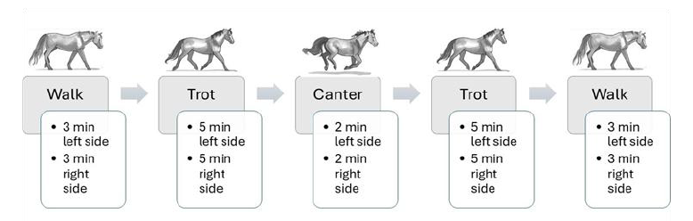
Eight horses in the control group (CG) underwent no exercise and remained in the pasture in the same environment. They were collected and measured for the same analyses at the same time points as the exercise group (EG) subjected to groundwork with a lunge rein (Fig.1). The horses were exercised three times per week on alternate days; for example, four horses were exercised on Mondays, Wednesdays, and Fridays, while the other four were exercised on Tuesdays, Thursdays, and Saturdays, all during the morning hours. The study was conducted du ring July and August (wintertime), with an average temperature of 12.3ºC and 60% relative humidity.
The horses were weighed weekly with a digital scale (model B650, Lider Balanças, Brazil). The experimenters assessed the horses' body condition score on a scale from 0 to 9 and evaluated the crest neck score through triple-blind palpation of fat deposits before and after the experiment, using a scale from 0 to 5 (Carter et al., 2009).
During the exercise, the horses were closely monitored using a modern heart rate and GPS made by Polar Electro, based in Lake Success, NY, USA. The system recorded heart rate, speed, and distance data every second. After the exercise, the data was analyzed using Polar Flow Software, which Polar Electro also developed.
The intervals between R wave peaks (RR) were monitored using the heart rate monitor with a precision of 1 ms. To analyze heart rate variability (HRV), Kubios HRV Standard software (Version 3.0.2) developed by the Biomedical Signal Analysis Group at the University of Kuopio in Finland was used, ensuring the accuracy of the data by removing sessions with less than 30 measurements. The Polar heart rate monitor was previously validated to assess horse heart rate variability (Mott et al., 2021). This analysis involves calculating various parameters such as mean RR interval, standard deviation (SDNN) of RR intervals, root mean square (RMSSD) of successive differences of RR intervals, and NN50 (number of successive RR interval pairs that differ more than 50 ms). To analyze time-varying changes, methods such as calculating the PNSi (Parasympathetic nervous system index based on mean R-R, RMSSD, and high frequency (HF) power) and SNSi (Sympathetic nervous system index calculated based on mean heart rate, Baevsky's stress index, and low frequency (LF) power) are used.
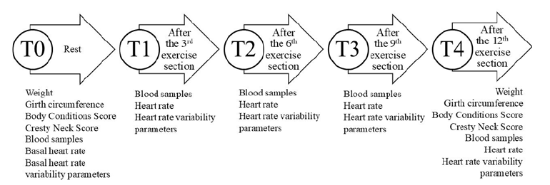
Blood samples were taken before exercising (T0) and after 1 hour of each groundwork session using 5mL vacutainer tubes with and without anticoagulant (ethylenediaminetetraacetic acid). The plasma was used to analyze GST and TBARS, and the serum was used to explore ROS and BChE. Blood samples were centrifuged for 10 minutes at 2,000 × g, then stored at −80 °C in 1.5-mL microcentrifuge tubes until analysis. Fig. 2 provides details of the time used to collect the samples.
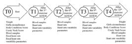
Illustration of collecting and analyzing time data in each moment. The control group was sampled at the same time points.
The serum was tested for the BChE (butyrylcholinesterase) enzyme using a modified version of the spectrophotometric method developed by Ellman et al. (2016). A 2mL reaction mixture containing 100mM potassium phosphate buffer (pH 7.5) and 1.0mM DTNB (5,5'-Dithiobis-2-Nitrobenzoic Acid) was used. The method is based on the formation of the yellow anion, 5,50-dithiol-bis-acid nitrobenzoic, which is measured by absorbance at 412nm during 2 minutes of incubation at 25°C. The enzyme was pre-incubated for 2 minutes, and the reaction was initiated by adding 0.8mM butyrylthiocholine iodide (BSCh). For the in vitro assay, varying concentrations of quercetin were added to a final volume of 2 mL. The absorbance was read on a spectrophotometer at 412 nm before and after incubation for 2 minutes at 25°C. All samples were run in duplicate or triplicate, and enzyme activity was expressed in µmol BSCh/min/mg of protein.
Lipid oxidation was quantified using the TBARS method described by Draper et al. (1993). To prevent further oxidation, plasma was mixed with 10% trichloroacetic acid and 50 ppm of butylated hydroxytoluene. The mixture was then incubated in a 90°C water bath for 30 minutes, cooled to room temperature using cold water, and centrifuged at 3,000 × g and 4 °C for 10 minutes. The resulting extract was reacted with a 0.02 mM TBA (thiobarbituric acid) solution at 90°C for 30 minutes, followed by cooling to room temperature and centrifugation at 10,000 × g at room temperature for 10 minutes. The absorbance was measured at 532 nm, and the TBARS levels were expressed as nMol MDA (malondialdehyde)/mg of protein.
Aliquots of 50µL of serum were used to perform a fluorescence assay to measure ROS (Myhre et al., 2003). The assay involved adding the serum to a medium containing Tris-HCl buffer (0.01mM, pH 7.4) and 2'-7'-dichlorofluorescein-diacetate of DCFH DA (dichloro-dihydro-fluorescein diacetate, 1 mM). After adding DCFH-DA, the medium was incubated in darkness for 1 hour. Fluorescence measurement was performed by exciting the sample at 488nm and measuring the emission at 525nm, with both slit widths set at 1.5nM. The results were expressed as DCFH-DA Fluorescence, using a standard curve of oxidized dichlorofluorescein to determine the oxidized dichloro-fluorescein.
A transparent 96-well plate was used to conduct the GSH analysis. The following steps were taken in sequence: 20μL of serum or plasma was added to the plate, followed by 20μL of distilled water or Milli-Q H2O, 10μL of GSH (100mM), and 240μL of a previously prepared system. The plate was then incubated at 37°C for 2 minutes. After that, the substrate 1-chloro-2,4-dinitrobenzene (CDNB) was added, and the reading was initiated in kinetic mode at λ=340 nm for 30 minutes with a 30-second interval at a temperature of 37°C. The system used for the activity was prepared with 20mL of 0.1 M potassium phosphate buffer (TFK) pH 7.5 (100 mM), supplemented with 0.0226 g of EDTA (2.5mM), and 10.5mL of distilled H2O or Milli-Q H2O. Finally, the enzyme activity was expressed in nanomoles of CDNB conjugated per minute per milligram of protein (Habig and Jakoby, 1981). The Coomassie Blue method (Bradford, 1976) was used to determine the protein concentrations, using bovine serum albumin as a standard.
The data was checked for normality using the Shapiro-Wilk normality test. The bidirectional repeated measures ANOVA test was conducted to analyze the interaction between groups and across time points. To identify which groups or time points differed, the Tukey post-hoc test was performed. Spearman r was calculated among the SNSi, PNSi, TBARS, ROS, BW, BCS, and CNS. To determine the strength of the correlation, we followed the Rule of Thumb (Hinkle et al., 2003). We considered values between 0.90 and 1 to have a very high correlation, 0.70-0.89 to have a high correlation, 0.50-0.69 to have a moderate correlation, 0.30-0.49 to have a low correlation, and less than 0.30 to have little to no correlation. The statistical analyses were performed using the commercial statistics package GraphPad Prism (GraphPad Prism 8 Software Inc., La Jolla, California, USA). We considered a P-value less than 0.05 significant for all analyses.
RESULTS
The exercise group experienced significant reductions in body weight, girth circumference, body condition score, and cresty neck score at the end of the experiment (T4) compared to the beginning (T0). In contrast, the control group did not show significant changes in these measurements. The differences between CG and EG at T4 were significant, demonstrating the impact of the exercise regimen on the measured parameters (see Table 1).
Table 2 shows the mean ± standard deviation values of biochemical parameters (TBARS, ROS, GST, and BChE) compared between two groups: the control group (CG) and the exercise group (EG) at different time points (T0-T4).
For TBARS, the CG did not show significant change over time (p=0.778). However, the EG displayed significant variations (p<0.001), with a noticeable increase at T2 and T3. Significant differences between the groups were observed at T2 and T3 (p<0.001), indicating an impact of the exercise on TBARS levels. Regarding ROS, the control group remained stable throughout the study period (p=0.887). In contrast, the exercise group experienced significant fluctuations (p<0.001), with a marked increase at T1 followed by decreases at T2, T3, and T4. Significant differences between the groups were observed at T1, T2, T3, and T4 (p<0.05). For GST, the control group showed no significant variation over time (p=0.897). However, the exercise group exhibited significant change (p=0.032), particularly an increase at T1 and T2. Significant differences between the groups were observed at T1 and T2 (p<0.05). Finally, for BChE, neither the control group (p=0.789) nor the exercise group (p=0.663) showed significant differences over time. Furthermore, no significant differences were found between the groups at any time (p>0.05).
Table 3 shows the mean ± standard deviation values of heart rate parameters and autonomic regulation indices (PNSi and SNSi) compared between different groups (control group [CG] and exercise group [EG]) and time points (T0-T4).
For heart rate (HR), the control group did not show a significant difference over time (p=0.887). On the other hand, the exercise group demonstrated significant change (p<0.001), with substantial increases observed from T1 to T4. A significant difference was observed between the groups at T1, T2, T3, and T4 (p<0.001), highlighting the impact of exercise on HR. Regarding maximum heart rate (HRmax), the control group remained stable across time points (p=0.823). However, the exercise group showed significant variations (p=0.007), with notable increases from T1 to T4. Significant differences between the groups were detected at T1, T2, T3, and T4 (p<0.001). For the parasympathetic nervous system index (PNSi), the control group showed no significant changes over time (p=0.728). In contrast, the exercise group experienced significant fluctuations (p<0.001), with an initial increase at T1 followed by a decrease at T3 and another increase at T4. Significant differences between the groups were evident at T1, T2, T3, and T4 (p<0.001). Lastly, the sympathetic nervous system index (SNSi) showed no significant variations over time for the control group (p=0.632). However, the exercise group displayed significant changes (p<0.001), with a marked increase at T3 followed by a decrease at T4. Significant differences between the groups were noted at T1, T2, T3, and T4 (p<0.001).
The correlation matrix in Table 4 displays the connections between oxidative metabolism markers (TBARS and ROS), autonomic regulation indices (PNSi and SNSi), and body fat parameters (weight, BCS, and CNS) in overweight horses undergoing aerobic exercise.
TBARS exhibited a strong positive correlation with SNSi, indicating that higher oxidative markers are linked to increased sympathetic nervous system activity. TBARS also showed strong negative correlations with body weight, body condition score, and cresty neck score, suggesting that oxidative marker decreases as body fat parameters reduce. ROS was strongly positively correlated with PNSi, implying a significant relationship between reactive oxygen species and parasympathetic nervous system activity. However, ROS did not correlate significantly with SNSi, BW, BCS, or CNS. PNSi did not correlate significantly with SNSi, BW, BCS, or CNS, indicating that parasympathetic nervous system activity is relatively independent of these parameters. SNSi had strong negative correlations with BW, BCS, and CNS, highlighting that sympathetic nervous system activity increases as body fat parameters decrease. Body Weight exhibited strong positive correlations with BCS and CNS, indicating that weight is closely associated with overall body condition and neck fat deposition. BCS was strongly and positively correlated with CNS, reflecting that the overall body condition score is a good indicator of neck fat accumulation.
DISCUSSION
The adverse impact of excessive weight on horses' performance has been widely recognized in the equine community. This includes an increased workload and the release of inflammatory proteins, which can harm the overall health of the animals (Pratt-Phillips and Munjizun, 2023). Our research on overweight horses revealed promising outcomes from a 12-session groundwork program. This program resulted in notable decreases in body weight, thoracic circumference, and body condition scores, indicating a positive response to the intervention.
This study's significant reduction in body weight, thoracic circumference, body condition, and cresty neck scores (CNS) indicates a positive response to aerobic exercise in overweight horses. The literature shows that elevated adiposity in equines is associated with metabolic dysfunctions, including insulin resistance and equine metabolic syndrome (EMS), as reported by Harris et al. (2020) and Reynolds et al. (2019). Adiposity control through exercise appears essential for preventing these conditions, and your study's results reinforce the importance of aerobic exercise in reducing body fat in horses predisposed to such dysfunctions.
The body enters a state of increased energy demand during physical exercise, particularly under high-intensity and prolonged duration. This heightened demand produces reactive oxygen species (ROS), known as free radicals, as a byproduct of incomplete oxygen reduction. The aerobic system is the primary metabolic pathway responsible for oxygen consumption, primarily within the mitochondria of cells. Approximately 85-90% of the oxygen produced during this process is utilized for energy production, while the remaining 10-15% is consumed by various oxidase and oxygenase enzymes or involved in direct oxidation reactions (Antunes Neto et al., 2018). Oxidative stress occurs when a significant imbalance exists between prooxidants and antioxidants, wherein prooxidants outweigh antioxidants. This imbalance can harm cellular functions, potentially leading to various health issues. Understanding this relationship is essential for comprehending the impact of exercise on the body’s oxidative balance.
In our study, we closely examined the levels of ROS in overweight horses during a structured aerobic exercise regimen. Our observations revealed a significant increase in ROS levels during the first week of training, indicative of an expected oxidative response to the new physical demands placed on the animals. However, as training progressed into the subsequent weeks, specifically starting in the second week, we noted that ROS levels began normalizing. This normalization suggests that the horses develop adaptive mechanisms to enhance their antioxidant defenses over time.
We also tracked levels of glutathione, a crucial antioxidant, and TBARS, a marker of lipid peroxidation. During the second and third weeks of training, we observed a concurrent rise in glutathione and TBARS levels. This increase in glutathione indicates a metabolic adaptation to the ongoing training. It suggests these horses were coping with oxidative stress and improving their overall antioxidant capacity. This pattern is particularly interesting because it contrasts sharply with the effects of high-intensity and prolonged exercise, which often leads to a disruption of oxidative balance due to excessive ROS generation. Instead, our findings highlight how controlled moderate aerobic exercise can stimulate beneficial adaptations in the antioxidant defense system, enhancing cellular resilience against oxidative damage. Overall, our research supports the conclusions of Antunes Neto et al. (2018) and highlights the importance of moderate-intensity exercise regimens for maintaining a healthy balance between the generation of reactive oxygen species (ROS) and antioxidant defense, particularly in horses with elevated body fat levels. By reducing the risk of oxidative damage, we can promote better health outcomes and performance in this group of animals.
Our findings are consistent with the research of Smarsh and Williams (2017), which reported similar results regarding lipid oxidation in trained and untrained yearlings. Additionally, the significant increase in plasma TBARS concentration observed during the second week of exercise may be due to the gradual accumulation of peroxides from lipid peroxidation, which decomposes into secondary oxidation products like MDA. This complex interaction among ROS, GSH, and lipid peroxidation demonstrates the dynamic physiological responses of horses to intensive training and provides valuable insights into their adaptive mechanisms.
Regular physical activity induces beneficial metabolic stress, termed "eustress," which triggers physiological changes that improve the body’s ability to handle stress over time (Caplin et al., 2021). Our study aligns significantly with these concepts, particularly regarding autonomic adaptation and oxidative stress. The exercise program in overweight horses resulted in an initial increase in reactive oxygen species (ROS) levels during the first training weeks, followed by a return to baseline levels, suggesting an adaptive response to initial oxidative stress. Similarly, sympathetic nervous system (SNS) activity increased early on and was subsequently balanced by restored parasympathetic activity, reflecting beneficial physiological adaptations. The stable levels of butyrylcholinesterase (BChE) observed throughout the training indicate that the exercise regimen effectively induced eustress rather than chronic stress, thereby avoiding excessive inflammatory or oxidative burdens (Matei et al., 2022). These findings underscore the importance of structured exercise in enhancing autonomic and metabolic health, contributing valuable insights into equine exercise physiology.
Butyrylcholinesterase, an enzyme found in the blood, has been identified as a potential stress biomarker associated with the autonomic nervous system in horses (Contreras-Aguilar et al., 2019a, 2019b). This enzyme is involved in the regulation of acetylcholine, which plays a crucial role in the interaction between the nervous and immune systems as an anti-inflammatory agent. BChE acts as a controller of acetylcholine activity through a negative feedback loop. When BChE levels decrease in the bloodstream, acetylcholine activity can increase, leading to a more robust systemic anti-inflammatory response and vice versa (Borovikova et al., 2000). The study found that BChE levels in horses remained consistent throughout the collection periods, suggesting that the animals could adapt to exercise without exceeding oxidative or inflammatory limits.
Evaluating the stress response related to exercise in horses is essential for effectively monitoring their training protocols and overall well-being. Our study reflected this approach using heart rate and heart rate variability indicators of autonomic regulation. These measures allowed for precise quantification of the sympathovagal balance, showing an increase in sympathetic nervous system activity in the early stages of training, followed by a gradual rebalancing with the parasympathetic nervous system recovery in the final weeks. Such findings align with existing research that employs HR and HRV as reliable parameters to assess the effects of exercise on autonomic modulation in horses (Coelho et al., 2022). These metrics offer insight into the physiological impact of exercise and serve as valuable tools for ensuring optimal health and adaptability in equine training regimes.
Although the parameters that constitute the indices of the Sympathetic Nervous System, such as R-R, RMSSD, and HF, and Parasympathetic Nervous System, such as HR, stress index, and LF, did not change over a few weeks (except for HR, as expected), the index themselves were altered. Specifically, there was a significant increase in sympathetic activity during the second and third weeks, followed by a decrease in parasympathetic activity. However, by the fourth week, parasympathetic activity had returned to its initial levels (T1). This shift in dominance from one system to another has been previously observed in horses undergoing training (Nyerges-Bohak et al., 2021; Coelho et al., 2022, 2023; Siqueira et al., 2023).
The study corroborates several existing research findings. The production of reactive oxygen species (ROS) due to aerobic exercise aligns with studies discussing the increased metabolic demand during physical activity, as described by authors like Antunes Neto et al. (2018), who examine the effects of exercise and diet on oxidative stress in horses. The increase in TBARS as an indicator of lipid peroxidation and its positive correlation with sympathetic nervous system activity, alongside the negative correlation with body fat parameters, is consistent with research on adaptive responses to exercise and the relationship between oxidative stress and the autonomic nervous system. Studies such as Stefanon et al. (2019) and Siqueira et al. (2014) highlight similar mechanisms in endurance horses, emphasizing the role of exercise in increasing sympathetic activity and oxidative adaptations. Additionally, the stability of butyrylcholinesterase and glutathione levels, which indicates adaptation to exercise without excessive inflammatory stress, supports references like Contreras-Aguilar et al. (2019b) and Matei et al. (2022), who explore the role of BChE and the antioxidant system in managing inflammatory and oxidative stress in equines during physical activity. These references theoretically support the findings observed in this study, showing that the physiological responses described are consistent with current scientific literature on the topic.
These findings underscore the importance of comprehensive management strategies in horse welfare and performance optimization. Attentiveness to factors such as body weight, condition, and stress levels is pivotal in minimizing oxidative stress and sympathetic nervous system activation in horses. Implementing tailored nutrition, exercise regimes, and stress-reduction practices contributes to equine health and performance, particularly in disciplines such as equine sports and exercise physiology. In summary, the intricate relationships observed in this study warrant further research into the underlying mechanisms and potential implications for equine health and performance. This emphasizes the need for ongoing investigation in this critical area of equine management.
CONCLUSIONS
This study reveals that a 12-session groundwork exercise regime effectively reduced weight, thoracic circumference, and body condition scores in overweight horses. The transient increase in reactive oxygen species (ROS) during the initial training phase and subsequent normalization, along with elevated GSH and TBARS levels, indicated positive metabolic adaptations without inducing oxidative stress.
The dynamic changes in autonomic nervous system activity, with an initial increase in sympathetic nervous system activity, followed by restored parasympathetic balance, further highlight beneficial physiological responses to exercise. Stable butyrylcholinesterase levels support the absence of excessive oxidative or inflammatory stress.
The correlations between oxidative stress markers, ANS indices, and body fat parameters suggest that exercise-induced fat loss is linked to oxidative metabolism and autonomic function changes. These findings emphasize the importance of integrated management strategies for optimizing equine health and performance, including nutrition, exercise, and stress-reduction practices. In summary, structured exercise programs improve the overweight horses' health and physiological resilience. Further research should explore these adaptations' underlying mechanisms and long-term implications for equine welfare and performance.
REFERENCES
- ANTUNES NETO, J.M.F.; RIVERA, R.J.B.; CALVI, R.G. et al. Effects of diet and exercise on body weight, oxidative stress, and immune response in horses. J. Equine Vet. Sci., v.69, p.22-27, 2018.
- BAMFORD, N.J.; POTTER, S.J.; HARRIS, P.A.; BAILEY, S.R. Effect of increased adiposity on insulin sensitivity and adipokine concentrations in horses and ponies fed a high-fat diet, with or without a once daily high glycemic meal. Equine Vet. J., v.48, p.368-373, 2016.
- BOROVIKOVA, L.V.; IVANOVA, S.; ZHANG, M. et al. Vagus nerve stimulation attenuates the systemic inflammatory response to endotoxin. Nature, v.405, p.458-462, 2000.
- BRADFORD, M.M. A rapid and sensitive method for quantitating microgram quantities of protein utilizing the principle of protein-dye binding. Anal. Biochem., v.72, p.248-254, 1976.
- CAPLIN, J.; FOLTZ, S.L.; GARNHAM, L.; ELLIS, A.D. Regular physical activities enhance physical and psychological health in horses: a review of the evidence. J. Equine Vet. Sci., v.104, p.e103689, 2021.
- CARTER, R.A.; GEOR, R.J.; STANIAR, W.B. et al. Apparent adiposity assessed by standardised scoring systems and morphometric measurements in horses and ponies. Vet. J., v.179, p.204-210, 2009.
- COELHO, C.S.; SILVA, A.S.B.A.; SANTOS, C.M.R. et al. Training effects on the stress predictors for young Lusitano horses used in dressage. Animals, v.12, 2022.
- COELHO, C.S.; SODRÉ, T.R.P.; SOUSA, L.N. et al. Analysis of stress predictors in Vaquejada horses running with different interval rest points. Stresses, v.3, p.858-867, 2023.
- CONTRERAS-AGUILAR, M.; ESCRIBANO, D.; MARTÍNEZ-SUBIELA, S. et al. Changes in saliva analytes in equine acute abdominal disease: a biochemistry approach. BMC Vet. Res., v.15, p.1-9, 2019a.
- CONTRERAS-AGUILAR, M.; HENRY, S.; COSTE, C. et al. Changes in saliva analytes correlate with horses’ behavioral reactions to an acute stressor: a pilot study. Animals, v.9, p.e993, 2019b.
- DRAPER, H.H.; SQUIRES, E.J.; MAHMOODI, H. et al. A comparative evaluation of thiobarbituric acid methods for determining malondialdehyde in biological materials. Free Radic. Biol. Med., v.15, p.353-363, 1993.
- ELLMAN, G.L.; COURTNEY, K.D.; ANDRES JR., V.; FEATHER-STONE, R.M. A new and rapid colorimetric determination of acetylcholinesterase activity. Biochem. Pharmacol., v.7, p.88-95, 2016.
- ELZINGA, S.; WOOD, P.; ADAMS, A.A. Plasma lipidomic and inflammatory cytokine profiles of horses with equine metabolic syndrome. J. Equine Vet. Sci., v.40, p.49-55, 2016.
- GORDON, M.E.; JERINA, M.L.; RAUB, R.H. et al. The effects of dietary manipulation and exercise on weight loss and related health indices in horses. Comp. Exerc. Physiol., v.6, p.33-42, 2009.
- HABIG, W.H.; JAKOBY, W.B. Assays for differentiation of glutathione S-transferases. Detoxication and drug metabolism: conjugation and related systems. Methods Enzymol., v.77, p.398-405, 1981.
- HARRIS, P.A.; BAMFORD, N.J.; BAILEY, S.R. Equine metabolic syndrome: evolution of understanding over two decades: a personal perspective. Anim. Prod. Sci., v.60, p.2103-2110, 2020.
- HINKLE, D.E., WIERSMA, W., JURS, S. G. Applied statistics for the behavioral sciences. Boston, MA: Houghton Mifflin, 5 ed., 756p., 2003.
- HENDRIX, J.; NIJS, J.; ICKMANS, K. et al. The interplay between oxidative stress, exercise, and pain in health and disease: potential role of autonomic regulation and epigenetic mechanisms. Antioxidants, v.9, p.1166, 2020.
- JANSSON, A.; GUNNARSSON, V.; RINGMARK, S. et al. Increased body fat content in horses alters metabolic and physiological exercise response, decreases performance, and increases locomotion asymmetry. Physiol. Rep., v.9, p.e14824, 2021.
- MARTIN-GIMENEZ, T.; BLAS, I.; AGUILERA-TEJERO, A. et al. Endocrine, morphometric, and ultrasonographic characterization of neck adiposity in Andalusian horses. Domest. Anim. Endocrinol., v.56, p.57-62, 2016.
- MATEI, D.; CATALINA, L.; ILIE, O. et al. Effects of exercise training on the autonomic nervous system with a focus on anti-inflammatory and antioxidant effects. Antioxidants, v.11, p.350-361, 2022.
- MOORE, J.L.; SICILIANO, P.D.; PRATT-PHILLIPS, S.E. Effects of diet versus exercise on morphometric measurements, blood hormone concentrations, and oral sugar test response in obese horses. J. Equine Vet. Sci., v.78, p.38-45, 2019.
- MOTT, R.; DOWELL, F.; EVANS, N. Use of polar V800 and actiheart 5 heart rate monitors to assess heart rate variability in horses. Appl. Anim. Behav. Sci., v.241, p.10541-10547, 2021.
- MOUSQUER, M.A.; CURSIO, B.R.; WENDT, C.G. et al. Perfil metabólico e escore de condição corporal de cavalos Crioulos finalistas da prova do Freio de Ouro. Sêmin. Ciênc. Agr., v.44, p.1947-1960, 2023.
- MYHRE, O.; ANDERSEN, J.M.; AARNES, H.; FONNUM, F. Evaluation of the Probes 2′,7′-dichlorofluorescin diacetate, luminol, and lucigenin as indicators of reactive species formation. Biochem. Pharmacol., v.65, p.575-1582, 2003.
- NYERGES-BOHAK, Z.; NAGY, K.; ROZSA, L.; POTI, P.; KOVACS, L. Heart rate variability before and after 14 weeks of training in Thoroughbred horses and Standardbred trotters with different training experience. PLoS One, v.16-26, p.e0259933, 2021.
- OTT, E.C.; CAVINDER, C.A.; WANG, S. et al. Oxidative stress biomarkers and free amino acid concentrations in the blood plasma of moderately exercised horses indicate adaptive response to prolonged exercise training. J. Anim. Sci., v.100, p.1-9, 2022.
- PRATT-PHILLIPS, S.; MUNJIZUN, A. Impacts of adiposity on exercise performance in horses. Animals, v.13, p.666, 2023.
- REYNOLDS, A.; KEEN, J.A.; FORDHAM, T.; MORGAN, R.A. Adipose tissue dysfunction in obese horses with equine metabolic syndrome. Equine Vet. J., v.51, p.760-766, 2019.
- SALIM, S. Oxidative stress and the central nervous system. J. Pharmacol. Exp. Ther., v.360, p.201-205, 2017.
- SHONO, S.; GIN, A.; MINOWA, F.; OKUBO, K.; MOCHIZUKI, M. The oxidative stress markers of horses - the comparison with other animals and influence of exercise and disease. Animals, v.10, p.617, 2020.
- SIQUEIRA, R.F.; TEIXEIRA, M.S.; PEREZ, F.P.; GULART, L.S. Effect of lunging exercise program with Pessoa training aid on cardiac physical conditioning predictors in adult horses. Arq. Bras. Med. Vet. Zootec., v.75, p.545-553, 2023
- SIQUEIRA, R.F.; WEIGEL, R.A.; NUNES, G.R.; MORI, C.S.; FERNANDES, W.R. Oxidative profiles of endurance horses racing different distances. Arq. Bras. Med. Vet. Zootec., v.66, p.455-461, 2014.
- SMARSH, D.N.; WILLIAMS, C.A. Oxidative stress and antioxidant status in Standardbreds: effect of age and training in resting plasma and muscle. J. Equine Vet. Sci., v.48, p.93-102, 2017.
- STEFANON, B.; PIGATTO, M.; FOGACCI, L. et al. Oxidative stress and inflammatory biomarkers in endurance horses. J. Equine Vet. Sci., v.83, p.102803, 2019.