 Experimental hourglass bariatric surgery with gastro-jejunal bypass (cberadg-y): description of the technique
ABSTRACT
Bariatric techniques are classified as restrictive, malabsorptive, or mixed, and seven are most used worldwide. Among these, the most popular are sleeve gastrectomy and Roux-en-Y gastric bypass, which are indicated for treating type 2 diabetes mellitus (T2DM) associated with obesity. The research describes an experimental bariatric surgery called hourglass with gastro-jejunal bypass (CBERADG-Y). The surgery consisted of dividing the stomach into two communicating compartments with pyloric closure and latero-lateral gastro-jejunal anastomosis between the caudal sac and the last jejunal quarter in Landrace pigs as an experimental model. Twelve obese castrated male Landrace pigs, with an average live weight of 110 kilograms, were operated on and divided equally into two groups: experimental (GE) and control (GC), with pre- and post-operative pain management and post-surgery dietary management. Among the results, the average surgery time was 75 minutes for the EG and 35 minutes for the CG, with the resulting weight loss derived from fat mass for the bariatric group, where Body Mass Index (BMI) was used as an evaluation variable. At the end of the experiment, it was concluded that hourglass bariatric surgery with gastro-jejunal bypass is a new, effective, and efficient surgical technique for reducing BMI in obese Landrace pigs.
Keyword:
bariatric surgery; gastro-jejunal bypass; metabolic surgery
RESUMO
As técnicas bariátricas são classificadas como restritivas, malabsortivas ou mistas, sendo sete as mais utilizadas em todo o mundo. Entre elas, as mais populares são a gastrectomia vertical e o bypass gástrico em Y de Roux, indicadas para o tratamento do diabetes mellitus tipo 2 (DM2) associado à obesidade. Esta pesquisa descreve uma cirurgia bariátrica experimental denominada cirurgia em ampulheta com bypass gastrojejunal (CBERADG-Y). A cirurgia consistiu na divisão do estômago em dois compartimentos comunicantes, com fechamento pilórico e anastomose gastrojejunal laterolateral entre o saco caudal e o último quarto do jejuno em suínos da raça Landrace, como modelo experimental. Doze suínos machos castrados e obesos da raça Landrace, com peso vivo médio de 110kg, foram operados e divididos igualmente em dois grupos: experimental (GE) e controle (GC), com manejo da dor pré e pós-operatória e manejo dietético pós-cirúrgico. Entre os resultados, o tempo médio de cirurgia foi de 75 minutos para o grupo experimental (GE) e de 35 minutos para o grupo controle (GC), com a perda de peso resultante derivada da massa gorda no grupo bariátrico, em que o índice de massa corporal (IMC) foi utilizado como variável de avaliação. Ao final do experimento, concluiu-se que a cirurgia bariátrica em ampulheta com bypass gastrojejunal é uma técnica cirúrgica nova, eficaz e eficiente para a redução do IMC em suínos Landrace obesos.
Palavras-chave:
cirurgia bariátrica; bypass gastrojejunal; cirurgia metabólica
INTRODUCTION
Obesity syndrome is a global pandemic causing morbidity and mortality in humans. Data from the World Health Organization (WHO) shows that approximately one billion adults and children are morbidly obese and more than two billion are overweight. (Fujisaka et al., 2023). Among the recommended therapies for this condition is bariatric/metabolic surgery, which provides the most robust option for the treatment of obesity compared to other currently available strategies, including lifestyle modification, pharmacotherapy, and endoscopic therapy (Shikora et al., 2022; Khan et al., 2016; Papamargaritis and Le Roux, 2021). There are about 50 surgical procedures with advantages and disadvantages (Shikora et al., 2022).
Bariatric techniques are classified as restrictive, malabsorptive, or mixed, and worldwide, seven are most used, two of which are the most popular (sleeve gastrectomy and Roux-en-Y gastric bypass), which are indicated for treating patients with DM2 associated with obesity (Shikora et al., 2022; Leitón Chavez and Vargas Lòpez, 2016). Sleeve gastrectomy (SG) is the most widely used surgery and since 2013 has become the preferred weight loss procedure, even above Roux-en-Y gastric bypass (RYGB) (Khorgami, 2017).
Bariatric surgeries have a high potential for causing certain universal postoperative complications such as leaks, bleeding, obstruction, and infection. In addition, there are unique long-term problems such as intestinal obstruction due to internal mesenteric hernias, stenosis, peptic and esophageal ulcers, and esophageal intussusception. However, the most common concerns in long-term follow-up are dumping syndrome, nutritional deficiencies due to inadequate levels of vitamins and minerals, iron deficiency anemia, and the irreversibility of the techniques, which would be limiting in pediatric patients (Shikora et al., 2022).
The objective of this article is to describe a new bariatric technique, called the hourglass with gastro-jejunal bypass, represented by the acronym CBERADG-Y.
ETHICAL ASPECTS
The choice of this experimental species was based on the Helsinki Declaration reviewed in 2013, and the number and management of animals was approved and supervised by the Ethics Committee of the Faculty of Veterinary Medicine and Animal Science of University of Córdoba, Colombia, through Act No. 009 of November 30, 2021.
MATERIALS AND METHOD
Twelve obese Landrace pigs, castrated males, with an average live weight of 110 kilograms and 6.5 months of age, were selected and divided equally into GC and GE groups. The animals chosen came from two litters from the pig breeding program at University of Córdoba, Colombia, and were managed individually in two-square-meter pens connected to each other under controlled conditions for five months.
To achieve obesity, the pigs were fed for four months from two months of age with a commercial finishing diet (Italcol® Levante 1 pig feed) enriched with 10 % commercial vegetable oil (Megaceite S.A.S) and 15 % molasses, based on the work of Hernández et al. (2016). At the start of the study, all animals were in good health based on clinical and paraclinical examination. The experimental phase began when the pigs were 6.5 months old and lasted one month, with BMI assessment on day 0 and day 30 post-surgery. The EG consisted of six pigs that underwent CBERADG-Y, and the CG consisted of six pigs that underwent celiotomy. All animals in the study were withdrawn from energy supplementation during the month of experimentation.
Berástegui experimental farm, the research site, is in a rural area of the municipality of Ciénaga de Oro, Córdoba, Colombia, with an average temperature of 29°C, relative humidity of 45-65%, altitude of 22 meters above sea level, and average annual rainfall of 1200mm.
To calculate BMI, weight in kilograms was divided by the square of the occipito-coccygeal length in centimeters (adapted from the publication by Plua et al., 2022). Measurements were recorded at the start of the study, on day 15, and on day 30 after surgery. The experimental bariatric technique was recorded by photographic sequence and videos according to surgical times.
This is a descriptive, cross-sectional, convenience, non-probabilistic study. Descriptive statistics were used to analyze the BMI data, yielding numerical results that express the behavior of the variable per individual and per group in the sample.
The animals were fasted from food and water for eight and ten hours, respectively. The pre-anesthetic protocol consisted of an analgesic (flunixin meglumine at 1.1mg/kg bw/im) and an antiemetic (metoclopramide at 0.5mg/kg bw/im). Azaperone (4mg/kg bw/im) was used as a tranquilizer, and xylazine (0.5mg/kg bw/iv) and ketamine (2mg/kg bw/iv) were mixed for induction, while maintenance was achieved with isoflurane delivered in oxygen. Anesthesia was supervised by Julio E. Cuervo, an anesthetist at the veterinary clinic of the Faculty of Veterinary Medicine and Animal Husbandry at University of Córdoba.
The experimental technique is classified as restrictive or malabsorptive because, with the compartmentalization of the stomach, the gastric capacity of the first receiving sac is reduced by approximately 20% in order to generate an early and prolonged feeling of satiety with a much smaller amount of food than is usually ingested, and because, on the other hand, the chyme reaches the last quarter of the small intestine (distal jejunum and ileum) more quickly, with a consequent reduction in BMI. All surgeries were performed by the author of the experimental technique, who is a professor of surgery and junior researcher according to the MinCiencias classification in Colombia.
After surgery, all pigs were given flunixin meglumine (1.1mg/kg bw/im/12 hours/3 days) for pain control. Oxytetracycline (5mg/kg bw/24 hours/3 days) was chosen for antibiotic therapy. There was no restriction on water intake, and feeding began between 16- and 24-hours post-surgery with the daily ration according to the manufacturer's instructions, which was divided into three portions administered every four hours starting at 8:00 a.m.
Development of the technique: Experimental hourglass bariatric surgery with gastrojejunal bypass (CBERADG-Y). Slaughtering operations were then used for the anatomical examination of the abdominal viscera in situ and ex situ, measurements of the length of the small intestine were taken, and the surgical technique was practiced with anatomical specimens to acquire manual dexterity (Figures 1 and 2).
The surgical technique began with the design shown in Fig. 1.

Schematic representation of experimental hourglass-shaped bariatric surgery with gastro-jejunal bypass (CBERADG-Y) in obese adult Landrace pigs. 1. Cardiofundic pouch, 2. Communicating duct (transit channel), 3. Somatopyloric pouch, 4. Gastro-jejunal diversion, 5. Pyloric closure, 6. Biliopancreatic enteric segment, 7. Common or confluent enteric segment, 8. Ileum, 9. Cecum (9), 10. Greater and lesser omentum, X: sites of insertion of the jejunal serosa to the serosa of the somatopyloric pouch.

A. Landrace pig stomach specimen for identification of anatomical regions, where C = cardia, F = fundus, Cu = body, P = pyloric antrum, and EP = pyloric sphincter. B. Simulation of the experimental hourglass surgery with gastro-jejunal bypass on anatomical Landrace pig stomach specimens with the terminology used to describe the surgical technique.
The animals that made up CG underwent celiotomy, and their intestines and stomach were manipulated for 15 minutes to simulate surgical stress with postoperative management similar to that of the EG.
RESULTS
The surgical approach to expose the stomach was based on the technique described by Toro (2015), Fossum and Hedlund (2013), and Martínez et al. (2014) who describe the Billroth II gastrojejunostomy technique with and without modification. The abdominal anatomical examination of the pig was based on the report by Baschar (2020).
According to the results obtained, there were no deaths due to the experimental technique, which is considered bariatric/metabolic, initially designed to induce weight loss at the expense of body fat mass, but which influenced zoometric parameters such as weight, abdominal circumference, occipito-coccygeal length, and body mass index. These results were published in a scientific article by Martinez et al. (2014). The proposed technique would be classified as mixed and is based on the reorganization of the digestive tract, where the stomach's reception capacity was reduced, with the fundus being the first reception site and the location of the oxyntic cells responsible for producing ghrelin, hydrochloric acid, and intrinsic gastric factor. The theory was postulated that the physical distension caused by food in the first gastric receptor sac would possibly generate negative feedback on ghrelin levels, with the consequent feeling of early and prolonged satiety with a much smaller amount of food than usual, resulting in a reduction of ⅓ of the volume consumed before surgery in the GE; because consumption went from 3kg of food/day to only 1kg, while the GC maintained its consumption of 3kg/day.
In addition, the rapid arrival of chyme to the last quarter of the small intestine (distal jejunum) could influence the microbiota and intestinal epithelial histophysiology and indirectly influence organs that affect digestion and metabolism. One of the effects would be on the release and levels of one of incretins that influence satiety and insulin levels, such as GLP-1.
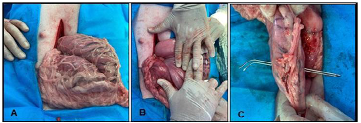
The surgical technique is presented in a series of photographs showing the gastric phases (Figures 3-6) and the gastro-jejunal anastomosis phase (Figure 7), described below:

First surgical step of experimental hourglass-shaped bariatric surgery with gastro-jejunal bypass (CBERADG-Y) in obese adult Landrace pigs, where A: Intraoperative exposure of the stomach of an obese Landrace pig and identification of its parts to initiate gastric compartmentalization B: Digital measurement from the greater curvature of the stomach to determine the needle insertion height. C: Needle insertion for placement of the U-shaped sutures responsible for stomach compartmentalization.

Second surgical step of experimental hourglass-shaped bariatric surgery with gastro-jejunal bypass (CBERADG-Y) in obese adult Landrace pigs, where intraoperative gastric compartmentalization in Landrace pigs showing the three U-shaped pins made with a 20 Fr Foley catheter. Where C-F = cardio-fundic sac and S-P=somato-pyloric sac.

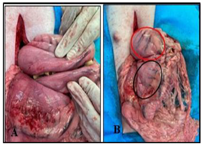
Third surgical step of experimental hourglass-shaped bariatric surgery with gastro-jejunal bypass (CBERADG-Y) in obese adult Landrace pigs, where: A. Manual approximation of the sero-muscular folds of the two gastric compartments, to invaginate the 20 Fr Foley catheter. B. Folds sutured with simple stitches making the 20 Fr Foley pins invisible.

Fourth surgical step of experimental hourglass-shaped bariatric surgery with gastro-jejunal bypass (CBERADG-Y) in obese adult Landrace pigs, where: A. Pegged with a 20 Fr Foley catheter to block the pyloric outlet. B. Omentalization of the peg.

Sixth surgical step of experimental hourglass-shaped bariatric surgery with gastro-jejunal bypass (CBERADG-Y) in obese adult Landrace pigs, where the stomach and small intestine are ex situ to initiate the gastro-jejunal anastomosis A. The length of the enterotomy and gastrotomy is determined using the numerical scale of the 10mL syringe as a measuring instrument, which measures the distance from the mesenteric edge to the anti-mesenteric edge. B. Gastrotomy of approximately five centimeters.
The procedure began with an incision of the skin and subcutaneous tissue in the epigastric area. Once the linea alba was visualized, an incision-puncture was made with a No. 22 scalpel until the abdominal cavity was reached, and the incision was enlarged with Metzenbaum scissors. The scissors were used in a semi-closed position, making progressive movements along the linea alba.
For the gastric phase, the stomach was located and exposed by grasping the body of the organ with gauze mounted on the thumb, index, and middle fingers. Once exposed, it was isolated with surgical towels moistened with 0.9% saline solution, and manual compartmentalization into two communicating sacs was initiated with the placement of three simple "U" stitches with pins (2.5cm) made from 20 Fr Foley catheters. To determine the height at which the third stitch would be placed, an estimate was made based on the greater curvature in the direction of the lesser curvature, using the surgeon's fingers as a reference, equivalent to four fingers (Fig. 3B). The multilayer stitches with 1-gauge polypropylene were perpendicular to the greater curvature and parallel to the blood vessels. To facilitate the passage of the thread edges, a U-shaped needle with an eyelet at each tip designed by the author was used (Fig. 3C).
To prevent the pins from irritating or damaging the serosa of adjacent viscera, the gastric surface proximal to the surgeon and adjacent to the pins was imbricated. The folds of the stomach that formed in the area were approximated with a simple continuous stitch or with three (3) simple stitches with 2-0 polydioxanone (Figures 5A and 5B).
The sac with the smallest capacity is made up of the cardia and fundus portions and was called the cardio-fundic sac (red circle in Fig. 5B) with an approximate capacity of 20% of the total stomach, and the sac with the largest capacity is made up of the body and pyloric portion and is called the somato-pyloric sac (black circle in Fig. 5B) with an approximate capacity of 80%.
The connecting passage between the new compartments was located near the lesser curvature, and its approximate diameter corresponded to the diameter of the small intestine. Intraoperatively, its permeability was verified by passing a 20 Fr/30mL Foley catheter balloon filled to maximum capacity with air, with a circumferential measurement of 11cm. To completely block the pyloric passage, which is where the gastric phase begins, a U-shaped suture was placed in a manner similar to that described for the creation of the two gastric sacs, and the peg was then omentalized to prevent irritation and possible adhesions (Figures 6A, 6B).
For the second surgical phase (gastrojejunal anastomosis) and to reduce manipulation of the viscera to be incised, four reference points were placed with 3-0 polyamide on the anti-mesenteric surface of the small intestine and on the free surface of the omental insertion of the somatopyloric sac.
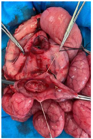
First, an incision-puncture was made in the center of the somato-pyloric sac of the stomach until the gastric lumen was reached. The center of the sac corresponds to the intersection of two imaginary lines, one drawn from the midpoint of the greater curvature to the midpoint of the lesser curvature, and the second line drawn from the midpoint of the dividing fold of the two sacs to the pylorus. The incision-puncture was enlarged with Metzembaum scissors until reaching the approximate equivalent of the measurement from the mesenteric to the antimesenteric edge of the small intestine, approximately five centimeters, using a 10mL disposable syringe (Figures 7A, 7B).
Next, the luminal contents were displaced using a digital milking maneuver in the selected intestinal portion and clamped cranially and caudally to the site identified for the incision. The jejunal incision-puncture at the anti-mesenteric edge was enlarged with Metzenbaum scissors, considering the same considerations as above (approximately the distance from the mesenteric edge to the anti-mesenteric edge of the small intestine, equivalent to five centimeters) (Fig. 8).
The incised anatomical sites were approximated for anastomosis, using 3-0 PDS and simple continuous sutures, starting with two simple reference points placed at the ends of the incisions. The stitches began at the edges distal to the surgeon and ended at the edges proximally to the surgeon. Once the tissue synthesis was complete, a leak test was performed to verify that there was no leakage of contents into the abdominal cavity, and omentalization was optional. The surgery ended with serosal fixation of the jejunum to the serosa of the somato-pyloric sac, considering an intestinal portion approximately 10 cm caudal and cranial to the gastro-jejunal anastomotic site. The fixation was made to the highest possible portion of the sac (Fig. 9).
Next, visceral lavage was performed with 0.9% saline solution at body temperature and the viscera were reintroduced. The abdominal anatomical planes were closed in the opposite direction to the incision, using 0 and 3-0 gauge polylactide 910 and simple continuous stitches for the muscular and subcutaneous planes, respectively, and 2-0 silk for the skin closure with simple stitches.

Seventh surgical step of experimental hourglass-shaped bariatric surgery with gastro-jejunal bypass (CBERADG-Y) in obese adult Landrace pigs, where The pig's stomach and small intestine are ex-situ, and the approximation of the two incised sites to begin the gastro-jejunal anastomosis is shown.

Eighth surgical step of experimental hourglass-shaped bariatric surgery with gastro-jejunal bypass (CBERADG-Y) in obese adult Landrace pigs, where; the pig's stomach and small intestine are ex situ and the serosal fixation of the jejunum to the serosa of the somato-pyloric sac (arrows), site of gastro-jejunal bypass (Dg-y), somato-pyloric sac (Ss-p), sf: cardio-fundic sac.
Some considerations regarding the surgical technique are:
-
The experimental technique began with the gastric phase to avoid unnecessary traction on the viscera, which is more fixed and heavier when compared to the small intestine.
-
The anatomy of the Landrace pig places the stomach deep in the cranial abdomen, forcing the surgeon to work on the organ almost in situ.
-
The gastric phase began with pyloric closure to reduce the risk of enteric reflux and thus contamination.
-
A double-pointed needle with a cutting edge and an eyelet at each end was designed to facilitate the placement of U-shaped stitches in the stomach (Figure 8).
-
The simple stitches are secured with a Foley catheter (2.5cm) to reduce the possibility of the knot migrating into the gastric lumen, which can cause mucosal ulcers due to continuous friction resulting from gastric motility.
-
The communicating passage between the gastric pouches is located at a high point on the assumption that food reaches the somatopyloric pouch driven more by peristalsis than by gravity, which could promote stimulation and mixing with gastric secretions.
-
The location of the gastric neo-ostium was made at a point closer to the lesser curvature and further from the greater curvature with the theoretical assumption of reducing the possibility of enteric reflux, for which it was complemented with serous fixation of the jejunum to the serosa of the somato-pyloric sac. In addition, it was postulated that the neo-ostium at a higher anatomical point could reduce the emptying time into the small intestine and promote the mixing of the contents derived from the cardio-fundic sac with gastric secretion, and that both theories ultimately seek to reduce the risks of complicating gastro-enteric ulcers.
-
To identify the jejunal portion of the bypass, it is necessary to begin by identifying the ascending colon and cecum, which lead to the ileum, the starting point for measuring the last quarter of the small intestine.
-
The small intestine of pigs is very thin compared to that of dogs or cats, which requires very delicate handling with minimal traction to avoid tearing.
-
Anesthesia induces relaxation of the abdominal muscle component but not of the parietal peritoneum, which easily detaches from its insertion once the celiotomy is enlarged.
-
The dark coloration of the small intestine after a certain period of exposure was resolved by further opening the parietal peritoneum, which was the cause of vascular compression.
-
For enterotomy, it is necessary to place four reference points at 9, 3, 12, and 6 o'clock. This step will prevent unnecessary injury to the anti-mesenteric surface and vascular bundle, because the empty jejunum and its thin wall cause the mesenteric and anti-mesenteric surfaces to be very close together.
-
The gastrojejunal anastomosis was closed with simple continuous stitches using 3-0 monofilament suture material, with the thread passing approximately 2 mm from the tissue edge and about 2 mm between needle passages.
-
To reduce manipulation and surgery time, the viscera were handled as much as possible with reference points using 9-10 polyglactin or 3-0 silk.
-
High intra-abdominal pressure is an important consideration to avoid including the intestine at the time of celiorrhaphy. This step was overcome by beginning the closure of the abdomen at the ends with the placement of two "X" stitches that also served to pull the wall, with the aid of a mosquito forceps.
-
The average duration of the surgeries was 75 minutes, which, together with preoperative fasting, exposure, and visceral manipulation, caused dehydration in the patient. Therefore, it was decided to perform visceral lavage with 0.9% saline solution at body temperature. In addition, prior to celiorrhaphy, 1 liter of 0.9% SS was infused intraperitoneally for the purpose of rehydration, facilitating visceral rearrangement, reducing the risk of fibrin adhesion, and diluting possible gastrointestinal residues.
-
Once recovered from anesthesia, the pigs had access to water in their pens. Feeding began between 16 and 24 hours post-surgery.
Some of the theoretical advantages of surgical techniques are:
-
It may be partially reversible in cases where it induces secondary anatomical or physiological deficiencies over time, especially in young patients, where the prevalence of obesity is increasing. This age group could be considered ideal for the proposed surgery.
-
Patients who undergo surgery clinically regain their appetite within 24 hours after surgery, with lower quantitative consumption of food on the fifth day after surgery (1/3 of the usual amount) and apparent satiety until the end of the experiment.
-
The physical reduction of the first gastric pouch allows for rapid filling with less food.
-
The non-excision of portions of the stomach possibly maintains the capacity for synthesis of precursors or hormones that influence gastrointestinal physiology and hemodynamics.
-
Both gastric pouches can be accessed endoscopically if evaluation is necessary.
-
There are few incisions (two) in the digestive tract and a single anastomosis site, which is latero-lateral with less luminal exposure time and less tissue manipulation.
-
The absence of incisions to compartmentalize the stomach reduces the chances of scar stenosis, allowing the stomach to accommodate food intake and contract without restriction.
-
The rapid recovery of patients after undergoing this technique allows it to be proposed as an outpatient surgery.
-
Gastrotomy for gastrojejunal anastomosis is performed below the insertion of the Latarget nerve and is complemented by gastrointestinal serosal suture 10 cm cranially and caudally from the anastomosis to minimize possible motility alterations, reduce biliary reflux, and secondary marginal ulcers.
-
The gastrointestinal anatomical redesign does not appear to cause motility disorders (no clinical signs of dysphagia, reflux, malabsorption syndrome, or constipation were observed).
-
The connecting and visceral peritoneum do not suffer damage beyond that caused by the temporary dehydration inherent in visceral exposure, so signs of peritonitis are minimal and the risk of mesenteric incarceration with or without intestinal volvulus is minimized.
-
Postoperative management with medication lasted a maximum of three days (analgesia and antibiotic therapy) with a return to ambulation after anesthesia recovery.
-
Open surgery does not require a learning curve, specialized instruments, or robust equipment.
-
The anatomical planes and surgical steps are easy to recognize and interpret.
-
There was no pig mortality attributable to the experimental surgical technique.
The results for the BMI variable are shown below in a group range (Table 1).
When comparing the groups, it was determined that the CG increased its BMI by 17%, while the EG reduced it by 6%, demonstrating that experimental bariatric surgery has a positive influence on this variable, but NOT energy supplementation restriction or surgical stress.
DISCUSSION
Bariatric/metabolic interventions are currently more focused on their applicability in obese patients with type 2 diabetes mellitus due to the various benefits achieved in pancreatic and hepatic histophysiology, with improvements in systemic insulin activity and, therefore, in the restoration of euglycemic levels, and secondarily, to the construction of knowledge that defines their applicability in non-obese diabetic patients (Martinez et al., 2024; Medina, 2018).
Studies cited in the review by Pacheco et al. (2019) have amply demonstrated that weight loss reduces morbidity and mortality and that bariatric surgery is currently the most effective treatment modality for achieving sustained weight reduction.
The development of this bariatric surgery is not intended to standardize a new technique, but rather to develop and contribute new knowledge that will help understand and improve the condition of obese patients without metabolic disease, with the ultimate goal of preventing the possible changes that accompany the condition, which, according to Amezquita et al. (2022), has a high morbidity/mortality rate. In addition, it is necessary to develop a technique that, over time, does not predispose patients to other secondary alterations, including deficiencies, because there is a possibility that it will be indicated in increasingly younger patients (Papamargaritis and Le Roux, 2021).
A new surgical technique should be easy to perform, not require a steep learning curve, reduce operating time (Parmar and Mahawar, 2018), not require specialized instruments or robust equipment, be reversible or partially reversible, and have minimal complications such as reflux, ulcers, leaks, stenosis, or occlusion; the organs involved should be easy to recognize and allow for minimal manipulation; the surgery should have minimal postoperative complications attributable to the incisional technique, with minimal anastomosis and no mesenteric opening, as occurs with Roux-en-Y gastric bypass or B-P bypass (Toro, 2015), which achieves sustainable weight loss over time and indirectly prevents or stops complications associated with obesity, such as degenerative liver disease, and, if present, possibly slows it down, as stated by Holst et al. (2022). These general conditions make a bariatric surgical technique effective and efficient, which, from our intraoperative perspective and during the study period, is achieved by CBERADG-Y.
Currently, bariatric surgery is the most effective treatment modality for achieving sustained weight loss and demonstrable reductions in mortality. In addition, it has been shown to be effective in improving and even remitting comorbidities in obese individuals, and there is evidence of its superiority over medical treatment. The indication for bariatric surgery in humans is given when the BMI is >40 kg/m²or BMI >35kg/m²with major comorbidities or BMI >30kg/m²with dysmetabolic syndrome or DM2 that is difficult to control (Pacheco et al., 2019).
The BMI variable behaved similarly to the results published by Antonio (2019) in his study of bariatric surgery in adolescents, so it is presumed that fat mass had the greatest influence on weight loss in the pigs in the EG. The tendency of the pigs in the EG to lose weight during the month of the study suggests that experimental bariatric surgery influenced this variable and that the results may be similar to those reported by Parmar and Mahawar (2018). These results are similar to one of the variables in Hernández (2016) and encourage the idea that CBERADG-Y could have a positive influence on weight control.
Furthermore, according to the data provided by the CG, discontinuing the supplement was not sufficient to reduce BMI. From this, it can be inferred that the CBERADG-Y had a greater influence on body condition than the type of food supplement, because the CG maintained its behavior and obese body condition until the end of the study. According to Stinson (2018), higher energy consumption from fat and carbohydrates leads to higher energy intake, which is clinically expressed by higher food consumption and subsequent weight gain, as recorded in the CG, which had the highest BMI.
The weight loss achieved in Landrace pigs after experimental surgery (CBERADG-Y) opens the door to longer-term research that will allow for a better understanding of the postoperative anatomy and physiology or mechanism(s) by which one of the objectives of the technique (weight loss) was achieved, as well as answer questions such as: Is the weight loss sufficient and does it remain stable over time with minimal complications? How long after surgery does weight loss stabilize? How is weight loss distributed in percentage terms according to the five-compartment classification? Can CBERADG-Y help reduce the comorbidity associated with overweight and obesity? Could CBERADG-Y be used in adolescent patients who are overweight or obese without comorbidities? Among others.
After experimental bariatric surgery, there is an expectation of significant weight reduction, mainly at the expense of fat mass and a very small or limited decrease in lean mass, since excessive loss of the latter can have different consequences on metabolism, as lean body mass is an important component that is closely related to glycemic regulation, weight loss maintenance, and therefore the long-term success of the surgery. An uncontrolled decrease generally affects people's quality of life due to functional deterioration and loss of strength (Plua et al., 2022).
The tendency of pigs in the EG to lose fat mass, according to subjective assessment and weight during the study month, suggests that experimental bariatric surgery influenced this variable and that the results may be similar to those reported by Parmar and Mahawar (2018), who achieved weight loss with maintenance over time when they performed gastric bypass with an anastomosis or mini-gastric bypass in adult humans, where weight loss at six, 12, 24, and 60 months was 60, 72, 78, and 76.6%, respectively. Similarly, Hernández (2016) report that duodeno-ileal bypass with sleeve gastrectomy, officially known by its acronym SADI-S, is an effective technique because it achieves a weight loss percentage of over 90% two years after surgery, which is maintained until the fifth year (Hernández et al. 2016; Laferrère B. et al., 2011).
CONCLUSION
The clock-shaped surgery with gastro-jejunal bypass (CBRADG-Y) can be classified as a bariatric gastrointestinal technique, restrictive-malabsorptive (mixed) and in development, according to the anatomical structures involved, weight reduction, and follow-up or stage of development over time, respectively. Furthermore, it is an effective and efficient technique for inducing fat mass loss in obese Landrace pigs.
ACKNOWLEDGEMENTS
Thanks to the swine program of the FMVZ of the University of Cordoba-Colombia for providing the pigs, their feed and management.
REFERENCES
- AMEZQUITA, G.M.V.; BAEZA, L.; MATÍAS RIOS, E. et al Bariatric surgery in adolescents. Rev. Chil. Pediatr, v.90, p.17, 2022.
- ANTONIO, J.; COCKRELL, H.; KANG, H.S. et al A pilot study of laparoscopic gastric plication in adolescent patients with severe obesity. J. Pediatr. Surg, v.54, p.1696-1701, 2019.
- BASCHAR, H.A. Gastroduodenostomy with magnets: a novel surgical option for the palliative treatment of irreversible pyloroduodenal obstructions. 2020. 105f. Thesis (Doctor of Veterinary Science) - National University of La Plata, ARG.
- FOSSUM, T.W.; HEDLUND, C.S. Gastric and intestinal surgery. Vet. Clin. North Am. Small Anim. Pract., v.33, p.1117-1145, 2013.
- FUJISAKA, S.; WATANABE, Y.; TOBE, K. The gut microbiome: a core regulator of metabolism. J. Endocrinol, v.256, p.e220111, 2023.
- HERNÁNDEZ HURTADO L.; SÁNCHEZ-MARGALLO, F.M.; DE LA CRUZ VIGO, J.L. et al Changes on adipose tissue distribution after laparoscopic Roux-en-Y gastric bypass in obese göttingen minipig. Effects on glucose metabolism. Obes. Surg, v.26, p.3001-3006, 2016.
- HOLST, J.J.; MADSBAD, S.; BOJSEN-MØLLER, K.N. et al Mechanisms in bariatric surgery: Gut hormones, diabetes resolution, and weight loss. Surg. Obes. Relat. Dis., v.14, p.708-714, 2018.
- KHAN, S.; ROCK, K.; BASKARA, A. et al Trends in bariatric surgery from 2008 to 2012. Am. J. Surg, v.211, p.1041-1046, 2016.
- KHORGAMI, Z.; SHOAR, S.; ANDALIB, A. et al Trends in utilization of bariatric surgery, 2010-2014: sleeve gastrectomy dominates. Surg. Obes. Relat. Dis, v.13, p.774-778, 2017.
- LAFERRÈRE, B.; REILLY, D.; ARIAS, S. et al Differential metabolic impact of gastric bypass surgery versus dietary intervention in obese diabetic subjects despite identical weight loss. Sci. Transl. Med, v.3, p.80re2, 2011.
- LEITÓN CHAVEZ, A.; VARGAS LÒPEZ, V. Bariatric surgery as a treatment option for type 2 diabetes mellitus. Pathophysiological fundamentals. Rev. Clín. Esc. Med, v.6, n.2, 2016.
- MARTINEZ, M.M.; GÓMES, J.; MEYER, M.E. et al Digestive tract surgery in companion animals Colombia: University of Cordoba Publishing Fund, 2014. 156p.
- MARTINEZ, M.M.; PÉREZ, D.M.; LANCHEROS, V. Zoometry of obese pigs undergoing experimental bariatric surgery hourglass with gastro-jejunal bypass. Rev. MVZ, v.29, p.e3483, 2024.
- MEDINA ESPITIA, N.M. Changes in muscle mass and fat-free mass in individuals undergoing gastric sleeve and Roux-en-Y gastric bypass surgery: literature review. 2018. 76f. Monograph (Undergraduate) - Pontificia Universidad Javeriana, Bogotá, COL.
- PACHECO SÁNCHEZ, D.; PÉREZ-ROMERO, N.; DOMÍNGUEZ-GARCÍA, J.; DE LA CRUZ-MERINO, L. Update on bariatric/metabolic surgery. Nutr. Clin. Med, v.3, p.113-127, 2019.
- PAPAMARGARITIS, D.; LE ROUX, C.W. Do gut hormones contribute to weight loss and glycaemic outcomes after bariatric surgery? Nutrients, v.13, p.762, 2021.
- PARMAR, C.D.; MAHAWAR, K.K. One Anastomosis (Mini) gastric bypass is now an established bariatric procedure: a systematic review of 12,807 patients. Obes. Surg, v.28, p.2956-2967, 2018.
- PLUA, W.; VÁSQUEZ, H.A.; JARRIM, K. et al Impact of gastric bypass on the body mass index of diabetic adults in the first post-surgical year. Rev. Cient. Invest. Conoc., v.6, p.612-624, 2022.
- SHIKORA, S.A.; EDGERTON, C.; HARRIS, D.; BUCHWALD, H. Metabolic surgery. Curr. Probl. Surg, v.59, p.101059, 2022.
- STINSON, E.J.; PAOLO PIAGGI, P.; IBRAHIM, M. et al High fat and sugar consumption during ad libitum intake predicts weight gain. Obesity, v.26, p.689-695, 2018.
- TORO, L.Z. Experimental surgery for type 2 diabetes mellitus. Cir. Esp., v.31, p.750-756, 2015.
The research data are available within the article itself.

 Source: Mastoby et al. (2024).
 Source: Mastoby et al. (2024).
 Source: Mastoby et al. (2024).
 Source: Mastoby et al. (2024).
 Source: Mastoby et al. (2024).
 Source: Mastoby et al. (2024).
 Source: Mastoby et al. (2024).
 Source: Mastoby et al. (2024).
 Source: Mastoby et al. (2024).