 Uveitis in septic foals - serial cases report
ABSTRACT
Uveitis is the most common ocular alteration in septic foals. Despite its importance, its detection still is a challenge for equine clinicians, and survived foals can maintain ophthalmic sequelae if not briefly treated. This report aims to describe ophthalmic findings and the outcome in nine septic foals at Pelotas University Veterinary Hospital (HCV-UFPel). All patients were diagnosed with sepsis according to clinic and laboratorial parameters. The main uveitis findings were conjunctival hyperemia, fibrin and flare in anterior chamber, iris change of color, low intraocular pressure. Topically, the election treatment for all foals was two eye drops of Prednisolone 1%, with varied frequency, until recovery of ophthalmic parameters. There was 66% of favorable recuperation in total foals and 100% of visual recovery in survived foals, with complete ocular pathway transparency. This report shows that in the foals where systemic disease recovery occurred, a proper and early topical treatment of uveitis could lead to good visual prognosis.
Keywords:
ophthalmology; neonatology; systemic disease; anterior uveitis; iridocyclitis
RESUMO
A uveíte é a mais comum alteração ocular em potros sépticos. Apesar dessa importância, sua detecção ainda é um desafio para clínicos de equinos, e, caso não tratada brevemente, pode gerar sequelas oftálmicas permanentes em potros sobreviventes. Este relato objetiva descrever achados oftálmicos e a evolução clínica de nove potros sépticos atendidos no Hospital de Clínicas Veterinárias da Universidade Federal de Pelotas. Os pacientes foram diagnosticados com sepse, baseando-se em parâmetros clínicos e laboratoriais. Os principais achados da uveíte foram: hiperemia conjuntival, fibrina livre e flare na câmara anterior, mudança de cor da íris e baixa pressão intraocular. Topicamente, o tratamento de eleição consistiu em duas gotas de prednisolona 1% colírio, em frequências variadas, até a recuperação dos parâmetros oftálmicos. Obtiveram-se 66% de recuperação favorável em relação ao total dos potros, e 100% de recuperação visual nos potros sobreviventes, com completa transparência do eixo visual. O presente relato demonstra que, em potros que se recuperam clinicamente da sepse, um tratamento precoce e eficaz da uveíte pode levar à recuperação visual favorável.
Palavras-chave:
oftalmologia; neonatologia; doenças sistêmicas; uveíte anterior; iridociclite
INTRODUCTION
Sepsis is the most prevalent syndrome in equine neonatology, with 60% prevalence in hospitalized foals (Theelen et al., 2019). Since 2014, sepsis was defined as a condition where concomitantly infection and systemic inflammatory response syndrome exists (Palmer, 2014). The sepsis criterion consensus for clinical diagnosis in equine patients is the detection of more than one infection sites ante or post-mortem when performing a blood culture is not possible (Sheats, 2019).
Neonates can manifest ocular alterations in sepsis due to secondary disorders like dehydration, entropion, blepharitis, and corneal ulcers. However, iridocyclitis (anterior uveitis) and chorioretinitis (posterior uveitis) are the most important ocular manifestations because of haemato-ocular barrier breaks led by sepsis (Repiso et al., 2010). Anterior uveitis is the most frequent ophthalmologic alteration in septic foals, and its detection is an essential marker for bad prognosis of foal survival (Wong et al., 2018).
There are few reports of uveal manifestations in foal’s systemic diseases and there is controversy among bacterial agents related with uveitis in literature. The most frequent reported are the gram positives Rhodococcus equi and Streptococcus equi, as result of sepsis and respiratory diseases (Barratt-Boyes et al., 1991; Reuss et al., 2009; Leiva and Peña, 2016]. However, Repiso et al. (2010) related predominance of gram negatives as Escherichia coli obtained from blood culture of uveitis patients.
Equine neonatology still lacks studies that confirm the real pathophysiology of uveitis in sepsis. One hypothesis is that microorganisms directly penetrate the hemato-ocular barrier. Another is that vascular inflammatory alterations in the uvea arise indirectly from deposition of immunocomplexes, like in the adult equine recurrent uveitis. Either those two pathological ways were described in foals with Rhodococcus equi infection (Reuss et al., 2009), although there is not a conclusion witch one is prevalent.
Beyond the general health aspect of the neonate, the correct and early handling of uveitis allows a better visual prognosis for patient’s athletic future. For this reason, improving equine neonatology in addition to equine ophthalmology tools can yield valuable results.
This report aims to describe ophthalmic findings from serial cases of septic foals with uveitis admitted at the Veterinary Hospital of Pelotas University (HCV-UFPel) in 2023 and 2024.
ETHICAL ASPECTS
The research was submitted to the Ethics Committee on Animal Use of the Federal University of Pelotas and approved under the number 031677/2023-42; date February, 2023.
CASUISTRY
There were admitted at HCV- UFPel nine foals of Thoroughbred (n=2) and Crioulo (n=7) breeds (table 1). All patients were diagnosed with sepsis and developed uveitis as sequel. Sepsis score (0 to 4) was established according to Wong et al. (2018), using clinic and laboratorial parameters. The most relevant parameters used in this score criterion were leukopenia or leukocytosis, hyperfibrinogenemia, diarrhea or pneumonia, prematurity and dysmaturity, lactate, and the presence of uveitis.
On first hospitalization day samples were collected for hematological and biochemistry analysis (table 1). It was established for comparison only on the first day of hospitalization sample analysis.
The mean sepsis score was 2. Three foals had died after hospitalization with different sepsis scores, foals 2, 5 and 6. The most relevant results for laboratorial analysis were leukocytosis due to neutrophilia in foals 1, 2, 3 and 4, and lymphocytosis in foals 2, 3, 5 and 9. Regarding biochemistry, fibrinogen was elevated in foals 1, 2 and 4. More information in table 2.
Feces samples were submitted to analysis in cases of diarrhea. Foals number 3 and 4 were positive for Equine Rotavirus in PCR (G3 and G14 proteins), and negative for Salmonella typhymurium (gene A) and Equine Coronavirus (gene M). The other foals didn’t have pathogenic agents at results on feces analysis.
Ophthalmic exam was performed including biomicroscopy with portable slit lamp (SLP-Z, Apramed®); Schirmer tear test I (STT I) (Synn vet®); aplanation tonometry (Tono-pen Avia Vet - Reichert®); and ultrasonography in B mode (LOGIQ e - GE®, linear probe, 10MHz).
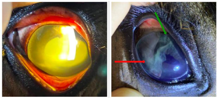
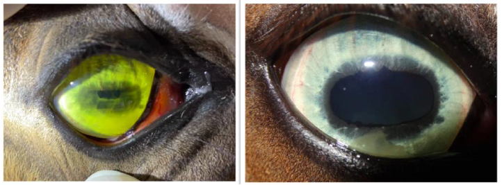
The main ophthalmic findings were conjunctival hyperemia, fibrin and flare in the anterior chamber (100% incidence), iris change of color (77,77% incidence), and low intraocular pressure (table 2; figure 1 and figure 2). All of them are related to bilateral anterior uveitis diagnosis. Rubeosis iridis also brought attention to both eyes of foal 4 (figure 2). By ultrasonography, foal 3 had hyperechogenic sedimentations at vitreous, compatible with posterior uveitis. The ocular surface and other anatomic structures didn’t have any alterations. In table 2, there is more specific information about ophthalmic findings.
Every patient received systemic treatment based on general conditions beyond ocular disease (diarrhea or pneumonia). Replacement and maintenance fluid therapy was instituted in cases according to the needs of each patient. All cases received Flunixin Meglumin as systemic anti-inflammatory therapy. The antibiotic systemic therapy was: Amicacin and Potassic Penicillin (case 1); Ceftriaxone (case 2); Doxiciclin (case 3, 7, 8 and 9); Gentamicin (case 4); Sulfadiazine with trimethoprim (case 8). Foals 5 and 6 did not receive any systemic antibiotics. This variation in antibiotic administration occurred due to different origin properties, veterinarians, field conditions, and previous treatments that those foals were submitted before ophthalmic examination.
Topically, the election treatment for all foals was eye drops of Prednisolone 1%, two drops per eye, with variations of days according to each patient response. The topical treatment was conducted until the eye showed absence of sediments (cells or protein) in the anterior chamber and recovery of transparency.
The outcome of ophthalmic complications was positive in cases 1, 3, 4, 7, 8 and 9 (66,66% of success). After a mean of 8,5 days on treatment, these foals showed absence of fibrin in anterior chamber and recovery of transparency, in addition to remaining visuals. Three foals that could be evaluated after recovery also had normal intraocular pressure. Ocular evolution data could not be collected for foals 2, 5, and 6, as they died soon after hospitalization.
Clinical and laboratory data from foals with uveitis due to sepsis at Veterinary Hospital of Federal University of Pelotas (HCV-UFPel)
Left image: Foal’s number 2 left eye on slit lamp exam showing flare and iris color change; Right image: Foal’s number 3 right eye with free fibrin on red arrow, and iris edema on green arrow.
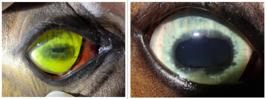
Left image: Foal’s number 4 right eye at the hospitalization day, showing flare, iris edema and iris color change (opaque yellow appearance). Right image: Same foal and eye after 3 days of topic and systemic treatment, still noted free fibrina and rubeosis iridis, and return to its blue iris.
DISCUSSION
This report describes cases of septic foals that developed bilateral anterior uveitis. It was not possible to perform blood culture due to technical limitations; however clinical and laboratorial data indicate sepsis in all foals according to Wong et al. (2018) and Sheats (2019) criterion (table 1). Although Rotavirus was found in two patients, it is known that sepsis can have opportunist bacterial infections or co-infections (Sheats, 2019).
Based on ultrasonographic findings, posterior uveitis was also suspected in Case 3. While sepsis is a recognized trigger for equine uveitis (Gilger et al., 2022), detailed descriptions of ophthalmic lesions in septic foals are limited in the literature.
All foals exhibited conjunctival hyperemia, a common but non-specific sign of uveitis. Other ocular inflammatory conditions can also affect the conjunctiva. Anterior chamber flare and free fibrin were the most consistent signal (100% incidence), indicating blood-aqueous barrier disruption. This finding is common in foals due to their high fibrin production (Repiso et al., 2010). Fibrin clearance averaged 8.5 days and occurred gradually during hospitalization.
Iris color changed in all cases. In Foal 4 (blue iris - figure 2), iridocyclitis-induced edema resulted in an opaque yellow appearance. In foals with brown irises, a dark green discoloration was observed. This color change likely reflects polymorphonuclear cell infiltration of the uvea in response to inflammation (Tarancón et al., 2019).
Low intraocular pressure (IOP) was another consistent finding. Although established normal foal IOP values are lacking for foals, the observed pressures were below the baseline for healthy adult horses (15 mmHg), suggesting uveitis. This decrease may be attributed to prostaglandin accumulation in the ciliary body and increased aqueous humor drainage via the uveoscleral pathway (Gilger et al., 2022). IOP tended to normalize following treatment.
Topical 1% prednisolone resulted in favorable uveitis outcomes in 66,66% of cases. This response rate may have been higher if more foals had survived sepsis. Prednisolone is often the preferred topical steroid due to its superior intraocular penetration and activity compared to dexamethasone (Matthews, 1999). While keratomycosis has been reported as a concern in other countries (Galera and Brooks, 2012), the authors' experience in Brazil suggests that topical steroid use in non-ulcerative cases does not typically induce fungal or bacterial ocular surface infections. This result shows that a proper and early topical treatment of uveitis in foals can lead to good visual prognosis.
Obviously, the systemic disease recovery of the survived foals corroborated the improvement of ocular condition. Although the systemic medications were targeting the general aspect of the foal, certainly the flunixin meglumin had a high contribution to ocular improvement. Flunixin and firocoxib both have a significant penetration into the aqueous barrier due to high solubility and bonding with plasma albumin (Hilton et al., 2011). However, the first appears to have a greater anti-inflammatory effect in uveal diseases (Gilger and Michau, 2004). About the systemic antibiotics, each one had a variation of intraocular penetration but it is presumed that the general treatment of sepsis stops new bacteremia and immunocomplex formation, collaborating indirectly to the ocular improvement.
CONCLUSION
Even septic foals with anterior or posterior uveitis could recover visual pathway. A complete and careful ophthalmic evaluation is fundamental and necessary in septic neonatal foals to reach a correct and timely diagnosis. It also guides a good therapeutic choice, both systemic and ocular, ensuring a better prognosis for visual recovery of the foal.
REFERENCES
- BARRATT-BOYES, S.M.; YOUNG, R.L.; CANTON, D.D.; MOHR, F.C. Streptococcus equi infection as a cause of panophthalmitis in a horse. J. Equine Vet. Sci., v. 11, p.229-231, 1991.
- GALERA, P.D.; BROOKS, D.E. Optimal management of equine keratomycosis. Vet. Med. Res. Rep., v.12, p.7-17, 2012.
- GILGER, B.C.; MICHAU, T.M. Equine recurrent uveitis: new methods of management. Vet. Clin. Equine Pract., v.20, p.417-427, 2004.
- GILGER, B.C.; DEGROOTE, R.; DEEG, C. Diseases of the uvea, uveitis, and recurrent uveitis. GILGER, B.C. (Ed.). Equine ophthalmology. [New Jersey]: Wiley, 2022. chap. 6, p.441-498.
- HILTON, H.G.; MAGDESIAN, K.G.; GROTH, A.D. et al. Distribution of flunixin meglumine and firocoxib into aqueous humor of horses. J. Vet. Intern. Med., v.25, p.1127-1133, 2011.
- LEIVA, M.; PEÑA, T. Ophthalmic diseases of foals. GILGER, B.C. (Ed.). Equine ophthalmology. [New Jersey]: Wiley, 2016. p.112-150.
- MATTHEWS, A. Equine recurrent uveitis‐an update. In Pract., v.21, p.370-376, 1999.
- PALMER, J. Update on the management of neonatal sepsis in horses. Vet. Clin. Equine Pract., v.30, p.317-336, 2014.
- REPISO, M.L.; BOSCH, L.M.; RUIZ, L.A.; et al. Uveal inflammation in septic newborn foals. J. Vet. Intern. Med., v.24, p.391-397, 2010.
- REUSS, S.M.; CHAFFIN, M.K.; COHEN, N.D. Extrapulmonary disorders associated with Rhodococcus equi infection in foals: 150 cases (1987-2007). J. Am. Vet. Medical Assoc., v.235, p.855-863, 2009.
- SHEATS, M.K. A comparative review of equine SIRS, sepsis, and neutrophils. Front. Vet. Sci., v.6, p.69, 2019.
- TARANCÓN, I.; LEIVA, M.; JOSE-CUNILLERAS, E. et al. Ophthalmologic findings associated with Rhodococcus equi bronchopneumonia in foals. Vet. Ophthalmol., v.22, p.660-665, 2019.
- THEELEN, M.J.; WILSON, W.D.; BYRNE, B.A. et al. Initial antimicrobial treatment of foals with sepsis: Do our choices make a difference? Vet. J., v.243, p.74-76, 2019.
- WONG, D.M.; RUBY, R.E.; DEMBEK, K.A. et al. Evaluation of updated sepsis scoring systems and systemic inflammatory response syndrome criteria and their association with sepsis in equine neonates. J. Vet. Intern. Med., v.32, p.1185-1193, 2018.
The research data are available within the article itself.