 Prodigiosin-conjugated silver nanoparticles target neuroimmune dysregulation and pro-inflammatory cytokines TNF-α, IL-1β/NF-kB pathways in an autistic model of rat pups
ABSTRACT
Autism spectrum disorder (ASD) is associated with neuroimmune dysregulation and elevated pro-inflammatory cytokines. This study explores the therapeutic potential of prodigiosin-conjugated silver nanoparticles (PG-AgNPs) in targeting these pathways in an autistic rat model. Autistic-like behavior was induced in rat pups through prenatal exposure to valproic acid (VPA) (600mg/kg). The treatment with PG-AgNPs (3mg/kg) has been applied for 7 days in rat pups after birth. Induction of autism by VPA decreased the level of 5-HT, DA, NE, GABA, glycine and taurine in cerebellum and cerebral cortex as well as reduction in brain cholesterol and antioxidants enzymes (GSH, SOD and CAT). However, it showed a significant increase in MAO, AChE, glutamate, aspartate, serine and lipid peroxidation. The inflammatory mediators NF-kB, TNF-α and IL-1β were increased and enhancing apoptotic markers (Bax, Bcl2) and reduce anti-apoptotic (Caspase-3) in brain tissue. PG-AgNPs treatment markedly reduced these inflammatory markers, restored neurotransmitter balance, and improved antioxidant defenses. Additionally, PG-AgNPs attenuated oxidative stress and modulated apoptotic markers. PG-AgNPs demonstrated significant neuroprotective, anti-inflammatory, and antioxidant effects in the autistic rat model, suggesting their potential as a nanotherapeutic approach for ASD. Further studies are needed to validate their long-term efficacy.
Keywords:
autism; valproate; PG-AgNPs; catecholamine; cytokines; pathways
RESUMO
O transtorno do espectro autista (TEA) está associado à desregulação neuroimune e ao aumento de citocinas pró-inflamatórias. Este estudo explora o potencial terapêutico de nanopartículas de prata conjugadas com prodigiosina (PG-AgNPs) no direcionamento dessas vias em um modelo de rato autista. Comportamento semelhante ao autismo foi induzido em filhotes de ratos por meio da exposição pré-natal ao ácido valproico (VPA) (600mg/kg). O tratamento com PG-AgNPs (3mg/kg) foi aplicado por sete dias, em filhotes de ratos após o nascimento. A indução do autismo por VPA diminuiu os níveis de 5-HT, DA, NE, GABA, glicina e taurina no cerebelo e no córtex cerebral, bem como a reduziu colesterol cerebral e as enzimas antioxidantes (GSH, SOD e CAT). No entanto, demonstrou um aumento significativo de MAO, AChE, glutamato, aspartato, serina e peroxidação lipídica. Os mediadores inflamatórios NF-kB, TNF-α e IL-1β foram aumentados, potencializando marcadores apoptóticos (Bax, Bcl2) e reduzindo a atividade antiapoptótica (Caspase-3) no tecido cerebral. O tratamento com PG-AgNPs reduziu significativamente esses marcadores inflamatórios, restaurou o equilíbrio neurotransmissor e melhorou as defesas antioxidantes. Além disso, as PG-AgNPs atenuaram o estresse oxidativo e modularam marcadores apoptóticos. As PG-AgNPs demonstraram efeitos neuroprotetores, anti-inflamatórios e antioxidantes significativos no modelo de rato autista, sugerindo seu potencial como uma abordagem nanoterapêutica para TEA. Estudos adicionais são necessários para validar sua eficácia a longo prazo.
Palavras-chave:
autismo; valproato; PG-AgNPs; catecolamina; citocinas; vias
INTRODUCTION
Autism spectrum disorder (ASD) is a neurodevelopmental disorder that is thought to be caused by exposure to a wide range of toxins or a change in a group of genetic factors. These etiological factors can interfere with neurobehavioral development, resulting in autistic symptoms. Autism is spreading at an alarming rate, with one in every 150 children affected worldwide. Autistic disorder, pervasive developmental disorder, and Asperger syndrome are the three types of ASD (Rossignol and Frye, 2012). This behavioral condition may arise from irregularities in various brain regions and neural pathways (Lord et al., 2000). A key characteristic of autism spectrum disorder (ASD) is impaired social reciprocity, alongside challenges in concentration, hyperactivity, communication difficulties, and repetitive behaviors (Gerlai and Gerlai, 2004). Individuals with ASD often encounter issues related to neurological conditions, including seizures (Rossignol and Frye, 2012).
Exposure to environmental factors during the prenatal or postnatal period, such as valproic acid (VPA), mercury, lead, viruses, air pollutants, toxins, thalidomide, and retinoic acid, can induce oxidative stress linked to autism (Nicolini et al., 2015). Valproate is a commonly prescribed anticonvulsant medication used to treat bipolar disorder. Numerous studies indicate that VPA may lead to behavioral issues, learning challenges, and various congenital malformations (Štefánik et al., 2015).
Anticonvulsant medication during pregnancy increased the risk of autism and sodium valproate is the drug most associated with autism. VPA's suppression of histone deacetylase (HDAC) and alterations in gene expression may account for some of the drug's teratogenic effects. Rodents exposed to VPA during pregnancy have been shown to develop behavioral traits similar to those seen in ASD, providing a strong animal model for understanding social cognitive impairment and potentially screening for the development of novel therapies for this condition (Kuwagata et al., 2009). Other possible effects of VPA include an increase in fatal oxidative stress, which disproportionately damages the brain compared to other fatal organs, or suppression of the folic acid system. Following these clinical reports, several researchers reported an animal model of autism by exposing pregnant rats to VPA. These research demonstrated neuron loss in the brainstem nuclei and cerebral cortex (Fenlon et al., 2015), and behavioral alternations consistent with or similar to reports in autistic children.
In recent years, nanotechnology has taken over most of the attention in the field of drug delivery, offering effective solutions to enhance the efficacy and specificity of therapeutic agents. NPs produced by biological techniques are far superior compared to those manufactured with physical and chemical techniques (Cheng et al., 2023). Silver nanoparticles (AgNPs) are absorbed more effectively, and their relatively small sizes allow them to cross membrane barriers and accumulate in tissues, leading to increased reactivity. These attributes make AgNPs an ideal vehicle for delivering bioactive compounds to the central nervous system (Liao et al., 2019).
It was revealed that biological pigments, including those synthesized by bacteria, can be employed for the green synthesis of nanoparticles (Singh et al., 2023). Prodigiosin (Pg), a secondary metabolite produced by Serratia marcescens, has demonstrated remarkable anti-inflammatory, antioxidant, and neuroprotective effects in preclinical studies. Its ability to modulate key signaling pathways, such as NF-κB and PI3K/Akt, suggests its potential to ameliorate neuroinflammation and oxidative stress, which are hallmarks of ASD (Hamada and Mohamed, 2024).
The present study aimed to investigate the possible ameliorative effect of prodigiosin-conjugated AgNP2 on the neurotoxicity in rat pups’ autistic models induced by prenatal exposure to VPA.
ETHICAL ASPECTS
University, and was approved under number (HU2021/Z/RKA0921-01). All animal procedures were conducted in accordance with the National Institutes of Health (NIH) Guidelines for the Care and Use of Laboratory Animals (8th edition).
MATERIAL AND METHODS
Sodium valproate (VPA) was purchased from Sigma Aldrich Co. PVT Ltd, USA and before used it was dissolved in NaCl solution. The treatment of VPA was IP at a single dose of (600mg/kg) (Al-Amin et al., 2015). Prodigiosin-conjugated AgNP2: Bacterial isolation, Preparation, extraction, purification and quantification of prodigiosin in addition to the formation of PG-conjugated AgNP2 and its characterization were performed in the Microbiology Department, Faculty of Science, Helwan University according to Farag et al. (2017) and El-batal et al. (2017).
Six adults male and twelve adult female rats, each weighing between 100-150g, were sourced from VACSERA in Cairo, Egypt. They were acclimatized for one week in a specific pathogen-free (SPF) environment, maintaining a temperature of 25±1°C and humidity at 55%. The rats were housed under controlled conditions at the Laboratory of Physiology, Faculty of Science, Helwan University, Cairo, with a 12-hour light/dark cycle, and had unlimited access to food and water.
To facilitate breeding, females were paired with males (two females per male) overnight. The following morning, females exhibiting sperm in their vaginal smears were considered pregnant, marking this as gestational day 0 (GD0). Three pregnant females received a single intraperitoneal injection of valproic acid (VPA) at a dosage of 600mg/kg on GD12.5. In contrast, the control group received physiological saline at the same time. Each pregnant female was housed individually until delivery.
The newborn rats remained with their mothers for 15 days before being separated by sex. The study continued with only male pups, which were divided into four groups of eight. Group 1 consisted of normal pups that received saline injections and served as the control group. Group 2 included normal pups treated with PG-AgNP2 at a dose of 3mg/kg (El-batal et al., 2017), via intraperitoneal injection. Group 3 comprised autistic model pups that received daily saline injections, while Group 4 included autistic model pups treated with PG-AgNP2 at the same dosage. All treatments began on postnatal day 15 and continued for seven days.
All procedures involving animals adhered to the National Institutes of Health (NIH) Guidelines for the Care and Use of Laboratory Animals, with approval from the Committee on Research Ethics for Laboratory Animal Care at Helwan University (approval number: HU/Z/010-19). At the start of the experimental phase, the rats were weighed and then euthanized via decapitation. Their brains were quickly removed from the skulls, blotted dry, and chilled. The brain tissues were dissected into cortex and cerebellum segments for monoamine and free amino acid assays and stored at -70°C. The remaining half of each brain was also kept at -70°C for further biochemical analysis.
The initial step in the high-performance liquid chromatography (HPLC) procedure for measuring monoamines in brain tissues (cerebellum and cerebral cortex) involved weighing and homogenizing the samples in a 1:10 weight/volume ratio of 75% aqueous HPLC-grade methanol. The homogenate was then centrifuged for 10 minutes at 3000 rpm, and the supernatant was promptly utilized for monoamine analysis after solid-phase extraction using a CHROMABOND NH2 phase column (Cat. No. 730031) to separate it from lipids and trace elements (Pagel et al., 2000).
The identification of free amino acid neurotransmitters such as GABA, Glycine, Taurine, Glutamate, Aspartate, and Serine in the cerebellum and cerebral cortex was performed using HPLC with the precolumn PITC derivatization method as established by Heinrikson and Meredith (Heinrikson and Meredith, 1984).
Acetylcholinesterase (AChE) activity in the brain was assessed using the colorimetric method outlined by Ellman (1961) Additionally, monoamine oxidase (MAO) activity was measured fluorometrically at excitation and emission wavelengths of 550nm and 404 nm, respectively, utilizing 5-hydroxytryptamine (500mM) as a substrate following the protocol by Dar et al. (2005).
The quantification of malondialdehyde (MDA), a biomarker for lipid peroxidation (LPO), was conducted according to the methodology described by Ohkawa et al. (1979). Nitric oxide (NO) levels were determined using the Griess reagent as per Green et al. (1982).
Glutathione (GSH) levels were assessed by reducing Elman's reagent (5,5′-dithiobis(2-nitrobenzoic acid; DTNB) with GSH to form a yellow compound. The absorbance of this reduced chromogen at 405nm is inversely proportional to its GSH content. Catalase (CAT) and superoxide dismutase (SOD) activities were estimated using techniques developed by Aebi (1984) and Nishikimi et al. (1972), respectively.
The amount of cholesterol in brain tissue samples was analyzed using a fluorometric enzymatic method with the Cholesterol Assay kit from Molecular Probe/Invitrogen, based on the approach described by Abulrob et al. (2005).
Nuclear factor kappa-B (NF-kB, P65; code #: CSB-E13148r) was estimated by ELISA kit supplies from CUSABIO life science, Wuhan, China, according to the manufacturer’s instructions. Meanwhile, tumour necrosis factor-α (TNF-α; Cat#; RTB00) and interleukin-1β (IL-1β; Cat#; RLB00) concentrations were measured using ELISA kits (R and D System, Minneapolis, MN, USA) in accordance with the manufacturer’s instructions.
According to the manufacturer’s instructions, a colorimetric caspase-3 assay kit (Sigma-Aldrich Co. USA) was used to examine brain tissue homogenates prepared in lysis buffer. By using ELISA kits, B cell lymphoma 2 (Bcl-2) and Bcl-2 associated X protein (Bax) levels in the tissue homogenate were determined (Life Span Bio-Sciences, Inc., Seattle, WA, USA).
The design of the experiment was purely random. Using the Statistical Package for the Social Sciences, data were presented as means±S.E. for the given number of independently performed experiments (SPSS 17.0 for Windows). The statistical significances within parameters were evaluated by one-way and multiple analysis of variation (ANOVA), where significant differences at p < 0.05.
RESULTS
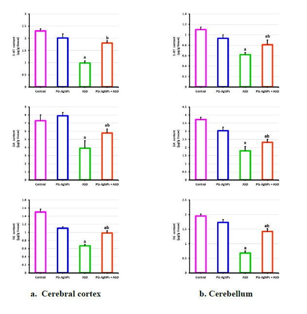
The current study aimed to evaluate the therapeutic effects of PG-AgNPs (3mg/kg) on a rat model of autism. As illustrated in Figure 1, prenatal exposure to a single intraperitoneal injection of valproic acid (600mg/kg on GD12.5) resulted in a significant reduction in serotonin (5-HT), dopamine (DA), and norepinephrine (NE) levels in both the cerebral cortex and cerebellum compared to the control group (p < 0.05). Conversely, postnatal administration of PG-AgNPs to autistic rat pups led to a notable increase in monoamine levels in these brain regions relative to the autistic models.
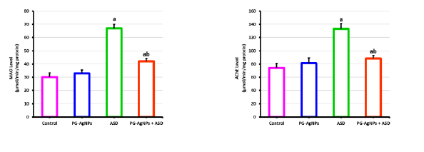
Specifically, dopamine and norepinephrine levels were significantly higher in the cerebral cortex following PG-AgNPs treatment compared to the autistic model. The observed decrease in monoamines within the ASD group indicates a disruption in monoaminergic neurotransmission, which was further evidenced by elevated activities of monoamine oxidase (MAO) and acetylcholinesterase (AChE) compared to controls. Remarkably, these parameters showed significant restoration (p < 0.05) following PG-AgNPs treatment, suggesting a strong neuro-modulatory effect of PG-AgNPs against autism-related neurotoxicity in rats (Fig. 2).
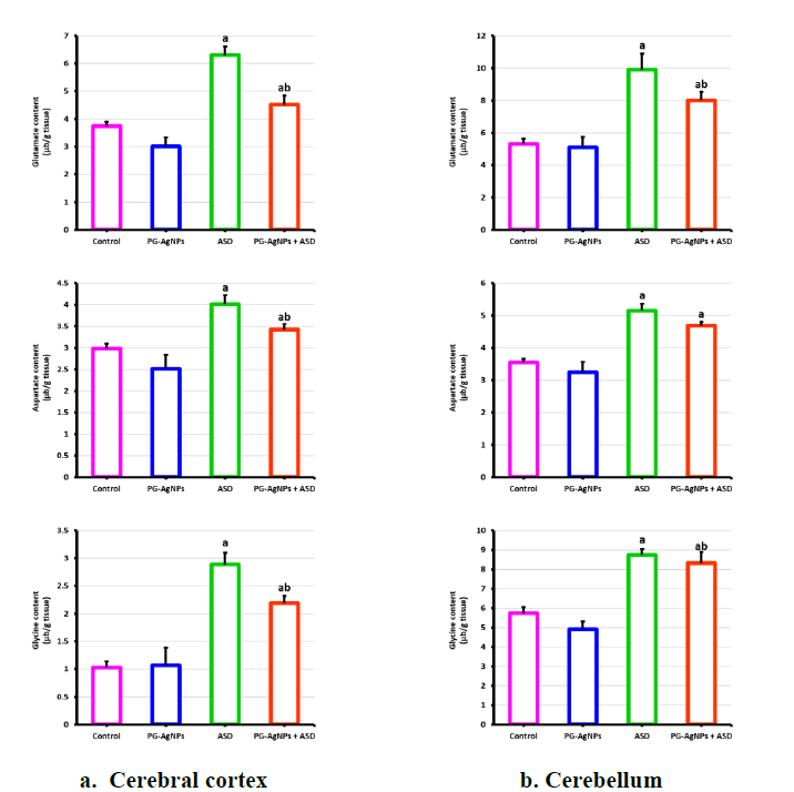
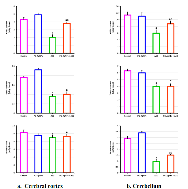
In the ASD rat model, excitatory amino acids such as glutamate, aspartate, and glycine were significantly elevated in both the cerebellum and cerebral cortex compared to controls (p < 0.05). Treatment with PG-AgNPs (3mg/kg) over seven days resulted in a substantial reduction of glutamate, aspartate, and glycine levels in both brain regions when compared to the ASD group (p < 0.05) (Fig. 3). Furthermore, Figure 4 illustrates a significant decrease in inhibitory amino acids (GABA, taurine, and serine) in animals prenatally exposed to valproate compared to controls. In contrast, PG-AgNPs treatment led to a significant increase in GABA concentrations in both examined areas relative to the ASD group.
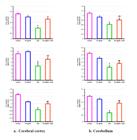
The effect of treatment with PG-AgNP2 (3mg/kg) for 7 days on the content of serotonin (5-HT), Dopamine (DA) and Norepinephrine (NE) in the cerebral cortex and cerebellum of autistic rat models. Data are expressed as means ± SE (8 animals/group). a: Significance change at p < 0.05 in comparison with the control group. b. Significance change at p < 0.05 in comparison with the autism syndrome disease model (ASD) group.
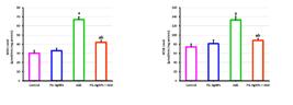
The effect of treatment with PG-AgNP2 (3mg/kg) for 7 days on the level of monoaminoxidase (MAO) and acetylcholinesterase (AChE) in brain tissue of autistic rat models. Data are expressed as means ± SE (8 animals/group). a: Significance change at p < 0.05 in comparison with the control group. b. Significance change at p < 0.05 in comparison with the autism syndrome disease model (ASD) group.
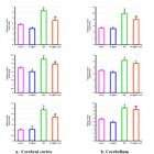
The effect of treatment with PG-AgNP2 (3mg/kg) for 7 days on the content of free excitatory (glutamate, aspartate and glycine) amino acids in the cerebral cortex and cerebellum of autistic rat models. Data are expressed as means ± SE (8 animals/group). a: Significance change at p < 0.05 in comparison with the control group. b. Significance change at p < 0.05 in comparison with the autism syndrome disease model (ASD) group.
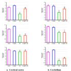
The effect of treatment with PG-AgNP2 (3mg/kg) for 7 days on the content of free inhibitory (GABA, taurine and serine) amino acids in the cerebral cortex and cerebellum of autistic rat models. Data are expressed as means ± SE (8 animals/group). a: Significance change at p < 0.05 in comparison with the control group. b. Significance change at p < 0.05 in comparison with the autism syndrome disease model (ASD) group.
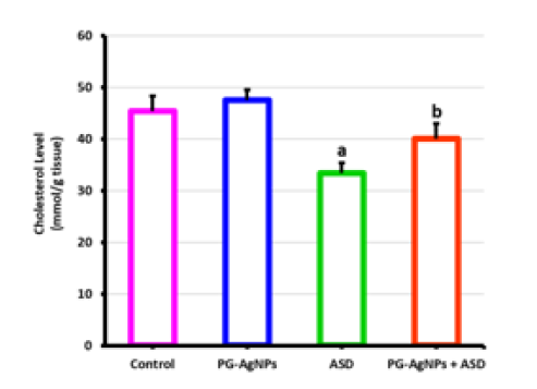
To assess neuronal lipid content within the autistic rat model, cholesterol levels were measured in brain tissue. The results indicated a significant decrease in brain cholesterol levels in the ASD group compared to controls. However, daily intraperitoneal injections of PG-AgNPs (3mg/kg) for seven days resulted in a significant increase in brain cholesterol content compared to the ASD group (p < 0.05) (Fig. 5).
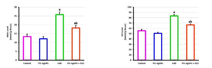
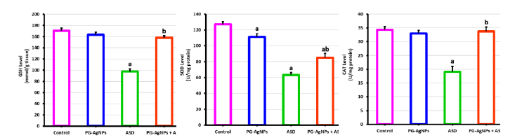
Induction of ASD by VPA induced oxidative stress, as indicated in significant elevation in MDA and NO (Fig. 6). On the other hand, GSH level (Fig. 7) and antioxidant enzymes SOD and CAT activities recorded a significant reduction at p < 0.05 as compared to control group. However, the MDA and NO levels decreased significantly in the autistic model after treatment with PG-AgNPs. Moreover, the treatment with PG-AgNPs markedly elevated the GSH level (p < 0.05) as compared to ASD group. Similarly, a significant increment in SOD and CAT activities was observed in autistic rat pups treated with PG-AgNPs.
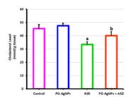
The effect of treatment with PG-AgNP2 (3mg/kg) for 7 days on the cholesterol content in brain tissue of autistic rat models. Data are expressed as means ± SE (8 animals/group). a: Significance change at p < 0.05 in comparison with the control group. b. Significance change at p < 0.05 in comparison with the autism syndrome disease model (ASD) group
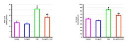
The effect of treatment with PG-AgNP2 (3mg/kg) for 7 days on the level of oxidative stress enzymes (MDA and NO) in brain tissue of autistic rat models. Data are expressed as means ± SE (8 animals/group). a: Significance change at p < 0.05 in comparison with the control group. b. Significance change at p < 0.05 in comparison with the autism syndrome disease model (ASD) group.
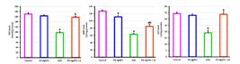
The effect of treatment with PG-AgNP2 (3mg/kg) for 7 days on the level of antioxidant enzymes (GSH, SOD and CAT) in brain tissue of autistic rat models. Data are expressed as means ± SE (8 animals/group). a: Significance change at p < 0.05 in comparison with the control group. b. Significance change at p < 0.05 in comparison with the autism syndrome disease model (ASD) group.
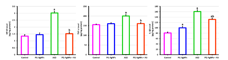
It is clear from the present study that autistic induction resulted in enhancement to inflammatory responses. This enhancement in inflammatory responses was very clear evidence in autism induction in our present study. This was indicated by the marked elevation (p < 0.05) in the brain proinflammatory cytokines, including TNF-α and IL-1β (Fig. 8) as compared to control group. The treatment with PG-AgNPs represents an anti-inflammatory property that was illustrated by a marked decrease in the levels of both studied proinflammatory cytokines (TNF-α and IL-1β). In addition, the induction of autism enhances the release of inflammatory mediators, as shown in significant increase in NF-kB (p < 0.05) in brain tissue as compared to control group. The treatment with PG-AgNPs reduced the activity of NF-kB significantly as compared to ASD.
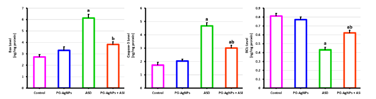
The levels of apoptotic markers Bax and Caspase-3 (Fig. 9), showed a marked increase in ASD group as compared to control group. on the other hand, Bcl2 levels were reduced significantly in autistic group (p < 0.05). Postnatal ip injection with (3mg/kg) PG-AgNPs restored the value of both apoptotic and antiapoptotic markers nearer to the control as compared to ASD group.
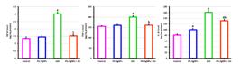
The effect of treatment with PG-AgNP2 (3mg/kg) for 7 days on brain neuroinflammatory cytokines markers of autistic rat models. Data are expressed as means ± SE (8 animals/group). a: Significance change at p < 0.05 in comparison with the control group. b. Significance change at p < 0.05 in comparison with the autism syndrome disease model (ASD) group.
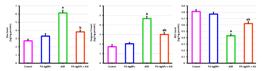
The effect of treatment with PG-AgNP2 (3mg/kg) for 7 days on brain level of apoptosis and antiapoptotic markers of autistic rat models. Data are expressed as means ± SE (8 animals/group). a: Significance change at p < 0.05 in comparison with the control group. b. Significance change at p < 0.05 in comparison with the autism syndrome disease model (ASD) group.
DISCUSSION
Autism Spectrum Disorder (ASD) is a complex neurodevelopmental condition that manifests through a range of behavioral impairments, including deficits in social interaction, communication challenges, and repetitive behaviors (Yin et al., 2024). While the exact causes of ASD remain unclear, a combination of genetic, environmental, and epigenetic factors is believed to contribute to its development. Given the increasing prevalence of ASD, there is a critical need for effective treatment strategies to mitigate its symptoms and improve the quality of life for affected individuals [Foley et al. (2012) and Apte and Kumar (2023)].
The induction of autism spectrum disorder (ASD) can influence the activity of monoamine oxidase (MAO) and acetylcholinesterase (AchE), potentially because of neuroinflammation and oxidative stress. In the brain, oxidative damage and the presence of inflammatory mediators can disrupt the normal functioning of these enzymes (Usui et al., 2023). This disruption impacts monoaminergic and cholinergic signaling pathways, ultimately affecting brain activity and contributing to the progression of autism. Research suggests that the decline in monoamine neurotransmitters observed in ASD animal models may result from the generation of reactive oxygen species (ROS) (Kuo and Liu, 2022). These ROS can inhibit enzymes responsible for monoamine synthesis, interfere with monoamine metabolism by accelerating their breakdown and removal, and block the reuptake of monoamines. Furthermore, the ROS can activate MAO, which increases the levels of hydroxyl radicals in the brain while reducing the concentrations of key neurotransmitters such as serotonin (5-HT), norepinephrine (NE), and dopamine (DA) (Montanari et al., 2022).
In this study, prenatal exposure to a single dose of VPA (600 mg/kg) on embryonic day 12.5 led to a notable reduction in the levels of serotonin (5-HT), dopamine (DA), and norepinephrine (NE) in the cerebellum and cerebral cortex. Previous studies have also documented similar decreases in serotonin levels, particularly in studies examining tryptophan metabolism and serotonin transporter activity in individuals with autism.
Hyperserotonemia is one of the neurochemicals finding in ASD, it is characterized by a 5-70% increase in blood serotonin levels (Esposito et al., 2024). In infants, as their blood-brain barrier is not fully developed, this allows serotonin produced elsewhere in the body to potentially enter the brain. Although it remains challenging to pinpoint the exact timing of peripheral serotonin exclusion in humans, it is generally accepted that the blood-brain barrier is not fully functional until the appearance of tight junction proteins, typically around two years of age in humans and by postnatal day 15 to 20 in rodents (Garbarino et al. (2019) and Liow et al. (2016)). This early window of serotonin entry into the brain could help in elevation in blood serotonin levels and consequently, increased serotonin levels might trigger negative feedback mechanisms, leading to the loss of serotonin terminals in the brain. This reduction in serotonin could contribute to the behavioral and cellular changes often associated with autism (Whitaker-Azmitia, 2005). Such mechanisms may also explain the observed reductions in cerebellar and cortical serotonin levels in the present study. Similarly, the decrease in dopamine levels identified in this study aligns with findings by Gątarek and Kałużna-Czaplińska (2022), who reported elevated levels of dopamine hydroxylase and homovanillic acid in autistic children, highlighting dopamine dysfunction as a potential contributor to autism symptoms. The observed decline in norepinephrine levels is also consistent with reduced dopamine, given that dopamine serves as a precursor for norepinephrine synthesis. Both neurotransmitters play essential roles in maintaining normal brain function, and their disruption may have significant implications for neurodevelopment (Kandel et al., 1995).
Research indicates that GABA is synthesized from glutamate through the action of the enzyme glutamic acid decarboxylase (GAD), which serves as the rate-limiting step in GABA production (Hussman, 2001). Many theories on amino acid neurotransmitters in autism suggest a suppression of the GABAergic system, leading to an overactivation of the glutamate system. This hyperactivity of glutamate may result in excitotoxicity, which can disrupt neuronal development (Bittigau and Ikonomidou, 1997). If this imbalance occurs during critical developmental periods, it could impair neuronal growth and connectivity. Excessive glutamatergic activity has been linked to various developmental abnormalities commonly observed in individuals with autism (Salem et al., 2023).
The application of metal nanoparticles has emerged as a promising advancement in the pharmaceutical field, as these therapeutic agents provide enhanced bioavailability, controlled delivery, and more precise targeting of tissues compared to conventional drug formulations. However, studies have shown that prolonged exposure to high doses of metal nanoparticles can lead to the accumulation of metals within cells. For instance, Patlolla et al. (2015), found that administering low doses of AgNP2 over a 7-day period did not result in significant metal accumulation or cellular toxicity, but instead improved the targeted delivery of drugs to cells. Prodigiosin-conjugated silver nanoparticles (PG-AgNPs) have demonstrated the ability to cross the blood-brain barrier (BBB), which is crucial for their therapeutic application in neurotoxicity. Research indicates that PG-AgNPs can effectively penetrate the BBB, thereby facilitating the delivery of therapeutic agents directly to the brain. For instance, a study highlighted that PG-AgNPs significantly reduced cadmium chloride-induced neurotoxicity in rat models by enhancing antioxidant levels and neurotransmitter concentrations in brain tissue. This suggests that PG-AgNPs not only navigate through the BBB but also exert protective effects against neuronal damage. Furthermore, the ability of silver nanoparticles to interact with brain microvascular endothelial cells has been documented, indicating their potential to influence BBB permeability positively. Overall, these findings support the notion that PG-AgNPs can traverse the BBB and provide neuroprotective benefits, making them a promising candidate for treating neurological disorders.
Interestingly, treatment with PG-AgNP2 restored monoamine levels in the brain tissue of rats, suggesting that PG-AgNP2 has a neuroprotective effect against the disruptions caused by autism induction. Many theories about neurotransmitter imbalances in autism propose that the GABAergic system is suppressed, resulting in an overactive glutamate system. Excessive glutamate activity can lead to excitotoxicity, which may disrupt normal neuronal development (MacFabe et al., 2008). When the glutamate system becomes overactive, it can impair neuronal growth and connectivity, particularly during critical developmental stages. This excessive glutamatergic activity has also been linked to autism spectrum disorder (ASD). These findings are similar with our observations of elevated glutamate and reduced GABA levels in animal models of autism. Treatment with PG-AgNP2 restored monoamine and free amino acid levels in the brain tissue of rats, further supporting the neuroprotective role of PG-AgNP2 against autism-related disturbances.
Reactive oxygen species (ROS) can damage brain biomolecules, contributing to neurodegenerative disorders and conditions like autism spectrum disorder (ASD). Teratogens such as valproic acid (VPA) increase oxidative molecules, including lipid peroxides, hydrogen peroxide, and hydroxyl radicals, while reducing antioxidant enzymes like superoxide dismutase (SOD) and glutathione (GSH) (Kaewngam et al., 2024). Studies link this oxidative imbalance to neuronal damage and behavioral abnormalities in ASD. In an autistic rat model, significant oxidative stress was observed, marked by elevated lipid peroxidation and decreased endogenous antioxidants (Liu et al., 2022). However, treatment with PG-AgNPs significantly restored redox balance by enhancing antioxidant protein levels and reducing ROS, malondialdehyde (MDA), and nitric oxide (NO) levels, demonstrating their neuroprotective and antioxidant properties (Chang et al., 2011).
Additionally, VPA exposure led to reduced brain cholesterol levels, likely due to elevated 7-dehydrocholesterol (7-DHC) levels and impaired 7-dehydrocholesterol reductase (DHCR7) activity (Lapenda et al., 2020). This enzyme is critical for cholesterol biosynthesis, requiring NADPH as a cofactor. Oxidative stress disrupts DHCR7 and other enzymes, reducing cholesterol production and impairing intracellular transport (Vallés and Barrantes, 2021). PG-AgNPs counteracted these effects by scavenging free radicals, restoring enzyme activity, and increasing high-density lipoprotein (HDL) cholesterol levels (Barua and Buragohain, 2024). These findings highlight the therapeutic potential of PG-AgNPs in mitigating oxidative damage and supporting brain function in ASD (Noori et al., 2024).
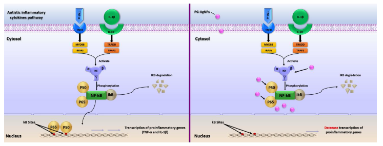
The present study reveals that prenatal exposure to valproic acid (VPA) significantly alters inflammatory cytokines, including NF-kB, TNF-α, and IL-1β, contributing to the development of autism spectrum disorder (ASD). Elevated levels of pro-inflammatory markers such as IL-6, TNF-α, and IL-1β suggest a neuroinflammatory response in ASD (Liu et al., 2017). NF-kB, a key transcription factor, regulates immune functions and inflammation by activating pro-inflammatory genes. It comprises proteins like p65 (RelA) and p105/p50 and remains inactive in the cytosol by interacting with IkB proteins (Kaltschmidt et al., 2022). Upon phosphorylation by IkB kinase (IKK), NF-kB translocates to the nucleus, binding DNA to promote inflammation-related gene transcription. TNF-α and IL-1β further activate NF-kB, amplifying inflammatory pathways. Chronic NF-kB activation, linked to the expression of cytokines, chemokines, and adhesion molecules, is implicated in neurodegenerative diseases. These findings highlight NF-kB’s central role in inflammation and its potential therapeutic targeting (Islan et al., 2022).
Prodigiosin demonstrates potent antioxidant and anti-inflammatory properties across various diseases. It effectively reduces stomach damage caused by HCl/ethanol by suppressing the activity of NF-kB and iNOS while enhancing the expression of HO-1 (Raish et al., 2021). Prodigiosin therapy enhanced the hippocampal antioxidant capacity and reduced inflammatory cytokine production in an aluminum chloride (AlCl3)-induced Alzheimer’s disease model by modulating the Nrf2/NF-kB pathway (Alsharif et al., 2023). It also promoted anti-inflammatory cytokine production while inhibiting pro-inflammatory cytokines. Mechanistically, prodigiosin lowered the RANKL/OPG ratio and downregulated the expression of RANKL, NF-kB p65, MMP13, and caspase-3 (Chen et al., 2023). In the present study, treatment with PG-AgNPs significantly inhibited NF-kB and the inflammatory cytokines TNF-α and IL-1β (Fig. 10).
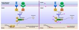
The effect of treatment with PG-AgNP2 on autistic inflammatory cytokines pathway: regulation of NF-kB signaling and transcription of proinflammatory genes (TNF-α and IL-1β). Nuclear factor-kappa B (NF-kB), Tumor Necrosis Factor alpha (TNF-α), Interleukin-1 beta (IL-1β), Tumor Necrosis Factor alpha Receptor (TNFR), Interleukin-1 Receptor (IL-1R), Myeloid differentiation primary response 88 (MYD88), Interleukin-1 receptor associated kinase (IRAKs), Tumor necrosis factor receptor type 1-associated DEATH domain protein (TRADD), Tumor necrosis factor receptor-associated factor (TRAF2), inhibitor of nuclear factor kappa-B kinase (IKK).
Autism induces apoptosis in a range of cells via disrupting the functions of protein kinase C, mitogen-activated protein kinase, and phospholipase C, as well as by blocking calcium-dependent ATPase or triggering the inositol triphosphate pathway (Malek et al., 2022). The new results indicate that while apoptosis-suppressing Bcl-2 reduced in brain tissue, apoptosis-causing genes (caspase-3 and Bax) increased at p < 0.05. The way autism increases Ca2+ entry into the mitochondria, which interferes with the mitochondria's normal metabolism and causes neuronal cell death and growth arrest, may help to explain our findings (Rossignol and Frye, 2012). There was decreased apoptosis in the brain tissue of rats given PG-AgNP2 in the present study. However, as evidenced by a decrease in the production of pro-apoptotic proteins (caspase-3 and Bax) and a rise in the expression of the anti-apoptotic protein Bcl-2, PG treatment reduced the loss of neuronal cells. Albrakati et al. (2021) found that PG kept depressed mice from dying, and our findings are in line with their findings. By suppressing Bax and caspase-3 and increasing Bcl-2, PG stopped the apoptotic cascade linked to stomach lesions brought on by injections of acidified ethanol (Qin et al., 2019).
CONCLUSION
In conclusion, it can be asserted that PG-AgNPs effectively mitigated the symptoms associated with valproic acid-induced autism. The beneficial effects observed with PG-AgNPs are likely due to their antioxidant and anti-inflammatory properties, suggesting their potential utility in the management of autism.
ACKNOWLEDGMENTS
This study was supported by Princess Nourah bint Abdulrahman University Researchers Supporting Project number (PNURSP2025R39), Princess Nourah bint Abdulrahman University, Riyadh, Saudi Arabia.
REFERENCES
- ABULROB, A.; SPRONG, H.; HENEGOUWEN, P.; STANIMIROVIC, D. The blood-brain barrier transmigrating single domain antibody: mechanisms of transport and antigenic epitopes in human brain endothelial cells. J. Neurochem., v.95, p.1201-1214, 2005.
- AEBI, H. Catalase in vitro. Methods Enzymol, v.105, p.121-126, 1984.
- AL-AMIN, M.M.; RAHMAN, M.M.; KHAN, F.R. et al Astaxanthin improves behavioral disorder and oxidative stress in prenatal valproic acid-induced mice model of autism. Behav. Brain Res., v.1, p.112-121, 2015.
- ALBRAKATI, A,; ALSHARIF, K.F.; AL OMAIRI, N.E. et al Neuroprotective efficiency of prodigiosins conjugated with selenium nanoparticles in rats exposed to chronic unpredictable mild stress is mediated through antioxidative, anti-inflammatory, anti-apoptotic, and neuromodulatory activities. Int. J. Nanomed, v.30 p.8447-8464, 2021.
- ALSHARIF, K.F.; ALBRAKATI, A.; AL OMAIRI, N.E. et al Neuroprotective efficacy of the bacterial metabolite, prodigiosin, against aluminium chloride-induced neurochemical alternations associated with Alzheimer's disease murine model: Involvement of Nrf2/HO-1/NF-κB signaling. Environ. Toxicol., v.38, p.266-277, 2023.
- APTE, M.; KUMAR, A. Correlation of mutated gene and signalling pathways in ASD. IBRO Neurosci. Rep, v.27, p.384-392, 2023.
- BARUA, N.; BURAGOHAIN, A. K. Therapeutic potential of silver nanoparticles (AgNPs) as an antimycobacterial agent: a comprehensive review. Antibiotics, v.20, p.1106, 2024.
- BITTIGAU, P.; IKONOMIDOU, C. Glutamate in neurologic diseases. J. Child Neurol., v.12, p.471-485, 1997.
- CHANG, C.C.; WANG, Y.H.; CHERN, C.M. et al Prodigiosin inhibits gp91(phox) and iNOS expression to protect mice against the oxidative/nitrosative brain injury induced by hypoxia-ischemia. Toxicol. Appl. Pharmacol., v.15, p.137-47, 2011
- CHEN, P.; WU, H.; BIAN, T. et al Prodigiosin improves acute lung injury in a rat model of rheumatoid arthritis via down-regulating the nuclear factor kappaB/nucleotide-binding domain, leucine-rich-containing family, pyrin domain-containing-3 signaling pathway. J. Physiol. Pharmacol., v.74, p.1, 2023.
- CHENG, X.; XIE, Q.; SUN, Y. Advances in nanomaterial-based targeted drug delivery systems. Front. Bioeng. Biotechnol., v.13, p.1177-1151, 2023.
- DAR, A.; KHAN, K.M.; ATEEQ, H.S. et al Inhibition of monoamine oxidase-A activity in rat brain by synthetic hydrazines: structure-activity relationship (SAR). J. Enzym. Inhib. Med. Chem., v.20, p.269-274. 2005.
- EL-BATAL, A.I.; EL-HENDAWY, H.H.; FARAAG, A.H. In silico and in vitro cytotoxic effect of prodigiosin-conjugated silver nanoparticles on liver cancer cells (HepG2). J. Biotech. Compu. Biol. Bionanotech., v.98, p.225-243, 2017.
- ELLMAN, G.L. Tissue sulfhydryl groups. Arch. Biochem. Bioph, v.82, p.70-77, 1961.
- ESPOSITO, D.; CRUCIANI, G.; ZACCARO, L. et al Systematic review on autism and hyperserotonemia: state-of-the-art, limitations, and future directions. Brain Sci., v.10, p.481, 2024.
- FARAG, A.H.; EL-BATAL, A.I.; EL-HENDAWY, H.H. Characterization of prodigiosin produced by Serratia marcescens strain isolated from irrigation water in Egypt. Nat. Sci., v.15 p.55-68, 2017.
- FENLON, L.R.; LIU, S.; GOBIUS, I. et al Formation of functional areas in the cerebral cortex is disrupted in a mouse model of autism spectrum disorder. Neural. Dev., v.3, p.10, 2015.
- FOLEY, A.G.; GANNON, S.; ROMBACH-MULLAN, N. et al Class 1 histone deacetylase inhibition ameliorates social cognition and cell adhesion molecule plasticity deficits in a rodent model of autism spectrum disorder. Neuropharmacology, v.63, p.750-60, 2012.
- GARBARINO, V.R.; GILMAN, T.L.; DAWS, L.C.; GOULD, G.G. Extreme enhancement or depletion of serotonin transporter function and serotonin availability in autism spectrum disorder. Pharmacol. Res, v.40, p.85-99, 2019.
- GĄTAREK, P.; KAŁUŻNA-CZAPLIŃSKA, J. Effect of Supplementation on levels of homovanillic and vanillylmandelic acids in children with autism spectrum disorders. Metabolites, v.12, p.423, 2022.
- GERLAI, R.; GERLAI, J. Autism: a target of pharmacotherapies? Drug Discov. Today, v.15, p.366-374, 2004.
- GREEN, L.C.; WAGNER, D.A.; GLOGOWSKI, J. et al Analysis of nitrate, nitrite, and [15N]nitrate in biological fluids. Anal. Biochem., v.126, p.131-138, 1982.
- HAMADA, M.A.; MOHAMED, E.T. Characterization of Serratia marcescens (OK482790)' prodigiosin along with in vitro and in silico validation for its medicinal bioactivities. BMC Microbiol., v.25, p.495, 2024.
- HEINRIKSON, R.L.; MEREDITH, S.C. Amino acid analysis by reverse-phase high-performance liquid chromatography: precolumn derivatization with phenylisothiocyanate. Anal. Biochem., v.136, p.65-74, 1984.
- HUSSMAN, J.P. Suppressed GABAergic inhibition as a common factor in suspected etiologies of autism. J. Autism Dev. Dis., v.31, p.247-248, 2001.
- ISLAN, G.A.; RODENAK-KLADNIEW, B.; NOACCO, N.; DURAN, N.; CASTRO, G.R. A promising biomolecule with many potential biomedical applications. Bioengineered, v.13, p.14227-14258, 2022.
- KAEWNGAM, S.; PRAJIT, R.; ANOSRI, T.; SUWANNAKOT, K. The effects of hesperidin on valproic acid-induced reduction in hippocampal neurogenesis through the antioxidant and apoptotic pathways in adult rats. Sci Rep, v.14, p.28864, 2024.
- KALTSCHMIDT, B.; HELWEG, L.P.; GREINER, J.F.; KALTSCHMIDT, C. NF-κB in neurodegenerative diseases: Recent evidence from human genetics. Front. Mol. Neurosci, v.2, p.15, 2022.
- KANDEL, E.R.; SCHWARTZ, J.H.; JESSELL, T.M. Essentials of neural science and behavior Norwalk: Appleton & Lange, 1995. p.296-297.
- KUO, H.Y.; LIU, F.C. Pathophysiological studies of monoaminergic neurotransmission systems in valproic acid-induced model of autism spectrum disorder. Biomedicines, v.10, p.560, 2022.
- KUWAGATA, M.; OGAWA, T.; SHIODA, S. et al Observation of fetal brain in a rat valproate-induced autism model: a developmental neurotoxicity study. Int. J. Dev. Neurosci., v.27, p.399-405, 2009.
- LAPENDA, J.C.L.; ALVES, V.P.; ADAM, M.L. et al Cytotoxic effect of prodigiosin, natural red pigment, isolated from Serratia marcescens UFPEDA 398. Indian J. Microbiol., v.60, p.182-195, 2020.
- LIAO, C.; LI, Y.; TJONG, S.C. Bactericidal and cytotoxic properties of silver nanoparticles. Int. J. Mol. Sci., v.20, p.449, 2019.
- LIOW, J.S.; ZOGHBI, S.S.; HU, S. et al (18)F-FCWAY, a serotonin 1A receptor radioligand, is a substrate for efflux transport at the human blood-brain barrier. Neuroimage, v.138, p.134-140, 2016.
- LIU, T.; ZHANG, L.; JOO, D. et al NF-κB signaling in inflammation. Signal Transduct. Target. Ther., v.2, p.17023, 2017.
- LIU, X.; LIN, J.; ZHANG, H. et al Oxidative stress in autism spectrum disorder-current progress of mechanisms and biomarkers. Front. Psychiatry, v.13, p.813304, 2022.
- LORD, C.; RISI, S.; LAMBRECHT, L. et al The autism diagnostic observation schedule-generic: a standard measure of social and communication deficits associated with the spectrum of autism. J. Autism Dev. Disord., v.30, p.205-23, 2000.
- MACFABE, D.F.; RODRÍGUEZ-CAPOTE, K.; HOFFMAN, J.E. et al A novel rodent model of autism: intraventricular infusions of propionic acid increase locomotor activity and induce neuroinflammation and oxidative stress in discrete regions of adult rat brain. Am. J. Biochem. Biotech., v.4, p.146-166, 2008.
- MALEK, M.; WAWRZYNIAK, A.M.; EBNER, M. et al Inositol triphosphate-triggered calcium release from the endoplasmic reticulum induces lysosome biogenesis via TFEB/TFE3. J. Biol. Chem., v.298, p.101740, 2022.
- MONTANARI, M.; MARTELLA, G.; BONSI, P. et al Autism spectrum disorder: focus on glutamatergic neurotransmission. Int. J. Mol. Sci., v.23, p.3861, 2022.
- NICOLINI, C.; AHN, Y.; MICHALSKI, B. et al Decreased mTOR signaling pathway in human idiopathic autism and in rats exposed to valproic acid. Acta Neuropathol. Commun., v.3, p.3, 2015.
- NISHIKIMI, M.; APPAJI, N.; YAGI, K. et al The occurrence of superoxide anion in the reaction of reduced phenazine methosulfate and molecular oxygen. Biochem. Biophys. Res. Commun., v.46, p.849-854, 1972.
- NOORI, A.S.; RAJABI, P.; SARGOLZAEI, J. et al Correlation of biochemical markers and inflammatory cytokines in autism spectrum disorder (ASD). BMC Pediatr., v.24, p.696, 2024.
- OHKAWA, H.; OHISHI, N.; YAGI, K. et al Assay for lipid peroxides in animal tissues by thiobarbituric acid reaction. Anal. Biochem., v.95, p.351-358, 1979.
- PAGEL, P.; BLOME, J.; WOLF, H.U. et al High-performance liquid chromatographic separation and measurement of various biogenic compounds possibly involved in the pathomechanism of Parkinson's disease. J. Chromatogr. B Biomed. Sci. Appl., v.746, p.297-304, 2000.
- PATLOLLA, A.K.; HACKETT, D.; TCHOUNWOU, P.B. et al Silver nanoparticle-induced oxidative stress-dependent toxicity in Sprague-Dawley rats. Mol. Cell. Biochem., v.399, p.257-268, 2015.
- QIN, S.; YIN, J.; HUANG, S. et al Astragaloside 4 protects ethanol-induced gastric mucosal injury by preventing mitochondrial oxidative stress and the activation of mitochondrial pathway apoptosis in rats. Front. Pharmacol., v.10, p.894, 2019.
- RAISH, M.; SHAHID, M.; BIN JARDAN, Y.A. et al Gastroprotective effect of sinapic acid on ethanol-induced gastric ulcers in rats: involvement of Nrf2/HO-1 and NF-κB signaling and antiapoptotic role. Front. Pharmacol., v.12, p.622815, 2021.
- ROSSIGNOL, D.A.; FRYE, R.E. Mitochondrial dysfunction in autism spectrum disorders: a systematic review and meta-analysis. Mol. Psychiatry, v.17, p.290-314, 2012.
- SALEM, F.E.H.; ABDEL-GABER, R.; AL-MEGRIN, W.A. et al Therapeutic potency ofCamellia sinensisextract on neurochemical and oxidative changes correlates to autistic disorder in rat pups model. Indian J. Anim. Res., v.57, p.1652-1661, 2023.
- SINGH, H.; DESIMONE, M.F.; PANDYA, S. et al Revisiting the green synthesis of nanoparticles: uncovering influences of plant extracts as reducing agents for enhanced synthesis efficiency and its biomedical applications. Int. J. Nanomed., v.18, p.4727-4750, 2023.
- ŠTEFÁNIK, P.; OLEXOVÁ, L.; KRŠKOVÁ, L. et al Increased sociability and gene expression of oxytocin and its receptor in the brains of rats affected prenatally by valproic acid. Pharmacol. Biochem. Behav., v.131, p.42-50, 2015.
- USUI, N.; KOBAYASHI, H.; SHIMADA, S. et al Neuroinflammation and oxidative stress in the pathogenesis of autism spectrum disorder. Int. J. Mol. Sci., v.24, p.5487, 2023.
- VALLÉS, A.S.; BARRANTES, F.J. Dysregulation of neuronal nicotinic acetylcholine receptor-cholesterol crosstalk in autism spectrum disorder. Front. Mol. Neurosci., v.14, p.744597, 2021.
- WHITAKER-AZMITIA, P.M. Behavioral and cellular consequences of increasing serotonergic activity during brain development: a role in autism? Int. J. Dev. Neurosci., v.23, p.75-83, 2005.
- YIN, W.; PULAKKA, A.; REICHENBERG, A. et al Association between parental psychiatric disorders and risk of offspring autism spectrum disorder: a Swedish and Finnish population-based cohort study. Lancet Reg. Health Eur., v.40, p.100902, 2024.